Abstract
Infectious bronchitis virus (IBV) causes an infectious respiratory disease of domestic fowl that affects poultry of all ages causing economic problems for the poultry industry worldwide. Although IBV is controlled using live attenuated and inactivated vaccines it continues to be a major problem due to the existence of many serotypes, determined by the surface spike protein resulting in poor cross-protection, and loss of immunogenicity associated with vaccine production. Live attenuated IBV vaccines are produced by the repeated passage in embryonated eggs resulting in spontaneous mutations. As a consequence attenuated viruses have only a few mutations responsible for the loss of virulence, which will differ between vaccines affecting virulence and/or immunogenicity and can revert to virulence. A new generation of vaccines is called for and one means of controlling IBV involves the development of new and safer vaccines by precisely modifying the IBV genome using reverse genetics for the production of rationally attenuated IBVs in order to obtain an optimum balance between loss of virulence and capacity to induce immunity.
Keywords: avian, coronavirus, homologous recombination, infectious bronchitis virus, infectious clone, poultry, reverse genetics, spike glycoprotein, vaccine, vaccinia virus
Modification of IBV Genome
Infectious bronchitis (IB) is an acute highly contagious respiratory disease of poultry that is prevalent throughout the world causing animal welfare issues and severe economic losses to the poultry industry worldwide.1-4 The etiological agent of IB is an avian coronavirus, infectious bronchitis virus (IBV), which belongs to the Gammacoronavirus genus, subfamily Coronavirinae, family Coronaviridae, order Nidovirales. IBV replicates primarily in epithelial cells of the respiratory tract causing IB characterized by nasal discharge, snicking, tracheal ciliostasis and rales in chickens,5 but it is also able to replicate in the epithelial cells of other organs such as the enteric tract, oviducts and kidneys.1-3,6 The main effects of an IBV infection are poor weight gain, renal disease, decreased egg production and poor egg quality resulting in major economic losses to poultry industries worldwide. The IBV genome consists of a 28 kb single-stranded RNA molecule of positive-sense polarity. The virion contains the four structural proteins; spike glycoprotein (S), small membrane protein (E), integral membrane protein (M) and nucleocapsid protein (N) which interacts with the genomic RNA. The S glycoprotein is a type I glycoprotein composed of three homopolymers that is responsible for binding to the target cell receptor and fusion of the viral and cellular membranes. The IBV S glycoprotein (1,162 amino acids) is cleaved into two subunits, S1 (535 amino acids, 90 kDa) comprising the N-terminal subunit of the S protein and S2 (627 amino acids, 84 kDa) comprising the C-terminal subunit. The S1 subunit incorporates the receptor-binding activity of the S protein and is responsible for inducing neutralizing and sero-specific antibodies.7,8 The ectodomain region of the S2 subunit contains a fusion peptide-like region9 and two heptad repeat regions involved in oligomerisation of the S protein10 and is required for entry into susceptible cells.11-13 The S2 subunit associates non-covalently with the S1 subunit and in addition to the ectodomain contains the transmembrane and C-terminal cytoplasmic tail domains.
IBV is currently controlled by the use of both live attenuated and inactivated boost vaccines. Neutralising antibodies, induced by the IBV S1 subunit, present in the respiratory tract are responsible for protecting against subsequent IBV infection and concomitant IB disease. However, amino acid variations in the S1 subunit have resulted in many different IBV serotypes requiring different vaccines due to lack of cross-protection. Commercial live attenuated vaccines are produced by multiple passages of virulent field isolates in embryonated domestic fowl eggs as a result of spontaneous mutations that cause attenuation of the virus. As a consequence, viruses that are attenuated by this approach have only a few mutations responsible for loss of virulence, and due to the nature of the procedure the attenuated viruses have different mutations that may affect their virulence and/or immunogenicity. Such a process requires a fine balance between loss of pathogenicity and retention of immunogenicity. Which mutations result in attenuation of pathogenicity is not known. A major drawback of this method is that once the virus is used to inoculate chickens the mutations that resulted in the attenuation of the vaccine virus may back-mutate resulting in a virulent virus; an undesirable consequence.1,14,15
Although the use of both live and attenuated IBV vaccines have played an important role in the successful expansion of the poultry industry, the existence and continual introduction of new IBV serotypes requires alternative strategies in order to circumvent the problem of poor cross-protection and for the production of safer vaccines. A new generation of IB vaccines is called for. One means of controlling IBV involves the development of new and safer vaccines by precisely modifying the IBV genome for the production of rationally attenuated IBVs to obtain an optimum balance between loss of virulence and capacity to induce immunity. Such vaccines would ideally be: (1) genetically stable, have a defined and uniform stable attenuated phenotype that is unable to back mutate to virulence; (2) have the potential for uniform immunogenicity, the loss of virulence should not affect immunogenicity; (3) flexibility, the modified genome can be manipulated to express different S genes or S1 subunits, allowing the same genetically defined vaccine to protect against differing serotypes and (4) allow administration in ovo. IBV vaccines generated by passage in embryonated eggs are highly virulent for embryos so in ovo application cannot be used. The IBV genome can be precisely modified through the use of a suitable reverse genetics system. We have developed such a reverse genetics system or “infectious clone” for the manipulation of the IBV genome and have modified an IBV genome by exchanging the S glycoprotein gene. The S genes were derived from virulent IBV strains and introduced into the genome of an attenuated IBV. The resultant recombinant IBVs were still avirulent but were able to act as vaccines for the protection of chickens against challenge with the parental virulent viruses from which the S genes were derived. These results have demonstrated that through the use of a reverse genetics system swapping the IBV S protein is a precise and effective way of generating genetically defined candidate IBV vaccines.
In our recent paper in PLoS ONE,16 we have shown that replacement of an IBV S glycoprotein from a pathogenic field isolate, IBV 4/91(UK), belonging to a different serotype as the receiver IBV (Beaudette) did not confer pathogenicity but did induce homologous protection. This confirmed our previous work in the Journal of Virology in which the S glycoprotein we used was derived from IBV M41 that belongs to the same serotype as the receiver virus IBV Beaudette.17,18
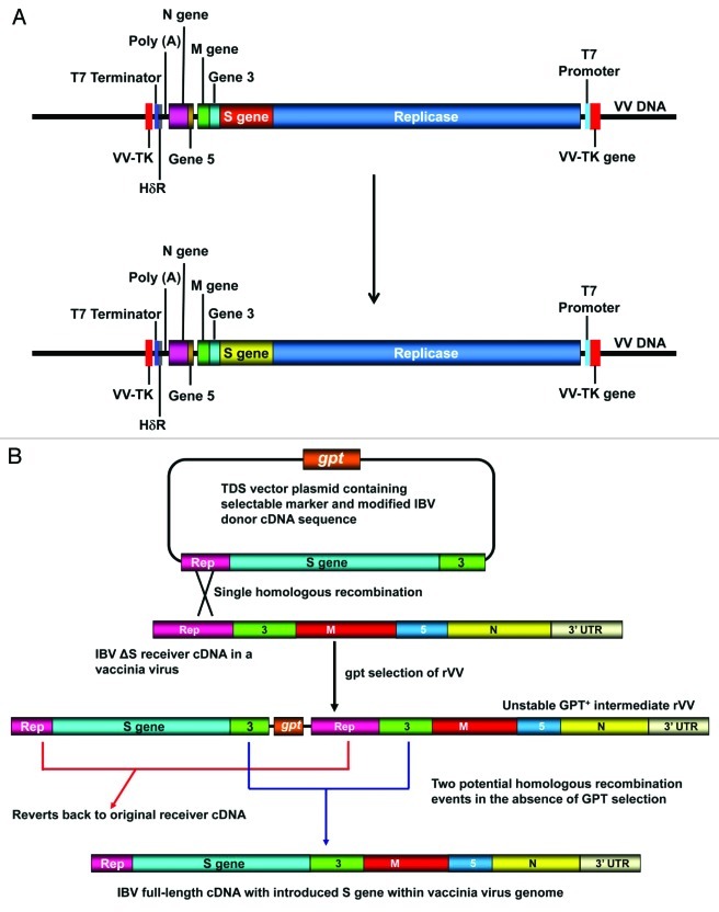
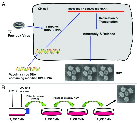
Our results involving replacement of the IBV S glycoprotein genes has demonstrated that the availability of our IBV reverse genetics system has opened up ways of modifying the IBV genome for the development of new genetically defined and intrinsically safer IBV vaccines. The reverse genetics system we use for modifying the IBV genome comprises of two processes. The first is outlined in Figure 1 and involves the direct modification of the IBV genome and the second process, outlined in Figure 2 centers on the recovery of infectious IBV. A full-length IBV cDNA, derived from the genomic RNA of the avirulent Beaudette strain, was sequentially assembled in vitro and ligated into a NotI site in the thymidine kinase (TK) gene of vaccinia virus19 (Fig. 1A) and is used as a template for modifying the IBV Beaudette genome. The IBV cDNA is under the control of a bacteriopahge T7 DNA dependent RNA polymerase promoter with a hepatitis delta virus ribozyme (HδR) sequence placed downstream of the coronavirus poly(A) tail followed by a T7 DNA dependent RNA polymerase termination sequence (Fig. 1A). The work described in our Journal of Virology17 and PLoS ONE16 papers described the replacement of the Beaudette S gene with the corresponding S gene sequences for the pathogenic IBV strains M41 and 4/91(UK), respectively, and is highlighted in Figure 1A.
Figure 1.
Modification of the IBV Beaudette genome. (A) Outlines the overall modification of the S gene in the full-length IBV Beaudette cDNA within the vaccinia virus genome. The positions of the T7 promoter and termination sequences are shown; the IBV cDNA is inserted within the vaccinia virus thymidine kinase gene. (B) Schematic diagram of the TDS process for inserting a heterologous S gene into a modified version of the full-length IBV cDNA that lacks the Beaudette S gene sequence. The new S gene sequence is inserted into the GPT selection plasmid flanked by Beaudette-derived sequence corresponding to sequences 5′ and 3′ to the deleted S gene sequence. A potential single-step homologous recombination event between the end of the replicase gene in the receiver IBV cDNA and the flanking sequence in the donor sequence in the GPT plasmid is shown. This results in a series of recombinant vaccinia viruses that are selected due to their GPT+ phenotype in the presence of MPA. Growth of a GPT+ recombinant vaccinia virus in the absence of MPA gives rise to two types of spontaneous intramolecular recombination events due to the presence tandem repeat sequences of the IBV cDNA in the recombinant vaccinia virus. This results in the generation of recombinant vaccinia viruses either with an IBV cDNA without an S gene (no modification) or a complete full-length IBV cDNA containing the heterologous S gene, the desired end product. Both recombination events result in the loss of the GPT gene. The IBV genes representing the structural and accessory genes are shown; a potential recombination event is indicated between the IBV replicase gene sequence common to both constructs.
Figure 2.
Recovery of infectious recombinant IBV. (A) Outlines the overall scheme for the recovery of infectious IBV from the recombinant vaccinia virus DNA containing the modified IBV cDNA. The vaccinia virus DNA is transfected into primary CK cells and infectious IBV RNA synthesized using T7 DNA dependant RNA polymerase expressed from a recombinant fowlpox virus. The T7-derived RNA is recognized as eukaryotic RNA and translated by the cellular machinery to produce the IBV replicase proteins that subsequently generate IBV genomic RNA and subgenomic mRNA leading to the assembly and release of infectious IBV virions. (B) Outlines the subsequent passage and recovery of the recombinant IBV in primary CK cells, in the case of BeauR-4/91(S) this was performed in 10-d-old embryonated eggs.16 Virus generated from P3 CK cells is analyzed for the presence of any modification and used for subsequent experiments.
In order to make alterations, such as exchange of the S gene, to the IBV Beaudette genome the IBV cDNA within the vaccinia virus genome is modified by homologous recombination using a vaccinia virus-based transdominant selection (TDS) method.20,21 The procedure, as described in our recent PLoS ONE paper, for inserting the S glycoprotein gene from IBV 4/91(UK) into the Beaudette genomic background, is summarized in Figure 1B. The first stage consisted of a single step homologous recombination event between the donor IBV cDNA sequence, the IBV 4/91(UK) S gene sequence, inserted into the E. coli guanine phosphoribosyltransferase (GPT) containing plasmid and the receiver IBV sequence, a modified version of the IBV Beaudette full-length genomic cDNA that lacked the Beaudette S gene sequence, in the vaccinia virus genome. The homologous recombination event occurred between one of the two Beaudette-derived sequences flanking either end of the heterologous 4/91(UK) S gene sequence, and the corresponding Beaudette sequence present 5′ and 3′ of the deleted S gene sequence in the receiver IBV cDNA. This resulted in the integration of the complete plasmid sequence into the receiver IBV cDNA allowing the selection of recombinant vaccinia viruses in the presence of mycophenolic acid (MPA). MPA is an inhibitor of purine biosynthesis, therefore only viruses expressing the E. coli GPT gene, which provides an alternative pathway for purine biosynthesis, are able to replicate in the presence of MPA and the alternative purine precursors xanthine and hypoxanthine. Randomly selected GPT+ viruses were then grown in the absence of MPA which resulted in a second internal recombination event between the tandem repeat IBV sequences causing the loss of the E. coli GPT gene (Fig. 1B). This second recombination step led to two possible outcomes; one event resulted in the original (unmodified) IBV sequence and the other, in the desired modification, the generation of an IBV cDNA containing the 4/91(UK) S gene sequence in the Beaudette cDNA that lacked the Beaudette S gene sequence (Fig. 1B). Recombinant vaccinia viruses that no longer expressed the GPT gene were isolated and sequence analysis used to identify those that contained the 4/91(UK) S gene sequence. Infectious IBV RNA was generated in situ by transfection of the vaccinia virus DNA containing the modified Beaudette cDNA into primary chick kidney (CK) cells previously infected with a recombinant fowlpox virus, rFPV-T7, expressing T7 RNA polymerase22 as outlined in Figure 2A. In this system infectious IBV RNA is produced from the T7 promoter immediately adjacent to the 5′ end of the IBV cDNA by the rFPV-T7-derived T7 RNA polymerase and terminates at the T7 termination sequence downstream of the HδR sequence, which autocleaves itself and the T7-termination sequence from the end of the poly(A) sequence, resulting in an authentic copy of the IBV genomic RNA. Cell supernatants from the transfected CK cells are filtered to remove any rFPV-T723 and potential recombinant IBVs are passaged three times in CK cells (Fig. 2B) to produce stocks of virus for sequence analysis to confirm the presence of the modified IBV sequence. We have also found that using the S glycoprotein from field isolates of IBV, such as 4/91(UK), that have not been adapted for growth on primary CK cells that we were unable to recover infectious virus using CK cells due to the fact that any potential virus was refractory for growth on CK cells.16 To circumvent this we have performed the initial rescue of the recombinant IBV in CK cells and instead of passaging any potential IBV the filtered supernatants on CK cells this was performed in 10-d-old embryonated eggs.16 The resultant recombinant IBVs, apart from any modification, are isogenic as they are derived from the same cDNA sequence. As indicated above we have successfully introduced two different heterologous S gene sequences into an IBV Beaudette genomic background and recovered infectious recombinant IBVs using our reverse genetics system. The recombinant IBVs were found to have the cell tropism associated with the heterologous S gene sequence16,17 and have been assessed as potential IBV vaccine candidates.
Assessment of Recombinant IBVs for Pathogenicity and Homologous Protection
Infection of 8-d-old specific pathogen free (SPF) chickens with either of the recombinant IBVs BeauR-M41(S) or BeauR-4/91(S) did not result in IBV-associated clinical signs, snicking, tracheal rales, wheezing and nasal discharge, by 10 d post-inoculation. In contrast, the pathogenic IBV strains M41 and 4/91(UK) resulted in clinical signs from three days post-infection. These results show that the replacement of the Beaudette S gene with a heterologous S gene from a virulent IBV strain, M41 or 4/91(UK), did not confer pathogenicity to the resulting recombinant viruses. This is an important finding for potential vaccine development because our results showed that although the S glycoprotein is a known virulence factor, with respect to receptor binding and responsible for tissue tropism, neither S glycoprotein derived from the two pathogenic IBVs conferred pathogenicity in vivo to the avirulent (Beaudette) receiver isolate of IBV.16,18
Three weeks after the primary inoculation with either BeauR-M41(S) or BeauR-4/91(S) the chickens were challenged with pathogenic IBV M41-CK or 4/91(UK), respectively. Clinical signs associated with a pathogenic IBV infection were not observed in the vaccinated chickens indicating that they had been protected against clinical disease when challenged with the homologous pathogenic virus;16,18 demonstrating that homologous protection had been induced by the appropriate recombinant IBV. Interestingly, results from prior inoculation of chickens with the recombinant IBV BeauR-4/91(S) and subsequent challenge with IBV M41, a different serotype of IBV to 4/91(UK), indicated that under experimental conditions BeauR-4/91(S) had induced some level of cross protection against M41 according to analysis of clinical signs.16 We were unable to isolate viable IBV or detect IBV-derived RNA from the tracheal cells of the vaccinated chickens following challenge with either pathogenic virus. This indicated that the recombinant viruses used to vaccinate the chickens prior to challenge had induced a protective response preventing the pathogenic viruses from successfully infecting the tracheal cells. Both virus and IBV-derived RNA was isolated from the tracheas of chickens that had not been vaccinated.
In conclusion, we have shown, using our IBV reverse genetics system,19-21 that replacement of the IBV Beaudette S glycoprotein with S gene sequences from the pathogenic IBVs M41 and 4/91(UK) did not confer virulence to the recombinant IBVs but in the resulting viruses, BeauR-M41(S) and BeauR-4/91(S), had tissue tropisms associated with the parental M41 and 4/91(UK) viruses. Chickens that were vaccinated with BeauR-M41(S) or BeauR-4/91(S) were found to be protected against clinical disease following challenge with M41 or 4/91(UK), whereas chickens vaccinated with Beaudette were not protected against challenge. The Beaudette isolate of IBV was attenuated after several hundred passages in embryonated hen’s eggs,24 which not only resulted in loss of virulence, but has also been implicated in loss of immunogenicity. Our results have shown that replacement of the Beaudette S gene with a heterologous gene from two different IBV serotypes resulted in recombinant IBVs, based on the Beaudette genome, that were able to act as potential vaccines for the protection of chickens following subsequent challenge with the parental pathogenic viruses. The swapping of the IBV S protein is a precise and effective way of generating genetically defined candidate IBV vaccines.
Acknowledgments
The authors would like to thank the following organizations for their financial support the Department of Environment, Food and Rural Affairs (DEFRA; www.defra.gov.uk/) project code OD0717, the Biotechnology and Biological Sciences Research Council (BBSRC; www.bbsrc.ac.uk/) and Intervet Schering-Plough UK.
Footnotes
Previously published online: www.landesbioscience.com/journals/biobugs/article/18983
References
- 1.Cavanagh D. Coronaviruses in poultry and other birds. Avian Pathol. 2005;34:439–48. doi: 10.1080/03079450500367682. [DOI] [PubMed] [Google Scholar]
- 2.Cavanagh D, Gelb J Jr. Infectious Bronchitis. In: Saif YM, ed. Diseases of Poultry. Iowa: Blackwell Publishing, 2008:117-35. [Google Scholar]
- 3.Jones RC. Viral respiratory diseases (ILT, aMPV infections, IB): are they ever under control? Br Poult Sci. 2010;51:1–11. doi: 10.1080/00071660903541378. [DOI] [PMC free article] [PubMed] [Google Scholar]
- 4.Sjaak de Wit JJ, Cook JKA, van der Heijden HMJF. Infectious bronchitis virus variants: a review of the history, current situation and control measures. Avian Pathol. 2011;40:223–35. doi: 10.1080/03079457.2011.566260. [DOI] [PMC free article] [PubMed] [Google Scholar]
- 5.Britton P, Cavanagh D. Avian coronavirus diseases and infectious bronchitis vaccine development. In: Thiel V, ed. Coronaviruses: Molecular and Cellular Biology. Norfolk, UK: Caister Academic Press, 2007:161-81. [Google Scholar]
- 6.Ambali AG, Jones RC. Early pathogenesis in chicks of infection with an enterotropic strain of infectious bronchitis virus. Avian Dis. 1990;34:809–17. doi: 10.2307/1591367. [DOI] [PubMed] [Google Scholar]
- 7.Koch G, Hartog L, Kant A, van Roozelaar DJ. Antigenic domains of the peplomer protein of avian infectious bronchitis virus: correlation with biological function. J Gen Virol. 1990;71:1929–35. doi: 10.1099/0022-1317-71-9-1929. [DOI] [PubMed] [Google Scholar]
- 8.Schultze B, Cavanagh D, Herrler G. Neuraminidase treatment of avian infectious bronchitis coronavirus reveals a hemagglutinating activity that is dependent on sialic acid-containing receptors on erythrocytes. Virology. 1992;189:792–4. doi: 10.1016/0042-6822(92)90608-R. [DOI] [PMC free article] [PubMed] [Google Scholar]
- 9.Luo Z, Weiss SR. Roles in cell-to-cell fusion of two conserved hydrophobic regions in the murine coronavirus spike protein. Virology. 1998;244:483–94. doi: 10.1006/viro.1998.9121. [DOI] [PMC free article] [PubMed] [Google Scholar]
- 10.de Groot RJ, Lujtjes W, Horzinek MC, van der Zeijst BAM, Spaan WJ, Lenstra JA. Evidence for a coiled-coil structure in the spike proteins of coronaviruses. J Mol Biol. 1987;196:963–6. doi: 10.1016/0022-2836(87)90422-0. [DOI] [PMC free article] [PubMed] [Google Scholar]
- 11.Tripet B, Howard MW, Jobling M, Holmes RK, Holmes KV, Hodges RS. Structural characterization of the SARS-coronavirus spike S fusion protein core. J Biol Chem. 2004;279:20836–49. doi: 10.1074/jbc.M400759200. [DOI] [PMC free article] [PubMed] [Google Scholar]
- 12.Guo Y, Tisoncik J, McReynolds S, Farzan M, Prabhakar BS, Gallagher T, et al. Identification of a new region of SARS-CoV S protein critical for viral entry. J Mol Biol. 2009;394:600–5. doi: 10.1016/j.jmb.2009.10.032. [DOI] [PMC free article] [PubMed] [Google Scholar]
- 13.Shulla A, Gallagher T. Role of spike protein endodomains in regulating coronavirus entry. J Biol Chem. 2009;284:32725–34. doi: 10.1074/jbc.M109.043547. [DOI] [PMC free article] [PubMed] [Google Scholar]
- 14.Hopkins SR, Yoder HW., Jr. Reversion to virulence of chicken passaged infectious bronchitis vaccine virus. Avian Dis. 1986;30:221–23. doi: 10.2307/1590639. [DOI] [PubMed] [Google Scholar]
- 15.McKinley ET, Hilt DA, Jackwood MW. Avian coronavirus infectious bronchitis attenuated live vaccines undergo selection of subpopulations and mutations following vaccination. Vaccine. 2008;26:1274–84. doi: 10.1016/j.vaccine.2008.01.006. [DOI] [PMC free article] [PubMed] [Google Scholar]
- 16.Armesto M, Evans S, Cavanagh D, Abu-Median AB, Keep S, Britton P. A recombinant avian infectious bronchitis virus expressing a heterologous spike gene belonging to the 4/91 serotype. PLoS ONE. 2011;6:e24352. doi: 10.1371/journal.pone.0024352. [DOI] [PMC free article] [PubMed] [Google Scholar]
- 17.Casais R, Dove B, Cavanagh D, Britton P. Recombinant avian infectious bronchitis virus expressing a heterologous spike gene demonstrates that the spike protein is a determinant of cell tropism. J Virol. 2003;77:9084–9. doi: 10.1128/JVI.77.16.9084-9089.2003. [DOI] [PMC free article] [PubMed] [Google Scholar]
- 18.Hodgson T, Casais R, Dove B, Britton P, Cavanagh D. Recombinant infectious bronchitis coronavirus Beaudette with the spike protein gene of the pathogenic M41 strain remains attenuated but induces protective immunity. J Virol. 2004;78:13804–11. doi: 10.1128/JVI.78.24.13804-13811.2004. [DOI] [PMC free article] [PubMed] [Google Scholar]
- 19.Casais R, Thiel V, Siddell SG, Cavanagh D, Britton P. Reverse genetics system for the avian coronavirus infectious bronchitis virus. J Virol. 2001;75:12359–69. doi: 10.1128/JVI.75.24.12359-12369.2001. [DOI] [PMC free article] [PubMed] [Google Scholar]
- 20.Britton P, Evans S, Dove B, Davies M, Casais R, Cavanagh D. Generation of a recombinant avian coronavirus infectious bronchitis virus using transient dominant selection. J Virol Methods. 2005;123:203–11. doi: 10.1016/j.jviromet.2004.09.017. [DOI] [PMC free article] [PubMed] [Google Scholar]
- 21.Armesto M, Casais R, Cavanagh D, Britton P. Transient dominant selection for the modification and generation of recombinant infectious bronchitis coronaviruses. Methods Mol Biol. 2008;454:255–73. doi: 10.1007/978-1-59745-181-9_19. [DOI] [PMC free article] [PubMed] [Google Scholar]
- 22.Britton P, Green P, Kottier S, Mawditt KL, Pe´nzes Z, Cavanagh D, et al. Expression of bacteriophage T7 RNA polymerase in avian and mammalian cells by a recombinant fowlpox virus. J Gen Virol. 1996;77:963–7. doi: 10.1099/0022-1317-77-5-963. [DOI] [PubMed] [Google Scholar]
- 23.Evans S, Cavanagh D, Britton P. Utilizing fowlpox virus recombinants to generate defective RNAs of the coronavirus infectious bronchitis virus. J Gen Virol. 2000;81:2855–65. doi: 10.1099/0022-1317-81-12-2855. [DOI] [PubMed] [Google Scholar]
- 24.Beaudette FR, Hudson CB. Cultivation of the virus of infectious bronchitis. J Am Vet Med Assoc. 1937;90:51–60. [Google Scholar]


