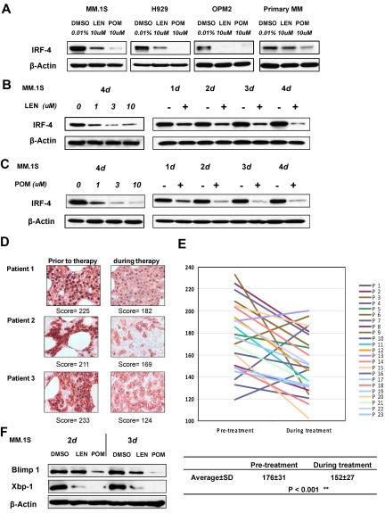
Figure 2.
IMiD compounds down-regulate IRF4 in MM cells in vitro and in lenalidomide-treated patients. (A) MM.1S, H929, OPM2, or primary myeloma cells were cultured with DMSO, pomalidomide, or lenalidomide at the indicated concentrations for 3 days. (B-C) MM.1S cells were incubated with lenalidomide (B) or pomalidomide (C) at different concentrations with a fixed time period of 4 days, or for different time periods with fixed concentration of 10μM, or with DMSO 0.01% as control. Cells were lysed, and cell lysates were analyzed for IRF-4 expression by Western blotting. β-actin expression was probed for loading control. (D) Double staining immunohistochemistry on paraffin-embedded marrow biopsy sections for IRF4 (black nuclear staining) and CD138+ (red staining) was performed before and during treatment with lenalidomide. Cells were rated for IRF4 reactivity as follows: 3 indicates strong; 2, moderate; and 1, negative/weak. The score for a sample is the sum of 100 cells (original magnification ×1000). (E) IRF4 scores of bone marrow biopsy samples, before and during treatment with lenalidomide. Lines connect scores from the same patients. (F) MM.1S cells were incubated with lenalidomide or pomalidomide for 2 or 3 days at 100μM. BLIMP1 and XBP1 expression was analyzed by Western blotting. β-actin expression was probed for loading control.

