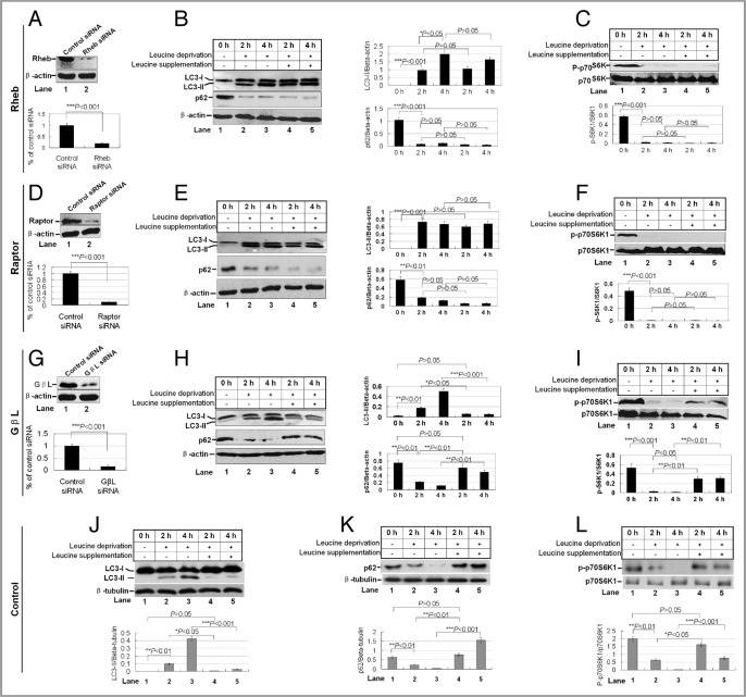
Figure 5.
The contribution of mTORC1 adaptors in leucine-mediated inhibition of autophagy and mTOR activity. HEK 293T cells were transduced with pGCsilencer™ RNAi vectors expressing shRNA against Rheb. (A, D and G) Western blotting for Rheb, Raptor and GβL expression (top) and quantification of these proteins compared with β-actin (bottom). (B, E and H) Leucine deprivation and supplementation on autophagy. HEK 293T cells were incubated in leucine-free medium in the absence or presence of 30 mM leucine for 2 h and 4 h. Cell lysates were analyzed by western blotting with the indicated antibodies (left), and the quantification of LC3-II/β-actin and p62/β-actin as the relative expression levels (right). Data are means ± SEM for at least three different experiments. (C, F and I) Leucine deprivation and supplementation on mTOR activity. The same cell lysates as described in (B, E and H) were probed with p-p70S6K and p70S6K antibodies (top) and quantified (bottom). (J–L). A control siRNA was used in the cells with leucine deprivation and supplementation. The protein levels of LC3-II, p62 and p-p70S6K were detected (top) and quantified (bottom).

