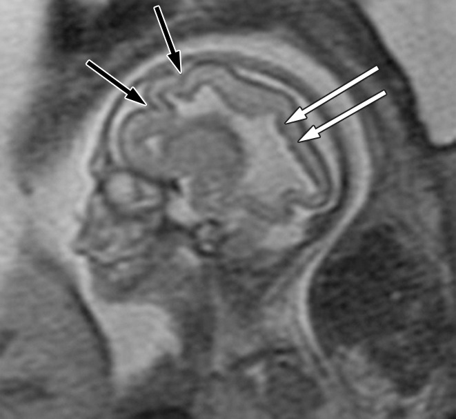
Figure 2a:

Polymicrogyria and PVNH detected with fetal MR imaging at 22 gestational weeks. (a) Sagittal single-shot fast SE T2-weighted image at 22 gestational weeks demonstrates multiple abnormal infoldings of the cortex, consistent with polymicrogyria (black arrows). PVNH appears as nodular areas along the wall of the lateral ventricular atrium, which protrude into the ventricular lumen (white arrows). (Reprinted, with permission, from reference 9.) (b) Postnatal axial fast spin-echo T2-weighted image at 2 days of age demonstrates bilateral frontal polymicrogyria, confirming fetal MR findings. (c) Fetal axial single-shot fast SE T2-weighted image demonstrates nodular area in the wall of the left lateral ventricle, which is isointense to germinal matrix and protrudes slightly into the ventricular lumen, and which was confirmed in the coronal plane, consistent with PVNH (arrows). (d) Postnatal axial spin-echo T2-weighted image at the level of the ventricular atria confirms the PVNH (arrow).
