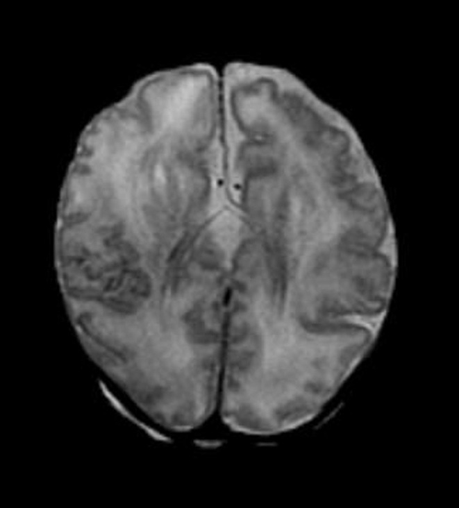
Figure 3d:

Diffuse polymicrogyria detected with fetal MR imaging at 36 gestational weeks. (a) Fetal axial single-shot fast SE T2-weighted image demonstrates bilateral abnormal cortical folding pattern characterized by too numerous sulci with abnormally small gyri, consistent with diffuse polymicrogyria. Both sylvian fissures extend too far posteriorly, which is often seen with polymicrogyria affecting the perisylvian areas (arrows). There is also callosal agenesis. (b) Axial single-shot fast SE T2-weighted image more superiorly again demonstrates diffuse polymicrogyria with abnormal posterior extent of the sylvian fissures (arrows). (c) Postnatal SE axial T2-weighted image corresponding to the same location as in a demonstrates diffuse polymicrogyria. (d) Postnatal SE axial T2-weighted image corresponding to b demonstrates abnormal posterior extent of the sylvian fissures bilaterally with diffuse polymicrogyria. (e) Fetal coronal single-shot fast SE T2-weighted image shows multiple abnormal infoldings, most prominent in the posterior left sylvian fissure (top arrow). Left cerebellar hypoplasia is also noted (bottom arrow). (Reprinted, with permission, from reference 9.) (f) Corresponding postnatal coronal three-dimensional spoiled gradient-recalled acquisition in the steady state T1-weighted image confirms numerous abnormal cortical infoldings, with apparent thickening of posterior left sylvian fissure due to multiple tiny cortical infoldings (arrow).
