Abstract
Summary
Background and objectives
Malnutrition, inflammation, atherosclerosis/calcification (MIAC) and endothelial dysfunction are the most commonly encountered risk factors in the pathogenesis of cardiovascular disease in ESRD patients. Epicardial adipose tissue (EAT) is the true visceral fat depot of the heart. The relationship between CAD and EAT was shown in patients with high risk of coronary artery disease. In this study, we aimed to investigate the relationship between EAT and MIAC syndrome in ESRD patients.
Design, setting, participants, & measurements
Eighty ESRD patients and 27 healthy subjects enrolled in this cross-sectional study. EAT and coronary artery calcification score were measured by a multidetector computed tomography (MDCT) scanner. Patients with serum albumin <3.5 mg/dl were defined as patients with malnutrition; those with serum C-reactive protein level >10 ng/dl (normal range, 0–5 ng/dl) had inflammation; and those with CACS >10 had atheroscleosis/calcification.
Results
Total CACS and EAT measurements were significantly higher in ESRD patients when compared with healthy subjects. There was a statistically significant relationship between EAT and CACS in ESRD patients (r = 0.48). EAT measurements were higher in PD patients than HD patients. Twenty-four of the patients had no component, 31 had one component, 17 had two components, and nine had all of the MIAC components. EAT was found to be significantly increased when the presence of MIAC components increased. EAT was positively correlated with age, body mass index, and presence of MIAC. These parameters were also found as independent predictors of increased EAT.
Conclusions
We found a relationship between EAT and components of MIAC syndrome in ESRD patients.
Introduction
Cardiovascular diseases (CVD) are the most common cause of mortality and morbidity in patients with ESRD receiving hemodialysis (HD) and peritoneal dialysis (PD) (1). Malnutrition, inflammation, atherosclerosis, endothelial dysfunction, coronary artery calcification (CAC), and left ventricular hypertrophy are the most commonly encountered risk factors in the pathogenesis of CVD in ESRD patients (2–4). Malnutrition, inflammation, atheroscleosis/calcification (MIAC) syndrome has been defined as the interaction between increased levels of proinflammatory cytokines, malnutrition, and atherosclerosis/calcification in ESRD patients (5,6). The presence of MIA components was found to be associated with increased mortality and morbidity in ESRD patients receiving PD (7) or HD (8). The coronary artery calcification score (CACS) in patients with ESRD reflects the severity of atherosclerotic vascular disease and predicts cardiovascular events (9,10). Epicardial adipose tissue (EAT) is the true visceral fat depot of the heart that accounts for approximately 20% of total heart weight, covers 80% of the cardiac surfaces, and is mostly in the grooved segments along the paths of coronary arteries (11–13). Recent studies showed a close relationship between coronary artery disease (CAD) and EAT using multidetector computed tomography (MDCT) and echocardiography in healthy subjects and patients at a high risk of CAD (14–17). In a recent study, the authors concluded that EAT acts as an extremely active organ that produces several bioactive adipokines as well as proinflammatory and proatherogenic cytokines such as tumor necrosis factor–α, monocyte chemotactic protein-1, IL-6, and resistin (16,18–21). Levels of most of these cytokines are also increased in ESRD patients (22–24). It is therefore reasonable to postulate that EAT is a source of inflammatory signals in patients with ESRD. Studies focusing on the association between the MIAC syndrome and EAT in ESRD patients are lacking. In this study, we investigated the relationship between EAT and MIAC components in ESRD patients.
Study Population and Methods
The study protocol was approved by the Medical Ethics Committee of Selcuk University (Meram School of Medicine, Konya, Turkey). Written informed consent was obtained from all of the subjects included in the study.
This was a cross-sectional study involving 80 ESRD patients (31 women, 49 men; mean age, 49 ± 14 years) receiving PD or HD for ≥6 months in the dialysis unit of Selcuk University and 27 healthy control subjects (14 women, 13 men; mean age, 54 ± 12 years) between February and June 2009. The Minitab 16 statistical program (Minitab, State College, PA) was used to determine sample size. The minimal sample volume was used to determine a difference of 20 cm3 in EAT with 80% power, and the 95% confidence interval was calculated to be 79.
Patients aged 18 to 70 years willing to participate in the assessment of CAC and EAT by MDCT were screened. A review of medical records (including information on age, gender, weight, duration of renal replacement treatment, medications, and primary disease of ESRD) was undertaken. Exclusion criteria were: (1) congestive heart failure; (2) active infection; (3) autoimmune disease; (4) secondary hyperparathyroidism; and (5) nephrotic-range proteinuria. Ninety-five patients were evaluated, and 15 patients were excluded from the study. Of these 15 patients, five patients had congestive heart failure (New York Heart Association class III–IV); four patients had active infection; three patients had secondary hyperparathyroidism; and three patients had autoimmune disease (including systemic lupus erythematosus and microscopic polyangitis). None of the patients included in the study had nephrotic-range proteinuria and arrhythmia on the basis of electrocardiography. The remaining 80 ESRD patients fulfilled the above criteria and were enrolled in the study. Twenty-seven age-matched and gender-matched healthy individuals referred from outpatient clinics of the Internal Medicine Department of Selcuk University were also enrolled as control subjects. They were subject to the same inclusion and exclusion criteria as the patients. HD patients were receiving thrice-weekly dialysis for a 4-hour period with a standard bicarbonate-containing dialysate bath using a biocompatible HD membrane (Polysulfone, FX-80 series, Fresenius, Germany). Dialysate flow rates were 500 ml/min, and blood-flow rates were 250 to 300 ml/min. The systolic BP (SBP) and diastolic BP (DBP) of patients and healthy subjects were measured in the upright sitting position after ≥5 minutes of rest using an Erka sphygmomanometer (PMS Instruments Limited, Berkshire, UK) with an appropriate cuff size. Two readings were recorded for each individual. The mean value of two readings was defined as the BP. Patients with SBP and DBP >140 and <90 mmHg, respectively, or who were already on antihypertensive treatment were assumed to be hypertensive.
Twenty-three patients were taking antihypertensive drugs (14 of them on angiotensin-converting enzyme inhibitors; eight receiving an angiotensin-II receptor blockers; and one receiving a calcium channel blocker and an angiotensin-converting enzyme inhibitor). Thirty-two patients were taking calcium containing phosphate binders.
Biochemical Analyses
Venous blood samples for biochemical analyses were drawn after an overnight fast before first exchange in PD patients and before the midweek session in patients receiving HD. All of the biochemical analyses, including those for total cholesterol, LDL cholesterol, HDL cholesterol, and plasma triglyceride concentrations, were undertaken using an oxidase-based technique by the Roche/Hitachi Modular System (Mannheim, Germany) in the Central Biochemistry Laboratory of the Meram School of Medicine.
MIAC Components
In this study, the serum levels of albumin and C-reactive protein (CRP), as well as CACS, were used to assess malnutrition, inflammation and atherosclerosis/calcification, respectively, just as in studies evaluating high cardiovascular mortality and morbidity in ESRD patients (25). Therefore, patients with a serum albumin of <3.5 mg/dl were defined as patients with malnutrition; those with a serum CRP level of >10 ng/dl (normal range, 0 to 5 ng/dl) had inflammation; and those with a CACS of >10 had atheroscleosis/calcification. The patients were classified according to the presence of MIAC components. Hence, patients without any component are classified as MIAC 0; those with one of the components are classified as MIAC 1; those with two of the components are classified as MIAC 2; and those with all of the components are classified as MIAC 3.
Evaluation of CACS and EAT
Unenhanced coronary computed tomography (CT) was quantified on retrospectively electrocardiography-gated cardiac CT using 64-slice MDCT (Sensation 64 Siemens Medical Solutions, Erlangen, Germany). The coronary CT protocol was: slice collimation, 64 × 0.6 mm; gantry rotation time, 0.33 seconds; pitch, 0.2; tube voltage, 120 kV; and tube current, 600 mA. If the heart rate was >65 beats per minute, heart rate control was achieved with a β-blocker. Multiplanar data reconstructions were obtained in standardized ventricular short-axis planes at the basal, midcavity, apical, and the horizontal long-axis plane with 3-mm slice thickness and 2-mm slice interval (26). To quantify CAC, all of the reconstructions were transferred to a personal computer-based workstation (Syngo CaScoring Wizard, Siemens Medical Solutions). CACS was defined as >2 contiguous pixels with Hounsfield units >130 as designed by Agatston et al. (27). All of the values of the left anterior descending coronary artery, circumflex coronary artery, and right coronary artery were added to calculate the CACS. In this study, CACS <10 was defined as the absence of atherosclerosis/calcification, and CACS >10 was defined as the presence of atherosclerosis/calcification. The rationale of using >10 as a cut off for CAC was that the functional deterioration of coronary arteries starts from low levels of CACS (e.g., >10) (38). To quantify EAT volume, all of the reconstructions were transferred to the personal computer-based workstation. A CT attenuation threshold between −200 and −20 Hounsfield units was used to isolate epicardial fat. Measurements of EAT and CACS were evaluated by two radiologists blinded to the study protocol. The interobserver variability was <10%.
Statistical Analyses
Statistical analyses were carried out using the Statistical Package for Social Sciences for Windows version 15.0 (SPSS, Chicago, IL). The parametric data are the means ± SDs, and the nonparametric data are the median values (interquartile range). The normal distribution of all variables was tested using the Kolmogorov–Smirnov test. Dichotomous variables were compared using the chi-squared test. Statistical differences between parametric data of two groups were analyzed using the t test. The Mann–Whitney U test was used to determine differences between nonparametric data. Statistical differences among parametric data of three groups were analyzed using the one-way ANOVA test, and the difference between subgroups was assessed with the post hoc Tukey test. Linear associations between continuous variables were assessed using the Spearman correlation test. Multivariate linear regression analyses were undertaken to determine independent associations among EAT and other variables. Age, body mass index (BMI), dialysis vintage, presence of MIAC, SBP, and diabetes mellitus (DM) were entered into the regression model as independent variables, and EAT was entered as a dependent variable. The backward elimination method was preferred in the stepwise regression analysis, and P > 0.1 was used as a criterion for elimination in this model. At the end of the fourth step, three variables including age, BMI, and the presence of MIA remained statistically significant in the model. P < 0.05 was considered significant for all tests.
Results
Baseline Characteristics of Patients
The baseline characteristics of 80 ESRD patients and 27 healthy subjects are shown in Table 1. The etiology of ESRD patients was diabetic nephropathy (n = 18), chronic glomerulonephritis (n = 12), hypertensive nephropathy (n = 23), polycystic kidney disease (n = 7), nephrolithiasis (n = 5), and unknown (n = 15). There were no differences with respect to the following variables between ESRD patients and healthy subjects: age, gender, BMI, predialysis levels of SBP and DBP, serum levels of LDL cholesterol, HDL cholesterol, triglyceride, and CRP.
Table 1.
The demographic and laboratory features of the ESRD patients and healthy subjects
| Parameters | Healthy Subjects (n = 27) (Mean ± SD) or Median (IQR) | ESRD Patients (n = 80) (Mean ± SD) or Median (IQR) | P |
|---|---|---|---|
| Age (years) | 54 ± 12 | 49 ± 14 | 0.09 |
| Men/women | 14/13 | 49/31 | 0.46 |
| BMI (kg/m2) | 26.2 ± 5.5 | 26.7 ± 5.1 | 0.63 |
| Dialysis vintage (months) | 56 ± 4 | ||
| SBP (mmHg) | 132 ± 22 | 138 ± 28 | 0.16 |
| DBP (mmHg) | 80 ± 11 | 86 ± 16 | 0.18 |
| Hemoglobin (mg/dl) | 12.5 ± 2.1 | 11.9 ± 1.7 | 0.06 |
| Albumin (g/dl) | 3.8 (3.5–4.0) | 3.7 (3.4–3.9) | 0.19 |
| AST (IU/L) | 21 ± 4 | 18 ± 6 | 0.06 |
| ALT (IU/L) | 16 ± 5 | 17 ± 7 | 0.63 |
| LDL cholesterol (mg/dl) | 94.3 ± 6.0 | 119.8 ± 35 | 0.33 |
| HDL cholesterol (mg/dl) | 37.3 ± 11.6 | 37.4 ± 14 | 0.97 |
| Triglyceride (mg/dl) | 122.1 ± 86 | 147 ± 94 | 0.05 |
| Calcium (mg/dl) | 8.9 ± 0.8 | ||
| Phosphorus (mg/dl) | 4.3 ± 1.0 | ||
| CRP (mg/dl) | 8.25 (3.75–14.5) | 9.8 (6.1–17.1) | 0.13 |
| PTH (pg/ml) | 353 ± 309 | ||
| Total CACS | 41 ± 112 | 144 ± 278 | 0.01 |
| Epicardial adipose tissue (cm3) | 121.5 ± 37.5 | 160 ± 76 | 0.02 |
AST, aspartate aminotransferase; ALT, alanine aminotransferase; PTH, parathyroid hormone; CACS, coronary artery calcification score; BMI, body mass index; SBP, systolic BP; DBP, diastolic BP; CRP, C-reactive protein; IQR, interquartile range.
Evaluation of CACS and EAT
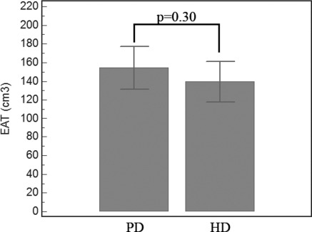
Total CACS and EAT measurements were significantly higher in ESRD patients compared with healthy subjects (Table 1; P = 0.01 and P = 0.02, respectively). There was a statistically significant relationship between EAT and CACS in ESRD patients (P < 0.0001, r = 0.48). When patients were classified according to their renal replacement therapy (HD and PD), EAT measurements were higher in PD patients than in HD patients (155.9 ± 69.0 versus 139.4 ± 62.7 cm3), but this difference was NS (P = 0.30) (Figure 1).
Figure 1.
Epicardial adipose tissue (EAT) in peritoneal dialysis (PD) and hemodialysis (HD) patients.
MIAC Components
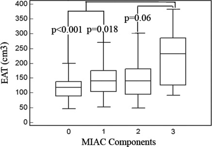
Patients were classified according to MIAC components. Twenty-four patients had none, 30 had one component, 17 had two components, and nine had all MIAC components. EAT measurements of the patients according to MIAC components are shown in Table 2. EAT was significantly increased if the number of MIAC components increased (Figure 2).
Table 2.
The relationship between EAT and MIAC components in ESRD patients
| Number of MIAC Components | Number of ESRD Patients (n = 80) | EAT (cm3) Mean ± SD |
|---|---|---|
| None | 24 (30%) | 121 ± 51 |
| One component | 30 (38%) | 145 ± 61 |
| Two components | 17 (21%) | 152 ± 75 |
| All components | 9 (11%) | 221 ± 101a |
One-way ANOVA test result was showed in the table. The P value of ANOVA test was 0.003. MIAC, malnutrition, inflammation, atherosclerosis/calcification; EAT, epicardial adipose tissue.
EAT difference between three components versus no components of MIAC, P = 0.01; EAT difference between three components versus one component of MIAC, P = 0.02; EAT difference between three components versus two components of MIAC, P = 0.06.
Figure 2.
The relationship between epicardial adipose tissue (EAT) and the components of malnutrition, inflammation, atherosclerosis/calcification (MIAC).
Linear Correlations
In the bivariate correlation analysis, EAT was positively correlated with age and BMI (r = 0.44, P = 0.001 and r = 0.44, P = 0.001, respectively). The multivariate linear regression analysis revealed that age, as well as the presence of MIAC and BMI, were independent predictors of increased EAT. Regression results are shown in Table 3.
Table 3.
The stepwise regression analysis to determine independent predictors of epicardial adipose tissue
| Parameters | Standardized Betaa | P | 95% CI |
|---|---|---|---|
| Step 1 (adjusted r2 = 0.34, P = 0.001b) | |||
| age (years) | 0.35 | 0.001 | 0.71 to 2.80 |
| BMI (kg/m2) | 0.31 | 0.002 | 1.65 to 7.02 |
| presence of MIAC | 0.19 | 0.05 | −1.52 to 87.64 |
| SBP (mmHg) | −0.07 | 0.43 | −0.65 to 0.28 |
| presence of DM | −0.02 | 0.82 | −41.47 to 32.93 |
| dialysis vintage (months) | 0.006 | 0.95 | −3.87 to 4.12 |
| Step 4 (adjusted r2 = 0.36, P = 0.001b) | |||
| age (years) | 0.34 | 0.001 | 0.72 to 2.75 |
| BMI (kg/m2) | 0.31 | 0.002 | 1.61 to 6.86 |
| presence of MIAC | 0.19 | 0.05 | −0.62 to 86.65 |
CI, confidence interval; BMI, body mass index; MIA, malnutrition, inflammation, atherosclerosis/calcification; DM, diabetes mellitus; SBP, systolic blood pressure.
Standardized beta means standardized regression coefficients of the variables.
P values are defined to model statistics for each step.
Discussion
There were five main findings of this study. First, EAT measured by MDCT was increased in ESRD patients when compared with healthy subjects. Second, EAT measurements were significantly correlated with the MIAC syndrome in ESRD patients. Third, there was a statistically significant relationship between EAT and CACS in ESRD patients. Fourth, EAT was significantly increased if the number of MIAC components was increased. Lastly, EAT was positively correlated with age, BMI, and MIAC in the stepwise analysis. This was the first study that evaluated the relationship between EAT and the MIAC syndrome in ESRD patients.
Several factors contribute to the high mortality seen in ESRD patients, but CVD remains the main cause of morbidity and mortality despite recent developments in renal replacement therapies (3,28). This can be attributed to many factors: advanced age, atherosclerosis, endothelial dysfunction, hypertension, anemia, hyperparathyroidism, chronic inflammation, DM (and its macrovascular and microvascular complications), left ventricular hypertrophy, malnutrition, and vascular calcification.
The MIAC syndrome was first defined in ESRD patients by Stenvinkel et al. (5,29). They concluded that malnutrition, inflammation, and atherosclerosis cause a vicious cycle and that proinflammatory cytokines play a central part in this process (7). CAC is part of the extended state of vascular calcification that can be detected even in the early decades of patients with ESRD (30). Wang et al. (6) showed an important association between malnutrition, inflammation, and atherosclerosis and valvular and vascular calcification in PD patients. In this study, CACS <10 was defined as the absence of atherosclerosis/calcification, and CACS >10 was defined as the presence of atherosclerosis/calcification. Caliskan et al. (38) investigated the correlation of CACs with coronary flow reserve (CFR) in HD patients. They concluded that patients with CACS of >10 had a significantly lower CFR. Hence, the functional deterioration of coronary arteries (decreased CFR) starts from low levels of CACS (e.g., >10). Taken together, these factors may contribute to premature CVD and the markedly increased mortality in patients with ESRD.
EAT and intra-abdominal visceral fat depots originate from the splanchnopleuric mesoderm (31). Mazurek et al. (16) concluded that, like abdominal visceral adipose tissue, EAT is also metabolically active because it can secrete proinflammatory cytokines and utilize free fatty acids (FFAs). Despite the smaller adipocyte size, EAT has a higher rate of uptake and secretion of fatty acids than other visceral fat depots. In health, EAT may act as a “buffering system” by scavenging excess FFAs that are toxic to the myocardium. However, under ischemic conditions, EAT may serve as a local energy source by providing FFAs for the increased metabolism of the myocardium (32,33). In a recent study, the authors concluded that EAT acts an extremely active organ that produces several bioactive adipokines, as well as proinflammatory and proatherogenic cytokines (tumor necrosis factor–α, monocyte chemotactic protein-1, IL-6, resistin, visfatin, omentin, leptin, plasminogen activator inhibitor-1, and angiotensinogen) (16,18–21). The levels of most of these proinflammatory cytokines were, in general, increased, and these cytokines were found to be associated with atherosclerosis and CAC in ESRD patients (22–24). In this study, increased EAT as measured by MDCT was related to CAC. We also found a close relationship between the components of MIAC and EAT. EAT measurements showed a significant increase with increasing components of the MIAC syndrome. EAT was highest among patients having all three components (221 ± 101 cm3) and lowest among those who do not have the MIAC syndrome (121 ± 51 cm3). This association might be attributed to increased levels of proinflammatory cytokines secreted by EAT.
When we classified ESRD patients according to renal replacement therapy, PD patients had similar EAT measurements when compared with HD patients. When comparing PD with HD, hypertriglyceridemia and obesity are commonly seen in PD patients (especially secondary to the use of high glucose ingredients of peritoneal dialysates). In contrast to the similar EAT values in the two groups, EAT was affected by more variables beyond BMI and hypertriglyceridemia.
Ix and Sharma (34) concluded that there is a link between obesity, CKD, and nonalcoholic fatty liver disease (NAFLD). Targher et al. (35) showed an association between NAFLD and CKD in patients with type 2 DM. However, in this study, there was no difference between ESRD patients and healthy subjects in terms of BMI and serum levels of aspartate aminotransferase and alanine transaminase. We therefore assumed that our patients did not have NAFLD.
EAT was found to be positively corraleted with age, BMI, and the presence of MIAC in the stepwise analysis. Studies have shown an important association between increased age, obesity, and the presence of the MIAC syndrome and cardiovascular mortality and morbidity in CKD populations (2–3,6). Our findings were consistent with those of other studies. Therefore, defining EAT could be valuable for further studies such as the determination of cardiovascular risk stratification in ESRD patients.
Despite the simplicity of evaluating EAT with echocardiography (36), EAT should be measured in three dimensions by MDCT: regional thickness, cross-sectional area, and total volume (33,37). Therefore, MDCT can be used to assess CAC and EAT in ESRD patients at a high risk of CVD. Echocardiography has been used for the measurement of EAT with a higher reproducibility and reliability in the general population, but there are no data about echocardiography for the assessment of EAT in ESRD patients (36). The lower cost, simplicity, and rapidity of echocardiography could make it the preferred measurement method for EAT.
Our study had two main limitations. First, this was a cross-sectional analysis of ESRD patients focusing on CAC and EAT. Second, the sample size was relatively small. This was not a prospective controlled study, so we cannot draw cause-and-effect relationships from our findings.
In conclusion, we found a relationship between EAT as defined by MDCT and components of the MIAC syndrome in ESRD patients. Further clinical and experimental studies are needed to determine the relationship between EAT and the MIAC syndrome.
Disclosures
None.
Acknowledgments
This study was supported by the Scientific Investigation and Project Foundation of Selcuk University Meram School of Medicine.
Footnotes
Published online ahead of print. Publication date available at www.cjasn.org.
References
- 1. United States Renal Data System: USRDS 2006 Annual Data Report: Atlas of End-Stage Renal Disease in United States. Bethesda, MD, National Institute of Health, National Institute of Diabetes and Digestive and Kidney Diseases, 2006 [Google Scholar]
- 2. Parfrey PS, Foley RN, Harnett JD, Kent GM, Murray D, Barre PE: Outcome and risk factors of ischemic heart disease in chronic uremia. Kidney Int 49: 1428–1434, 1996 [DOI] [PubMed] [Google Scholar]
- 3. Collins AJ. Cardiovascular mortality in end-stage renal disease. Am J Med Sci 325: 163–167, 2003 [DOI] [PubMed] [Google Scholar]
- 4. Parfrey PS, Foley RN, Harnett JD, Kent GM, Murray DC, Barre PE: Outcome and risk factors for left ventricular disorders in chronic uraemia. Nephrol Dial Transplant 11: 1277–1285, 1996 [PubMed] [Google Scholar]
- 5. Stenvinkel P, Heimburger O, Lindholm B, Kaysen GA, Bergstrom J: Are there two types of malnutrition in chronic renal failure? Evidence for relationships between malnutrition, inflammation and atherosclerosis (MIA syndrome). Nephrol Dial Transplant 15: 953–960, 2000 [DOI] [PubMed] [Google Scholar]
- 6. Wang AY, Woo J, Lam CW, Wang M, Chan IH, Gao P, Lui SF, Li PK, Sanderson JE: Associations of serum fetuin-A with malnutrition, inflammation, atherosclerosis and valvular calcification syndrome and outcome in peritoneal dialysis patients. Nephrol Dial Transplant 20: 1676–1685, 2005 [DOI] [PubMed] [Google Scholar]
- 7. Stenvinkel P, Chung SH, Heimburger O, Lindholm B: Malnutrition, inflammation, and atherosclerosis in peritoneal dialysis patients. Perit Dial Int 21[Suppl 3]: S157–S162, 2001 [PubMed] [Google Scholar]
- 8. Tonbul HZ, Demir M, Altintepe L, Guney I, Yeter E, Turk S, Yildiz A: Malnutrition-inflammation-atherosclerosis (MIA) syndrome components in hemodialysis and peritoneal dialysis patients. Ren Fail 28: 287–294, 2006 [DOI] [PubMed] [Google Scholar]
- 9. Haydar AA, Hujairi NM, Covic AA, Pereira D, Rubens M, Goldsmith DJ: Coronary artery calcification is related to coronary atherosclerosis in chronic renal disease patients: A study comparing EBCT-generated coronary artery calcium scores and coronary angiography. Nephrol Dial Transplant 19: 2307–2312, 2004 [DOI] [PubMed] [Google Scholar]
- 10. Raggi P, Boulay A, Chasan-Taber S, Amin N, Dillon M, Burke SK, Chertow GM: Cardiac calcification in adult hemodialysis patients: A link between end-stage renal disease and cardiovascular disease? J Am Coll Cardiol 39: 695–701, 2002 [DOI] [PubMed] [Google Scholar]
- 11. Shirani J, Berezowski K, Roberts WC: Quantitative measurement of normal and excessive (cor adiposum) subepicardial adipose tissue, its clinical significance, and its effect on electrocardiographic QRS voltage. Am J Cardiol 76: 414–418, 1995 [DOI] [PubMed] [Google Scholar]
- 12. Iacobellis G, Corradi D, Sharma AM: Epicardial adipose tissue: Anatomic, biomolecular and clinical relationships with the heart. Nat Clin Pract Cardiovasc Med 2: 536–543, 2005 [DOI] [PubMed] [Google Scholar]
- 13. Corradi D, Maestri R, Callegari S, Pastori P, Goldoni M, Luong TV, Bordi C: The ventricular epicardial fat is related to the myocardial mass in normal, ischemic and hypertrophic hearts. Cardiovasc Pathol 13: 313–316, 2004 [DOI] [PubMed] [Google Scholar]
- 14. Park MJ, Jung JI, Oh YS, Youn HJ: Assessment of epicardial fat volume with threshold-based 3-dimensional segmentation in CT: Comparison with the 2-dimensional short axis-based method. Korean Circ J 40: 328–333, 2010 [DOI] [PMC free article] [PubMed] [Google Scholar]
- 15. Djaberi R, Schuijf JD, van Werkhoven JM, Nucifora G, Jukema JW, Bax JJ: Relation of epicardial adipose tissue to coronary atherosclerosis. Am J Cardiol 102: 1602–1607, 2008 [DOI] [PubMed] [Google Scholar]
- 16. Mazurek T, Zhang L, Zalewski A, Mannion JD, Diehl JT, Arafat H, Sarov-Blat L, O'Brein S, Keiper EA, Johnson AG, Martin J, Goldstein BJ, Shi Y: Human epicardial adipose tissue is a source of inflammatory mediators. Circulation 108: 2460–2466, 2003 [DOI] [PubMed] [Google Scholar]
- 17. Eroglu S, Sade LE, Yildirir A, Bal U, Ozbicer S, Ozgul AS, Bozbas H, Aydinalp A, Muderrisoglu H: Epicardial adipose tissue thickness by echocardiography is a marker for the presence and severity of coronary artery disease. Nutr Metab Cardiovasc Dis 19: 211–217, 2009 [DOI] [PubMed] [Google Scholar]
- 18. Baker AR, Silva NF, Quinn DW, Harte AL, Pagano D, Bonser RS, Kumar S, Mc Ternan PG: Human epicardial adipose tissue expresses a pathogenic profile of adipocytokines in patients with cardiovascular disease. Cardiovasc Diabetol 5: 1, 2006 [DOI] [PMC free article] [PubMed] [Google Scholar]
- 19. Kremen J, Dolinkova M, Krajickova J, Blaha J, Anderlova K, Lacinova Z, Haluzikova D, Bosanska L, Vokurka M, Svacina S, Haluzik M: Increased subcutaneous and epicardial adipose tissue production of proinflammatory cytokines in cardiac surgery patients: Possible role in postoperative insulin resistance. J Clin Endocrinol Metab 91: 4620–4627, 2006 [DOI] [PubMed] [Google Scholar]
- 20. Cheng KH, Chu CS, Lee KT, Lin TH, Hsieh CC, Chiu CC, Voon WC, Sheu SH, Lai WT: Adipocytokines and proinflammatory mediators from abdominal and epicardial adipose tissue in patients with coronary artery disease. Int J Obes 32: 268–274, 2008 [DOI] [PubMed] [Google Scholar]
- 21. Fain JN, Sacks HS, Buehrer B, Bahouth SW, Garrett E, Wolf RY, Carter RA, Tichansky DS, Madan AK: Identification of omentin mRNA in human epicardial adipose tissue: Comparison to omentin in subcutaneous, internal mammary artery periadventitial and visceral abdominal depots. Int J Obes 32: 810–815, 2008 [DOI] [PubMed] [Google Scholar]
- 22. Yildiz A, Tepe S, Oflaz H, Yazici H, Pusuroglu H, Besler M, Ark E, Erzengin F: Carotid atherosclerosis is a predictor of coronary calcification in chronic haemodialysis patients. Nephrol Dial Transplant 19: 885–891, 2004 [DOI] [PubMed] [Google Scholar]
- 23. Stenvinkel P, Heimburger O, Paultre F, Diczfalusy U, Wang T, Berglund L, Jogestrand T: Strong association between malnutrition, inflammation, and atherosclerosis in chronic renal failure. Kidney Int 55: 1899–1911, 1999 [DOI] [PubMed] [Google Scholar]
- 24. Tintut Y, Patel J, Parhami F, Demer LL: Tumor necrosis factor-alpha promotes in vitro calcification of vascular cells via the cAMP pathway. Circulation 102: 2636–2642, 2000 [DOI] [PubMed] [Google Scholar]
- 25. Pecoits-Filho R, Lindholm B, Stenvinkel P: The malnutrition, inflammation, and atherosclerosis (MIA) syndrome: The heart of the matter. Nephrol Dial Transplant 11: 28–31, 2002 [DOI] [PubMed] [Google Scholar]
- 26. Abbara S, Desai JC, Cury RC, Butler J, Nieman K, Reddy V: Mapping epicardial fat with multi-detector computed tomography to facilitate percutaneous transepicardial arrhythmia ablation. Eur J Radiol 57: 417–422, 2006 [DOI] [PubMed] [Google Scholar]
- 27. Agatston AS, Janowitz WR, Hildner FJ, Zusmer NR, Viamonte M, Jr., Detrano R: Quantification of coronary artery calcium using ultrafast computed tomography. J Am Coll Cardiol 15: 827–832, 1990 [DOI] [PubMed] [Google Scholar]
- 28. Foley RN, Parfrey PS, Sarnak MJ: Clinical epidemiology of cardiovascular disease in chronic renal disease. Am J Kidney Dis 32[Suppl 3]: S112–S119, 1998 [DOI] [PubMed] [Google Scholar]
- 29. Stenvinkel P: Inflammatory and atherosclerotic interactions in the depleted uremic patient. Blood Purif 19: 53–61, 2001 [DOI] [PubMed] [Google Scholar]
- 30. London GM, Guerin AP, Marchais SJ, Metivier F, Pannier B, Adda H: Arterial media calcification in end-stage renal disease: Impact on all-cause and cardiovascular mortality. Nephrol Dial Transplant 18: 1731–1740, 2003 [DOI] [PubMed] [Google Scholar]
- 31. Ho E, Shimada Y: Formation of the epicardium studied with the scanning electron microscope. Dev Biol 66: 579–585, 1978 [DOI] [PubMed] [Google Scholar]
- 32. Marchington JM, Pond CM: Site-specific properties of pericardial and epicardial adipose tissue: The effects of insulin and high-fat feeding on lipogenesis and the incorporation of fatty acids in vitro. Int J Obes 14: 1013–1022, 1990 [PubMed] [Google Scholar]
- 33. Wang TD, LW, Chen MF: Epicardial adipose tissue measured by multidetector computed tomography: Practical tips clinical implications. Acta Cardiol 26: 55–68, 2010 [Google Scholar]
- 34. Ix JH, Sharma K: Mechanisms linking obesity, chronic kidney disease, and fatty liver disease: The roles of fetuin-A, adiponectin, and AMPK. J Am Soc Nephrol 21: 406–412, 2010 [DOI] [PMC free article] [PubMed] [Google Scholar]
- 35. Targher G, Chonchol M, Bertolini L, Rodella S, Zenari L, Lippi G, Franchini M, Zoppini G, Muggeo M: Increased risk of CKD among type 2 diabetics with nonalcoholic fatty liver disease. J Am Soc Nephrol 19: 1564–1570, 2008 [DOI] [PMC free article] [PubMed] [Google Scholar]
- 36. Iacobellis G, Willens HJ: Echocardiographic epicardial fat: A review of research and clinical applications. J Am Soc Echocardiogr 22: 1311–1319, 2009 [DOI] [PubMed] [Google Scholar]
- 37. Wang TD, Lee WJ, Shih FY, Huang CH, Chen WJ, Lee YT, Shih TT, Chen MF: Association of epicardial adipose tissue with coronary atherosclerosis is region-specific and independent of conventional risk factors and intra-abdominal adiposity. Atherosclerosis 213: 279–287, 2010 [DOI] [PubMed] [Google Scholar]
- 38. Caliskan Y, Demirturk M, Ozkok A, Yelken B, Sakaci T, Oflaz H, Unsal A, Yildiz A: Coronary artery calcification and coronary flow velocity in hemodialysis patients. Nephrol Dial Transplant 25(8): 2685–2690, 2010 [DOI] [PubMed] [Google Scholar]


