Fig. 6.
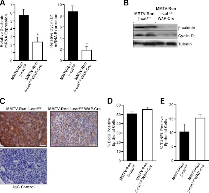
End-stage tumors from MMTV-Ron β-catF/F WAP-Cre mice maintain β-catenin loss. A, qRT-PCR analysis of β-catenin and cyclin D1 expression was performed on mRNA isolated from end-stage mammary tumors of control and MMTV-Ron β-catF/F WAP-Cre mice. *, P < 0.05. B, Western blot analysis was performed on mammary tumor lysates from control and MMTV-Ron β-catF/F WAP-Cre mice. The tumor lysates were analyzed for expression of β-catenin and cyclin D1. Tubulin was used as the loading control. C, Representative immunohistological images of end-stage mammary tumors from MMTV-Ron β-catF/F and MMTV-Ron β-catF/F WAP-Cre mice showing β-catenin expression are depicted. An IgG control of the MMTV-Ron β-catF/F image is provided to show secondary cross reactivity with an isotype control antibody. D, Quantification of BrdU incorporation to assess proliferation rates of mammary tumors in MMTV-Ron β-catF/F and MMTV-Ron β-catF/F WAP-Cre mice is shown. E, Quantification of TUNEL positive cells to assess the apoptotic rate in end-stage mammary tumors isolated from MMTV-Ron β-catF/F and MMTV-Ron β-catF/F WAP-Cre mice is shown. Scale bar, 50 μm.

