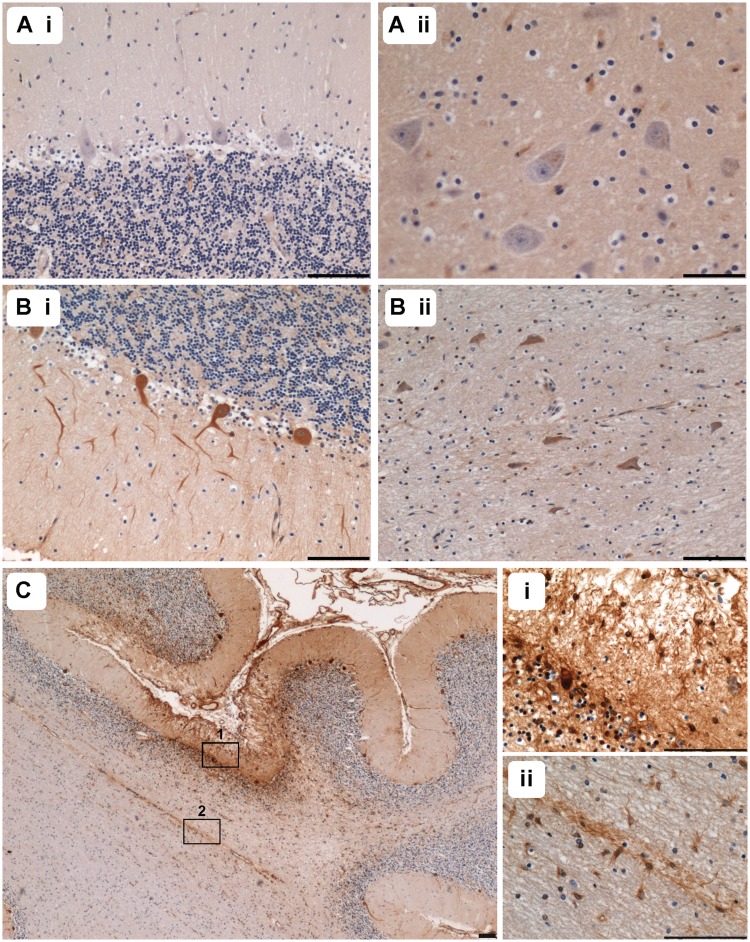
Figure 6.
Evidence of blood–brain barrier dysfunction was observed in many patients with extravasation of plasma proteins. Fibrinogen immunohistochemistry in control tissues demonstrated a lack of immunoreactivity in Purkinje cells [A(i)] and dentate nucleus neurons [A(ii)], while evaluation of a patient with m.8344A>G shows evidence of immunopositive Purkinje cells [B(i)], Patient 10) and dentate nucleus neurons [B(ii)]. Extravasation of fibrinogen was demonstrated in an ischaemic-like lesion in the cerebellar cortex of Patient 4 (C; fibrinogen). Examination at a higher magnification reveals evidence of Purkinje cell uptake [C(i)] and immunoreactivity within the molecular and granular cell layers is observed [C(i)]. In the adjacent white matter [C(ii)], a vessel showing immunoreactivity for fibrinogen is surrounded by immunopositive glial cells. Scale bars = 100 µm.

