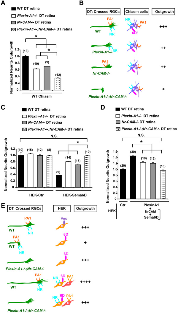
Figure 5. Nr-CAM and Plexin-A1 in crossed RGCs mediate effects of Sema6D on RGC outgrowth.
(A) Outgrowth of Plexin-A1−/−;Nr-CAM−/− DT explants is reduced on WT chiasm cells compared to WT, Plexin-A1−/− single or Nr-CAM−/− DT explants. (B) Summary of results in Figure 5A. Note that outgrowth of Plexin-A1−/− or Nr-CAM−/− DT explants are only partially reduced, but outgrowth of Plexin-A1−/−;Nr-CAM−/− DT explants are further reduced on WT chiasm cells. (C) Quantification of Nr-CAM−/−, Plexin-A1−/− and Plexin-A1−/−;Nr-CAM−/− outgrowth from DT retinal explants on Sema6D+ HEK cells. Note that Sema6D+ HEK cells only partially inhibit outgrowth of Nr-CAM−/− or Plexin-A1−/− DT explants, but neurite outgrowth is similar to controls in Plexin-A1−/−;Nr-CAM−/− DT explants. (D) Outgrowth of Nr-CAM−/−, Plexin-A1−/− and Plexin-A1−/−;Nr-CAM−/− DT retinal outgrowth on Sema6D+/Nr-CAM+ and Plexin-A1+ HEK cells. (E) Summary of results in Figures 5C and 5D. Note that Plexin-A1−/−;Nr-CAM−/− DT axons grew no differently on Sema6D+ HEK cells or on Sema6D+/Nr-CAM+ and Plexin-A1+ HEK cells than on HEK cells expressing empty vector. (n) = number of explants for each condition, * p<0.01

