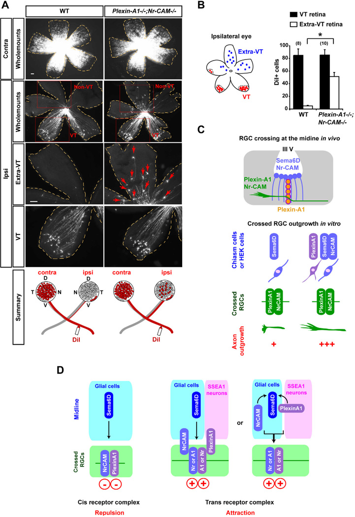
Figure 8. Non-VT RGCs are redirected ipsilaterally in Plexin-A1−/−;Nr-CAM−/− mutants.
(A) Representative E17.0 WT and Plexin-A1−/−;Nr-CAM−/− mutant retina whole mounts, showing retrogradely labeled RGCs contralateral (Contra) or ipsilateral (Ipsi) to DiI application in the optic tract. While DiI+ RGCs are restricted to the VT crescent after ipsilateral DiI application to the optic tract in wild type retina, RGCs were also observed in non-VT retina after ipsilateral optic tract labeling in Plexin-A1−/−;Nr-CAM−/− retina (arrows). (n ≥ 8 embryos for each condition). (B) Quantification of the ipsilateral projection from the VT and non-VT region in Plexin-A1−/−;Nr-CAM−/− mutants. Plexin-A1−/−;Nr-CAM−/− mutants show ~50 DiI+ RGCs in the non-VT region of ipsilateral retina, while WT display <5 DiI+ cells in non-VT regions. However, the number of DiI+ cells in VT regions are not significantly different between WT and Plexin-A1−/−;Nr-CAM−/− mutants. (C) Summary of interactions between Nr-CAM and Plexin-A1 on crossed RGCs and Sema6D substrates in vitro, and between Nr-CAM and Plexin-A1 on crossed RGCs and Nr-CAM/Sema6D (on midline radial glia) and Plexin-A1 (on SSEA-1+ neurons) in the chiasm in vivo. (D) Model of interactions between Nr-CAM and Plexin-A1 on crossed RGCs and Nr-CAM, Plexin-A1 and Sema6D on chiasm cells in vivo: changing the receptor complex in cis and reforming a receptor complex in trans between RGCs and chiasm cells implements RGC midline crossing. Scale bars, 100µm, (n) = number of explants for each condition, * p<0.01.

