Abstract
We report the success of oral cyclosporine therapy in a patient with severe vision-threatening vernal keratoconjunctivitis. A child presented with severe allergy which was not controlled with topical steroids, cyclosporine and mast cell stabilizers. Oral steroids were required repeatedly to suppress inflammation. Child showed a dramatic improvement and stabilization with oral cyclosporine therapy. Oral cyclosporine therapy can be tried in severe vision-threatening allergy refractory to conventional therapy.
Keywords: Allergy, cyclosporine, vernal keratoconjunctivitis
Vernal keratoconjunctivitis (VKC) is a chronic, recurrent bilateral inflammatory disorder of the conjunctiva and cornea that affects mostly young males. Treatment is tailored according to severity of disease.[1] We report a case of severe vision-threatening VKC who was refractory to conventional therapy and showed a dramatic improvement with oral cyclosporine therapy.
Case Report
A 6-year-old child was referred to our clinic in September 2006 with chronic allergic conjunctivitis since 1½ years of age. There was no family history of allergy and the child did not have systemic allergies or atopy. He was using prednisolone 1% and olopatadine 0.1% eye drops both three times a day since two months. On examination uncorrected visual acuity was 20/80 in the right eye and 20/60 in the left eye. Conjunctiva showed severe diffuse congestion and increased bulbar pigmentation. There were giant cobblestone papillae and thickening of the superior tarsal conjunctivae. There was severe annular limbal inflammation with gelatinous thickening and Horner-Trantas dots and peripheral corneal vascularization. Cornea showed severe diffuse punctuate erosions. Refraction was not possible due to severe photophobia. Intraocular pressure could not be measured due to photophobia and spasm. Fundus examination showed 0.6 cups with regular neuroretinal rim.
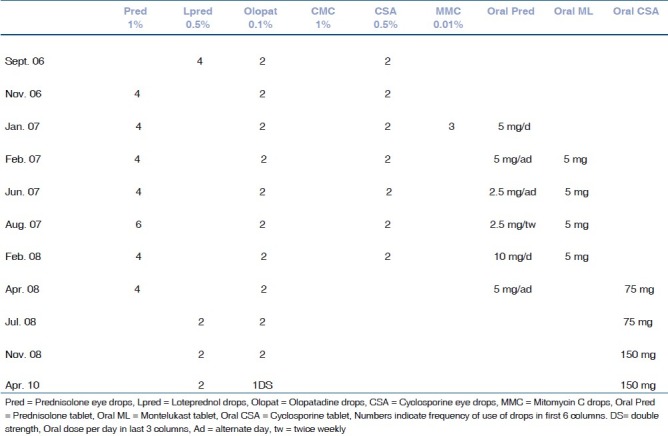
The child was advised loteprednol 0.5% drops four times a day, olopatadine 0.1% drops twice a day, carboxymethylcellulose 1% drops four times a day and cyclosporine eye drops 0.5% twice daily (prepared in lubricants) [Table 1]. The surface inflammation however continued. In November 2006, loteprednol 0.5% drops were stopped and the child was put back on prednisolone 1% eye drops four times a day due to worsening allergy. The child worsened on attempts to reduce the frequency of steroid drops. We tried higher concentrations of cyclosporine eye drops (1% and 2%), however the child did not tolerate them well and complained of a severe burning sensation.
Table 1.
Medical treatment summary

In January 2007, mitomycin C 0.01% drops were given three times a day for a week, but no improvement was observed. Oral prednisolone 5 mg/day was started and after three weeks of therapy there was a marked reduction of the corneal erosions and limbal inflammation. The oral steroids were reduced to 5 mg alternate day in February 2007, 2.5 mg alternate daily in June 2007 and 2.5 mg twice weekly in August 2007. Oral Montelukast 5 mg/day were also tried for one year.
In February 2008 the child developed a shield ulcer in his right eye. The oral steroids were stepped up to 10 mg/day and debridement of the ulcer was done under intravenous anesthesia. A bandage lens was put in twice but lost within two days each time. The shield ulcer was refractory to treatment and the child was referred to an immunologist for immunosuppressive therapy.
After baseline work up he was started on oral cyclosporine 75 mg/day (3 mg/kg body weight). The child showed a dramatic response and his shield ulcer healed rapidly. Oral steroids were slowly tapered and stopped over the next three months. Prednisolone 1% eye drops were replaced with loteprednol 0.5% eye drops twice daily.
In November 2008 the child developed a shield ulcer in his left eye. The ulcer healed over two weeks after a debridement was done and cyclosporine was stepped up to 150 mg/day (5 mg/kg body weight).
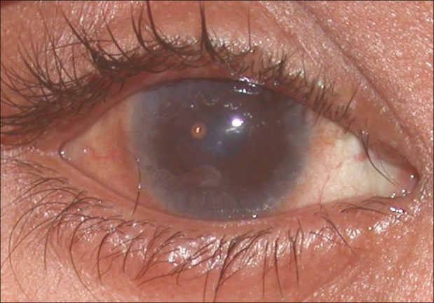
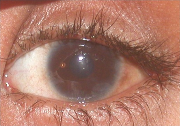
The child is on regular follow-up and is maintained on oral cyclosporine, topical loteprednol, olopatadine and lubricants. The child is significantly more comfortable now, there is a remission of inflammation and the ocular surface is stable, though it shows signs of a burned down inflammation [Figs. 1 and 2]. He however continues to have seasonal exacerbations [Table 1]. During the entire course of his treatment, the intraocular pressures were never high and no lens opacities were noted. Visual acuity at the last follow-up was 20/80 in right eye and 20/40 in the left eye.
Figure 1.

Right eye during remission shows burnt out inflammation. Annular limbal scarring, a central shield ulcer scar and diffuse subepithelial haze
Figure 2.

Left eye during remission shows burnt out inflammation. Limbal scarring more prominent at 5 o’clock, a central shield ulcer scar and diffuse subepithelial haze
Discussion
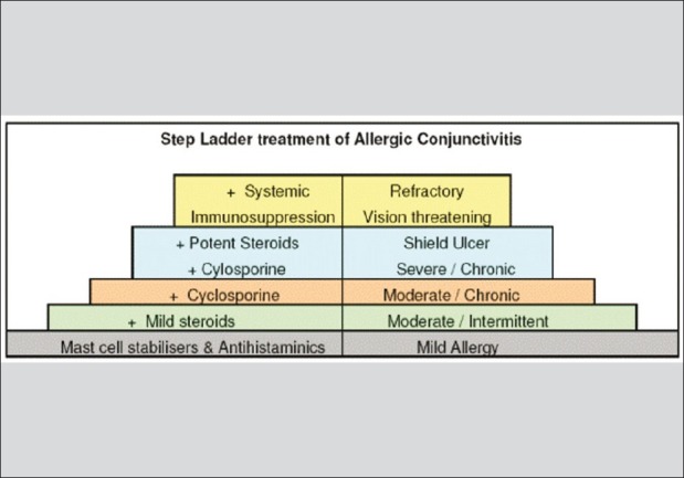
The management of vernal keratoconjunctivitis is determined by the severity of the disease. A step ladder pattern of treatment [Fig. 3] should be followed. Mast cell stabilizers are effective in mild disease (symptoms with conjunctival involvement alone).[1] In patients with moderate disease (papillae or limbal inflammation with punctate erosions) with intermittent or seasonal episodes, mild steroids are safe to use, however if the disease is chronic, cyclosporine drops (0.5–2%) may be steroid sparing and safer for long-term management.[2–4] In patients with severe disease (cobblestone papillae or limbal deficiency with coarse erosions or shield ulcers) potent steroids are indicated in addition to mast cell stabilizers, lubricants and cyclosporine. Though effective, side effects restrict their long-term use. Patients with refractory severe disease or vision-threatening allergy may benefit from systemic immunosuppressive therapy. Oral steroids are very effective; however, if required repeatedly second line immunosuppressives may be agents of choice.[5] Guidelines regarding systemic immunosuppressive therapy are not available in the literature except for an isolated case report of immunoglobulin therapy.[5]
Figure 3.

Step ladder treatment
Our patient had severe refractory and potentially vision-threatening allergy. We decided to try systemic cyclosporine therapy based on the mechanism of action of cyclosporine and the immunopathogenic mechanisms in VKC.
The immunopathogenic mechanism of VKC is complex and involves an IgE-mediated immediate hypersensitivity response as well as a delayed Th2 type of immune reaction. A Th2-driven mechanism with the involvement of mast cells, eosinophils, and lymphocytes has been suggested.[6,7] Th2 lymphocytes are responsible for both hyperproduction of IgE and for differentiation and activation of mast cells and eosinophils. Cyclosporine A is effective in controlling ocular inflammation by blocking Th2 lymphocyte proliferation and interleukin 2 (IL) production. It also inhibits histamine release from mast cells and basophils and, through a reduction of IL-5 production; it may reduce the recruitment and the effects of eosinophils on the conjunctiva.[1] Moreover, the therapeutic efficacy of cyclosporine in VKC, a conjunctival hyperproliferative disorder,[8] seems to be related to the drug's efficacy in reducing conjunctival fibroblast proliferation rate and IL-1β production.[1] Multiple studies on the efficacy of topical CsA (0.05–2%) for treating vernal keratoconjunctivitis have consistently shown a beneficial effect of the drug and its steroid-sparing effect.[2–4]
Our patient responded dramatically to oral cyclosporine and we were able to discontinue his oral steroids totally. We were also able to shift him onto milder steroid drops. After seven months of treatment there was a flare up and a recurrent shield ulcer, which promptly responded to stepping up of cyclosporine dose to 5 mg/kg. He has tolerated cyclosporine therapy very well with no major side effects and is being monitored regularly. His ocular surface has stabilized though he still has seasonal fluctuation of the surface inflammation.
References
- 1.Leonardi A. Emerging drugs for ocular allergy. Expert Opin Emerg Drugs. 2005;10:505–20. doi: 10.1517/14728214.10.3.505. [DOI] [PubMed] [Google Scholar]
- 2.Tesse R, Spadavecchia L, Fanelli P, Rizzo G, Procoli U, Brunetti L, et al. Treatment of severe vernal keratoconjunctivitis with 1% topical cyclosporine in an Italian cohort of 197 children. Pediatr Allergy Immunol. 2010;21:330–5. doi: 10.1111/j.1399-3038.2009.00948.x. [DOI] [PubMed] [Google Scholar]
- 3.Keklikci U, Soker SI, Sakalar YB, Unlu K, Ozekinci S, Tunik S. Efficacy of topical cyclosporin A 0.05% in conjunctival impression cytology specimens and clinical findings of severe vernal keratoconjunctivitis in children. Jpn J Ophthalmol. 2008;52:357–62. doi: 10.1007/s10384-008-0577-z. [DOI] [PubMed] [Google Scholar]
- 4.Kiliç A, Gürler B. Topical 2% cyclosporine A in preservative-free artificial tears for the treatment of vernal keratoconjunctivitis. Can J Ophthalmol. 2006;41:693–8. doi: 10.3129/i06-061. [DOI] [PubMed] [Google Scholar]
- 5.Derriman L, Nguyen DQ, Ramanan AV, Dick AD, Tole DM. Intravenous immunoglobulin (IVIg) in the management of severe refractory vernal keratoconjunctivitis. Br J Ophthalmol. 2010;94:667–9. doi: 10.1136/bjo.2009.165548. [DOI] [PubMed] [Google Scholar]
- 6.Maggi E, Biswas P, Del Prete G, Parronchi P, Macchia D, Simonelli C, et al. Accumulation of Th-2-like helper T cells in the conjunctiva of patients with vernal conjunctivitis. J Immunol. 1991;146:1169–74. [PubMed] [Google Scholar]
- 7.Calder VL, Lackie PM. Basic science and pathophysiology of ocular allergy. Curr Allergy Asthma Rep. 2004;4:326–31. doi: 10.1007/s11882-004-0079-0. [DOI] [PubMed] [Google Scholar]
- 8.Leonardi A, Borghesan F, DePaoli M, Plebani M, Secchi AG. Procollagens and inflammatory cytokine concentrations in tarsal and limbal vernal keratoconjunctivitis. Exp Eye Res. 1998;67:105–12. doi: 10.1006/exer.1998.0499. [DOI] [PubMed] [Google Scholar]


