Abstract
Purpose
To describe an alternative positioning technique for the fixation of pediatric medial epicondyle fractures which offers some significant advantages over traditional supine positioning.
Methods
At our institution, 27 patients with a displaced medial epicondyle fracture requiring open reduction and fixation were positioned prone for the procedure. The internally rotated operative arm lies on the hand table with the elbow in a natural flexed, pronated position. The elbow can be brought into extension and flexion for appropriate intraoperative radiographs. The fracture is then reduced with the arm in flexion and pronation, without having to pull excessively on the fragment. After reduction, the fragment is held easily in place for surgical fixation. A similar group of patients from the same time period positioned supine was also examined and compared to the patients who had the surgery prone.
Results
The average age of the 27 patients was 11.2 years (range 5.1–16.9 years). Indications for operative treatment were displaced medial epicondyle fracture (14), medial epicondyle fracture with associated elbow dislocation (12), and medial epicondyle fracture with ulnar nerve symptoms (1). At a mean of 4.5 months of follow up (1–11 months), 7 patients required the removal of hardware for screw irritation. There were no infections in the 27 surgeries and there were no other intraoperative or postoperative complications. Mild loss of flexion and extension was common in the group. Patients who had surgery in the supine position were similar with regards to patient demographics and postoperative complications, including the need for screw removal.
Conclusions
While displaced medial epicondyle fractures can be treated successfully with traditional positioning, placing patients prone for the fixation of pediatric medial epicondyle fractures offers some significant advantages over supine positioning.
Keywords: Medial epicondyle, Prone positioning, Technique
Summary
Medial epicondyle fractures constitute approximately 14 % of fractures involving the distal humerus and 11.5 % of all fractures in the elbow region [1–3]. Most often, this injury occurs in children between the ages of 9 and 14 years, with a peak incidence in the age range 11–12 years [1, 4, 5]. Treatment is generally nonoperative for nondisplaced or minimally displaced fractures. There is some controversy regarding the appropriate treatment in displaced fractures [1, 2, 4–14]. Traditional descriptions of the surgical technique to fix fractures of the medial epicondyle recommend supine positioning. We describe a technique using prone positioning that offers some significant advantages when fixing these fractures.
Technique
The patient is induced under general endotracheal anesthesia in the supine position. The operative table is positioned with gel bolsters aligned longitudinally to give support to the torso (Fig. 1). A hand table is attached to the bed on the injured side. A tourniquet is placed on the upper arm. The patient is then flipped to the prone position. The nonoperative arm is tucked down adjacent to the side of the body. The head is positioned such that the cervical spine is in a neutral position. Bony prominences of the lower extremity are well padded. The internally rotated operative arm lies on the hand table with the elbow in a natural flexed, pronated position (Fig. 2). The elbow can be brought into extension and flexion for appropriate intraoperative radiographs. The patient is prepped and draped in a standard fashion.
Fig. 1.
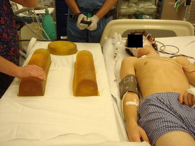
The operative table is positioned with gel bolsters aligned longitudinally to give support to the torso
Fig. 2.
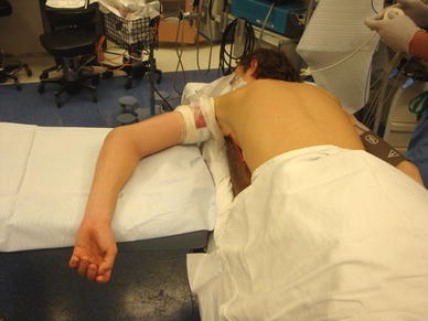
A hand table is attached to the operative side, and the arm is allowed to sit in a flexed pronated position with the shoulder internally rotated. The patient must have adequate internal rotation of the shoulder
The arm is exsanguinated with an Esmarch bandage. A longitudinal skin incision (approximately 4 cm) is centered over the medial epicondyle. Frequently, the displaced fragment can be palpable (Fig. 3). Dissection is carried deep and the displaced epicondyle is identified. The ulnar nerve is identified and protected. Any fracture hematoma is removed.
Fig. 3.
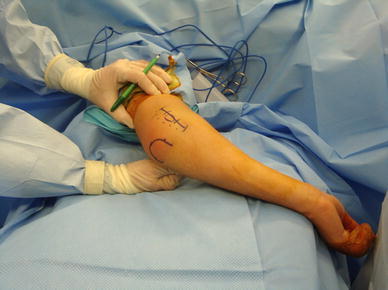
An approximately 4-cm incision is centered over the medial epicondyle
The fracture is then reduced with the arm in flexion and pronation, without having to pull excessively on the fragment. After reduction, it is possible to hold the fragment in place, and a 4.5-mm cannulated guide wire is placed into the center of the epicondyle fragment with a soft tissue guide. Anteroposterior and lateral fluoroscopic images can be obtained with movement of the arm (Fig. 4). The wire is passed up the medial column of the distal humerus, while avoiding the olecranon fossa by using fluroscopic guidance (Fig. 5). A second wire may be placed for rotational stability during drilling and screw placement. The length of the screw is determined by placing a measuring guide over the guide wire. The wire is then overdrilled. An appropriate length partially threaded 4.5-mm cannulated screw is selected and inserted over the guide pin. A washer is occasionally required, which helps distribute forces and prevent screw migration. Intraoperative radiographs should be used to confirm reduction and screw placement (Fig. 6). Elbow stability should be assessed. The wound is irrigated, and standard skin closure is carried out. The arm is splinted in 90° of flexion with a plan for early transition to a hinged elbow brace early active motion to help prevent stiffness.
Fig. 4.
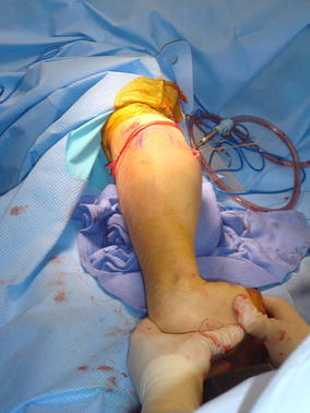
The elbow can be easily brought into extension to obtain anteroposterior radiographs of the elbow to assess pin positioning
Fig. 5.
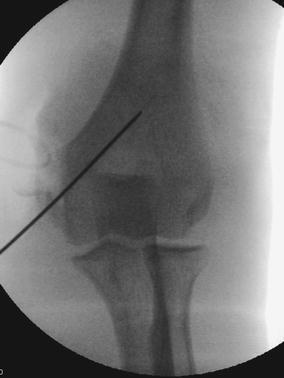
Anteroposterior radiograph of the distal humerus with the guide wire in place prior to screw placement
Fig. 6.
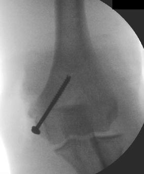
Final radiograph with screw in place and medial epicondyle reduced into its original position
Methods
We performed a CPT code search to identify all cases of open reduction and fixation of a medial epicondyle fracture at our institution from 2004 to 2011. We divided this group of patients into patients positioned prone and supine for the procedure. Positioning during this continuous period was surgeon-dependent, as some members within our group prefer prone while others prefer supine positioning. Twenty-seven patients were identified who had the procedure prone and 48 patients had the surgery in the supine position over the same time period. Data collected included demographic information, type of injury, as well as clinical data at follow up, including presence of pain, range of motion, surgical complications, and need for additional procedures.
Results
In the prone group, there were 16 male patients and 11 female patients. The average age of the patients was 11.2 years (range 5.1–16.9 years). Indications for operative treatment were displaced medial epicondyle fracture (14), medial epicondyle fracture with associated elbow dislocation (12), and medial epicondyle fracture with ulnar nerve symptoms (1).
In the 27 patients at a mean of 4.5 months of follow up (range 1–24 months), 7 patients required the removal of hardware for screw irritation. One additional patient has irritation over the screw but has elected to keep the screw in place. There were no infections in the 27 surgeries and there were no other intraoperative or postoperative complications. There were no anesthetic complications associated with the prone positioning.
Twenty-four patients have no pain at the latest follow up. Two patients have mild pain and one patient has mild pain with return to pitching. One patient has ulnar nerve symptoms which were present preoperatively. Two patients achieved less than 100° of flexion. One patient achieved only 95° of flexion and one patient only achieved 100° of flexion. Seven patients lost 5–20° of terminal flexion when compared to their contralateral elbow. Seventeen patients had a loss of terminal extension, while ten patients achieved full extension. Five patients had a loss of 20° of extension or more, including one patient who lost 45° of extension, and 12 patients lost between 5 and 20° of extension.
In the supine group, the demographics were similar to the prone group. The group included 26 male patients and 22 female patients. The average age of the patients was 12.3 years (range 6.8 years). Indications for operative treatment were displaced medial epicondyle fracture (32), medial epicondyle fracture with associated elbow dislocation (14), and medial epicondyle fracture with another associated extremity fracture (2).
In the 48 patients at a mean of 7.8 months of follow up (range 1–30 months), 12 patients required the removal of hardware for screw irritation. Two patients had mild transient ulnar nerve symptoms postoperatively. One patient developed a posterior shoulder contracture. Two patients had benign serosanguinous drainage postoperatively treated with local wound care but did not require irrigation and debridement. There were no infections in the 48 surgeries and there were no other intraoperative or postoperative complications.
Forty-seven patients have no pain at the latest follow up. One patient has mild pain with return to pitching. Thirty-three patients achieved full flexion. Two patients achieved less than 100° of flexion. One patient achieved only 90° of flexion and one patient only achieved 100° of flexion. Thirteen patients lost 5–20° of terminal flexion when compared to their contralateral elbow. Fourteen patients had a loss of terminal extension, while 34 patients achieved full extension. Six patients had a loss of 20° of extension or more, including one patient who lost 70° of extension, with the remainder losing between 20 and 30° of extension. Eight patients lost between 5 and 20° of extension.
Discussion
The medial epicondyle is a traction apophysis which begins to ossify around the age of 4–6 years, and fuses around the age of 15 years [1, 9]. The flexor mass (composed of the flexor carpi radialis, palmaris longus, and part of the pronator teres), as well as the ulnar collateral ligament, has its origin on the anterior aspect of the apophysis [1, 9, 14, 15].
The mechanism of injury for these fractures has been proposed to be either a direct blow or, more commonly, occurs after a fall on an outstretched arm with hyperextension of the wrist and a valgus moment about the elbow. The resultant force leads to avulsion of the medial epicondyle with the common flexor mass [1, 14]. Frequently, this fracture is associated with an elbow dislocation [13, 14].
Treatment is generally nonoperative for nondisplaced or minimally displaced fractures. There is some controversy regarding the appropriate treatment in displaced fractures [1, 2, 4–14]. Absolute indications for surgical treatment are incarceration in the joint or an elbow dislocation with ulnar nerve symptoms. Relative indications include ulnar nerve dysfunction and an elbow instability, especially in a patient with a displaced fracture who uses the arm for high demand activities [1]. In a recent systematic review of 14 studies, while the rate of union was 9.33 times higher in patients treated operatively, there was no difference with regard to pain or ulnar nerve symptoms in the two groups [11].
Some studies have supported surgical treatment in the setting of a displaced fracture. Hines et al. [4] surgically repaired 31 patients displaced more than 20 mm and demonstrated good to excellent results in these patients. Case and Hennrikus [6] treated 8 patients operatively with a displacement of 5 mm or more with good results. More recently, Louahem et al. reviewed the surgical treatment of 139 patients with displaced medial epicondyle fractures and reported excellent results in 130 cases and good results in 9 cases [13].
In contrast, several studies support nonoperative treatment. Josefsson and Danielsson treated displaced fractures (1–15 mm) nonoperatively and had good results at a mean of 11 years follow up, despite demonstrating a high nonunion rate [10]. Bede et al. [2] recommended nonoperative treatment based on their series of 50 patients treated operatively and nonoperatively, and only suggested operative treatment in cases where the fragment was incarcerated in the joint or when there was associated ulnar neuritis. Farsetti et al. [8] retrospectively compared two groups with medial epicondyle fractures displaced between 5 and 15 mm and demonstrated similar results between nonoperative and operative treatment. Nonunion may still occur in patients treated surgically [7].
Traditional descriptions of the surgical technique to fix fractures of the medial epicondyle recommend supine positioning. The arm is flexed and pronated to relax the common flexor wad [1, 6, 9, 14]. In order to have the medial epicondyle accessible, the arm must be externally rotated [14], which tends to force the forearm into supination and naturally places the flexor mass on stretch. In order to reduce the fracture in this position, the elbow must be flexed and the forearm must be pronated. This positioning can be awkward, and, frequently, a towel clip must be used to reduce the fracture given the residual tension of the flexor muscles [14]. In addition, there is a valgus moment placed on the arm in this position which may further impede reduction. Although K-wire fixation has been described [4], in general, cannulated screw fixation is employed [9, 14].
While displaced medial epicondyle fractures can be treated successfully with traditional positioning, we have found that prone positioning for the treatment of these fractures offers significant advantages. Although mentioned in one source [16], this has not been popularized or discussed in the current literature. In placing the patient prone, the incision is made over the arm in its natural resting position with minimal manipulation. Also, as the arm naturally sits in the flexed and pronated position, the flexor mass and, therefore, the fracture fragment is no longer under tension. In this position, placing a valgus moment across the elbow during attempted reduction is avoided. This aids in the reduction of the fragment and eliminates the need to hold tension while positioning the guide wire, as is often required in the supine position.
There are limitations to this technique. Specifically, this positioning is not possible in patients with significant shoulder pathology that prevents internal rotation of the shoulder. Further, surgeons who do not routinely operate on patients prone may be less comfortable in this position and may find the process of getting the patient safely positioned time consuming. Prone positioning does have some unique risks associated with it and an understanding of these risks and communication with the anesthesiologist is critical. There are mild but predictable changes in physiology, such as decreased cardiac output, as well as increased functional residual capacity when patients are positioned prone for surgery. There is also the risk of skin pressure injury, peripheral nerve injury, brachial plexus traction injury, and/or injury to the cervical spine due to improper positioning [17]. Finally, there is no difference in objective outcomes regarding this technique and its advantage is in the feel/ease of fracture reduction.
Conclusions
Positioning patients prone in the fixation of pediatric medial epicondyle fractures offers significant advantages over traditional supine positioning. There are minor, predictable differences with regard to anesthetic complications that must be recognized. Due to the anatomy of the medial epicondyle and the flexor mass attached to it, the medial epicondyle fracture fragment can be more easily reduced under less tension by positioning the patient prone. The complication rate is similar in patients who are positioned supine for the surgery, with approximately 25 % of patients requiring screw removal for screw irritation. Mild loss of range of motion is common in both groups.
Conflict of interest
The authors have no conflicts of interest to disclose related to this manuscript.
References
- 1.Beaty JH, Kasser JR. The elbow: physeal fractures, apophyseal injuries of the distal humerus, osteonecrosis of the trochlea, and T-condylar fractures. In: Beaty JH, Kasser JR, editors. Rockwood and Wilkins’ fractures in children. 7. Philadelphia: Lippincott Williams & Wilkins; 2010. pp. 566–577. [Google Scholar]
- 2.Bede WB, Lefebvre AR, Rosman MA. Fractures of the medial humeral epicondyle in children. Can J Surg. 1975;18(2):137–142. [PubMed] [Google Scholar]
- 3.Chessare JW, Rogers LF, White H, Tachdjian MO. Injuries of the medial epicondylar ossification center of the humerus. AJR Am J Roentgenol. 1977;129(1):49–55. doi: 10.2214/ajr.129.1.49. [DOI] [PubMed] [Google Scholar]
- 4.Hines RF, Herndon WA, Evans JP. Operative treatment of medial epicondyle fractures in children. Clin Orthop Relat Res. 1987;223:170–174. [PubMed] [Google Scholar]
- 5.Wilson NI, Ingram R, Rymaszewski L, Miller JH. Treatment of fractures of the medial epicondyle of the humerus. Injury. 1988;19(5):342–344. doi: 10.1016/0020-1383(88)90109-X. [DOI] [PubMed] [Google Scholar]
- 6.Case SL, Hennrikus WL. Surgical treatment of displaced medial epicondyle fractures in adolescent athletes. Am J Sports Med. 1997;25(5):682–686. doi: 10.1177/036354659702500516. [DOI] [PubMed] [Google Scholar]
- 7.Duun PS, Ravn P, Hansen LB, Buron B. Osteosynthesis of medial humeral epicondyle fractures in children. 8-year follow-up of 33 cases. Acta Orthop Scand. 1994;65(4):439–441. doi: 10.3109/17453679408995489. [DOI] [PubMed] [Google Scholar]
- 8.Farsetti P, Potenza V, Caterini R, Ippolito E. Long-term results of treatment of fractures of the medial humeral epicondyle in children. J Bone Joint Surg Am. 2001;83-A(9):1299–1305. doi: 10.2106/00004623-200109000-00001. [DOI] [PubMed] [Google Scholar]
- 9.Hennrikus WL. Open reduction and internal fixation of displaced medial epicondyle fracture using a screw and washer. In: Kocher MS, Millis MB, editors. Pediatric orthopaedic surgery. Philadelphia: Elsevier Saunders; 2011. pp. 37–44. [Google Scholar]
- 10.Josefsson PO, Danielsson LG. Epicondylar elbow fracture in children. 35-year follow-up of 56 unreduced cases. Acta Orthop Scand. 1986;57(4):313–315. doi: 10.3109/17453678608994399. [DOI] [PubMed] [Google Scholar]
- 11.Kamath AF, Baldwin K, Horneff J, Hosalkar HS. Operative versus non-operative management of pediatric medial epicondyle fractures: a systematic review. J Child Orthop. 2009;3(5):345–357. doi: 10.1007/s11832-009-0192-7. [DOI] [PMC free article] [PubMed] [Google Scholar]
- 12.Lee HH, Shen HC, Chang JH, Lee CH, Wu SS. Operative treatment of displaced medial epicondyle fractures in children and adolescents. J Shoulder Elbow Surg. 2005;14(2):178–185. doi: 10.1016/j.jse.2004.07.007. [DOI] [PubMed] [Google Scholar]
- 13.Louahem DM, Bourelle S, Buscayret F, Mazeau P, Kelly P, Dimeglio A, Cottalorda J. Displaced medial epicondyle fractures of the humerus: surgical treatment and results. A report of 139 cases. Arch Orthop Trauma Surg. 2010;130(5):649–655. doi: 10.1007/s00402-009-1009-3. [DOI] [PubMed] [Google Scholar]
- 14.Smith BG, Pierz KA. Open reduction and internal fixation of fractures of the medial epicondyle. In: Flynn JM, Wiesel SW, editors. Operative techniques in orthopaedic surgery. Philadelphia: Lippincott Williams & Wilkins; 2011. pp. 1042–1045. [Google Scholar]
- 15.Silberstein MJ, Brodeur AE, Graviss ER, Luisiri A. Some vagaries of the medial epicondyle. J Bone Joint Surg Am. 1981;63(4):524–528. [PubMed] [Google Scholar]
- 16.Morrissy RT, Weinstein SL. Open reduction and internal fixation of fractures of the medial epicondyle. In: Morrissy RT, Weinstein SL, editors. Atlas of Pediatric Orthopaedic Surgery. 4. Philadelphia: Lippincott Williams & Wilkins; 2006. [Google Scholar]
- 17.Edgcombe H, Carter K, Yarrow S. Anaesthesia in the prone position. Br J Anaesth. 2008;100(2):165–183. doi: 10.1093/bja/aem380. [DOI] [PubMed] [Google Scholar]


