Abstract
Context:
Accessory mental foramen is a rare anatomical variation. Even so, in order to avoid neurovascular complications, particular attention should be paid to the possible occurrence of one or more accessory mental foramen during surgical procedures involving the mandible.
Case report:
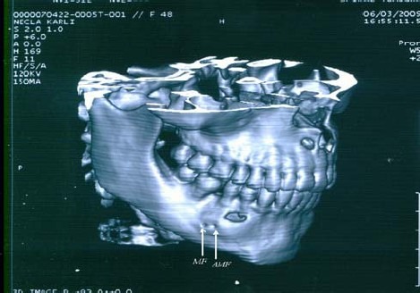
A 3-dimensional computed tomography (3D-CT) scan of a female patient revealed an accessory mental foramen on the right side of her mandible.
Conclusion:
A 3D-CT scan should be obtained prior to mandibular surgeries so that the presence of accessory mental foramen can be detected, and so that the occurrence of a neurosensory disturbance or hemorrhage can be avoided. Although this anatomical variation is rare, it should be kept in mind that an accessory mental foramen may exist.
Keywords: Mandible, 3D-CT, panoramic radiograph, clinical anatomy
Introduction
The mental foramen is located on the anterolateral aspect of the mandible, 13-15 mm superior to the inferior border of the mandibular body. The direction of the opening of the mental foramen is outward and upward in a posterior orientation[1]. The mental foramen is most usually single in human; when it is double or multiple, the additional foramen is termed accessory foramen. An accessory mental foramen is reported to be rare, with a prevalence ranging from 1.4 to 10 %[2,3,4].
Utmost care to the accessory mental foramen and nerve is essential during dental implant surgery and any surgical procedure involving the mandibular molar and premolar region. This care may reduce the rate of paralysis and hemorrhage in mental region, lower lip and gingiva from the mental foramen to the midline of the ipsilateral side.
Case Report
A female patient aged 48 years old was referred to the Department of Oral and Maxillofacial Surgery in the Faculty of Dentistry at Istanbul University with symptoms of pain and swelling localized on the anteromedial aspect of her right mandible. Intraoral examination revealed a swelling of the vestibule of the mouth, extending from the mesial side of the right canine to the distal side of the right second premolar.
Initial radiological examination on the panoramic radiograph depicted a poorly circumscribed expansible radiolucency. A biopsy was performed, and the pathologic diagnosis was odontogenic keratocyst. Prior to enucleation of the cyst with extraction of the involved tooth, a 3-dimensional computed tomography (3D-CT) image was requested (Fig. 1). Inspection of the CT image revealed the presence of double mental foramen. Of the mental foramina, the mesial one was defined as an accessory mental foramen by an experienced anatomist (H. A. B).
Fig. 1.

3D-CT scan of a 48-year old female patient depicting the accessory mental foramen. MF: Mental foramen; AMF: Accessory mental foramen.
Discussion
The mental foramen is incomplete until the 12th gestational week, when the mental nerve separates into several fasciculi at that site. It has been suggested that separation of the mental nerve earlier than the formation of the mental foramen could be a reason for the formation of the accessory mental foramen[5].
The incidence of accessory mental foramen varies between ethnic groups, and is reported as follows: 2.6% in French; 1.4% in American Whites; 5.7% in American Blacks; 3.3% in Greeks; 1.5% in Russians; 3.0% in Hungarians; 9.7% in Melanesians; and 3.6% in Egyptians[6]. Studies performed in a Japanese population showed that accessory mental foramen is less rare, with a prevalence ranging from 6.7 to 12.5% in Japan[7]. In a previous study of ours, we found a unilateral accessory mental foramen among 45 dry mandibles (2.22%)[8]. These reports reveal that non-Caucasians may have a higher incidence of accessory mental foramen than Caucasians.
Previous studies reported no gender differences[7,9]. The case in the present report is a female.
In the case reported on here, the foramina were located under the second premolar. This finding is consistent with the findings of our previous study in which the highest percentage (42.3%) of mental foramen were found under the second premolar[8].
Absence of mental foramen has also been reported. De Freitas found no mental foramen in 3 cases out of 2870 sides of 1435 dry skulls[10]. Instead of 2 cases cited by Freitas et al, No other published reports of the absence of mental foramen have been found.
Accessory mental foramina have been reported to be detected by macroscopic investigations on dry skulls, investigations with plane radiography, periapical radiography, and computed tomography. As far as we are concerned, unbiased radiological interpretation of an accessory mental foramen is possible only on CT images since the disadvantages of low image quality, low magnification, and distortion on the panoramic and periapical radiographs is a concern. We contend that if the mental foramen cannot be clearly identified on panoramic radiographs under ordinary exposure and viewing conditions, a 3D-CT should be utilized to determine the extent and location of the mental foramen prior to surgical procedures. However, if only a panoramic radiograph instead of a CT scan can be obtained, in order to improve visualization of the manndibular canal, the patient's head should be tilted 5° downward with reference to the Frankfort horizontal reference bar of the machine, as suggested by Dharmar[11].
Reports of neurosensory disturbances during surgical procedures involving the mandible are not rare; for instance, neurosensory disturbances are reported to range up to 12% in genioplasty[12]. In order to avoid neurovascular complications during implant placement, regional anesthesia, surgical correction of jaw deformities and periapical surgery, the probability of the existence of an accessory mental foramen should be kept in mind.
References
- 1.Haghanifar S, Rokouei M. Radiographic evaluation of the mental foramen in a selected Iranian population. Indian J Dent Res. 2009;20:150–152. doi: 10.4103/0970-9290.52886. [DOI] [PubMed] [Google Scholar]
- 2.Igarashi C, Kobayashi K, Yamamoto A, Morita Y, Tanaka M. Double mental foramina of the mandible on computed tomography images: a case report. Oral radiol. 2004;20:68–71. [Google Scholar]
- 3.Cagirankaya LB, Kansu H. An accesory mental foramen: a case report. J Contemp Dent Pract. 2008;9(1):98–104. [PubMed] [Google Scholar]
- 4.Kieser J, Kuzmanovic D, Payne A, Dennison J, Herbison P. Patterns of emergence of the human mental nerve. Archives of Oral Biology. 2002;47:743–747. doi: 10.1016/s0003-9969(02)00067-5. [DOI] [PubMed] [Google Scholar]
- 5.Naitoh M, Hiraiwa Y, Aimiya H, Gotoh K, Ariji E. Accessory mental foramen assessment using cone-beam computed tomography. Oral Surg Oral Med Oral Pathol Oral Radiol Endod. 2009;107:289–294. doi: 10.1016/j.tripleo.2008.09.010. [DOI] [PubMed] [Google Scholar]
- 6.Sawyer DR, Kiely ML, Pyle MA. The frequency of accessory mental foramina in four ethnic groups. Archives of Oral Biology. 1998;43(5):417–420. doi: 10.1016/s0003-9969(98)00012-0. [DOI] [PubMed] [Google Scholar]
- 7.Toh H, Kodama J, Yanagisako M, Ohmori T. Anatomical study of the accessory mental foramen and the distribution of its nerve. Okajimas Folia Anat Jpn. 1992;69:85–87. doi: 10.2535/ofaj1936.69.2-3_85. [DOI] [PubMed] [Google Scholar]
- 8.Kokten G, Buyukanten M, Balcioglu HA. Foramen mentalenin cap ve lokalizasyonunun kuru kemik ve panoramik radyografilerde. Istanbul Universitesi Dishekimligi Fakultesi Dergisi. 2004;38(10):65–71. [Google Scholar]
- 9.Naitoh M, Hiraiwa Y, Aimiya H, Gotoh K, Ariji E. Accessory mental foramen assessment using cone-beam computed tomography. Oral Surg Oral Med Oral Pathol Oral Radiol Endod. 2009;107(2):289–294. doi: 10.1016/j.tripleo.2008.09.010. [DOI] [PubMed] [Google Scholar]
- 10.de Freitas V, Madeira MC, Toledo Filho JL, Chagas CF. Absence of the mental foramen in dry human mandibles. Acta Anat (Basel) 1979;104(3):353–355. doi: 10.1159/000145083. [DOI] [PubMed] [Google Scholar]
- 11.Dharmar S. Locating the mandibular canal in panoramic radiographs. Int J Oral Maxillofac Implants. 1997;12:113–117. [PubMed] [Google Scholar]
- 12.Hwang K, Lee WJ, Song YB, Chung IH. Vulnerability of the inferior alveolar nerve and mental nerve during genioplasty: an anatomic study. J Craniofac Surg. 2005;16(1):10–14. doi: 10.1097/00001665-200501000-00004. [DOI] [PubMed] [Google Scholar]


