Abstract
Context:
The analysis of allergic drug reaction pathology may be difficult, especially if multiple histological reaction patterns are detected on review of hematoxylin and eosin (H&E) stained sections. In this case, we emphasize the value of adding immunohistochemistry (IHC) and multicolor direct immunofluorescence (DIF) as tools to improve the diagnosis of these complex disorders.
Patient and Methods:
Our patient is a twenty-year-old Caucasian female, who presented with a sudden onset of erythematous macules on the skin following administration of amoxicillin. Lesional tissue was examined by H & E and IHC, and perilesional tissue by DIF and IHC.
Results:
The H&E findings revealed diffuse dermal edema, and a mild, superficial, perivascular dermatitis with a mixed inflammatory infiltrate, consistent with an allergic drug eruption. The IHC and DIF studies revealed autoreactivity to sweat glands, nerves and dermal blood vessels, as well as dermal deposits of immune reactants such as fibrinogen and complement around the inflamed areas.
Conclusions:
Fibrin-fibrinogen degradation products have been shown in some cases of allergic disorders; thus, we encourage the effect further testing for these immunoreactants in biopsies from patients with possible allergic drug reactions.
Keywords: Drug eruption, multicolor immunofluoresence, immunohistochemistry
Introduction
Drug hypersensitivity results from interactions involving a pharmacologic agent and the immune system[1]. Drug hypersensitivity reactions represent only a small subset of all adverse drug reactions. Allergic reactions to medications represent a class of drug hypersensitivity reactions mediated by immunoglobulin E (IgE)[1]. Immune-mediated drug reactions may be classified in the Gell and Coombs classification system, a broadly accepted conceptual framework for understanding complex immune reactions[1]. However, some drug hypersensitivity reactions involve further, poorly understood mechanisms that are not easily classified.
Identifiable risk factors for drug hypersensitivity reactions include age, female gender, concurrent illnesses, current medications (including alternative therapy), and a previous hypersensitivity to related medications[1]. Drug hypersensitivity usually represents a clinical diagnosis, based on empirical data[1]. However, laboratory testing may be useful in the diagnosis, with skin testing providing the greatest specificity. Treatment is largely supportive, and includes discontinuation of the offending medication, symptomatic treatment, and patient instruction[1].
Patients with penicillin allergy should avoid carbapenems, and vigilance should be used in prescribing cephalosporins in these patients[2]. Reactions to radiocontrast media can be limited by pretreatment with prednisone, diphenhydramine, and either ephedrine or a histamine H2 -receptor antagonist[2].
Case Report
A twenty-year-old Caucasian female initially presented to an otolaryngologist with a sore throat and ear infection, and was prescribed amoxicillin following examination. After a three day course of amoxicillin, the patient complained of widespread pruritus, at which time her antibiotic treatment was discontinued. The following day, the patient suddenly presented with a morbilliform rash, including a few small, tense blisters. Skin lesions were located on the neck, chest, abdomen, arms, legs, and left hip. No palmoplantar, conjunctival, oral, or genital lesions were observed. No herpes virus infection was noticed. The patient confirmed associated weakness concurrent with the rash. Chest radiography findings were within normal limits. The patient denied the use of complementary and alternative medicine products.
Amoxicillin was discontinued prior to obtaining a lesional skin punch biopsy. At the same time, a second, perilesional skin punch biopsy was taken and placed in Michel's transport medium for direct immunofluorescence (DIF) studies. The patient then began a therapeutic regimen of diphenhydramine hydrochloride, 25 mg four times daily. Oral prednisone tablets were provided at an initial dose of 40 mg per day for 3 days, and subsequently reduced to 20 mg per day for 3 days, and followed by 20 mg every other day until the skin lesions resolved. The patient was also prescribed pramoxine/hydrocortisone ointment, which was applied topically four times per day until the lesions healed.
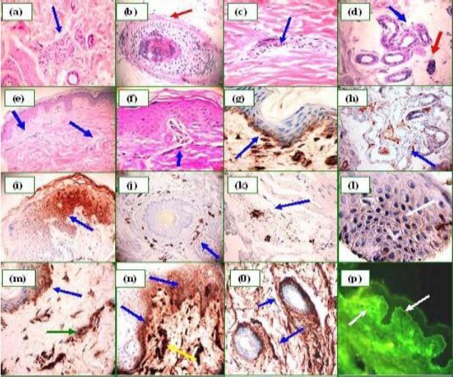
Examination of the H&E sections demonstrated a histologically unremarkable epidermis. No prominent interface or dermal periadnexal inflammation was identified (Fig. 1). A mild, superficial, perivascular dermal infiltrate of lymphocytes, histiocytes, and occasional eosinophils was appreciated. Neutrophils were rare. No acantholysis was appreciated. Focal, mild, perieccrine lymphocytic inflammation was noted; however, no significant eccrine gland necrosis was observed (Fig. 1).
Fig. 1.

H&E sections (a through d). a Displays a mild, perineural, lymphocytic infiltrate (blue arrow) (100X). b. Mild hair follicle spongiosis and a mild, perifollicular, lymphocytic infiltrate (red arrow) (200X). c. Mild dermal perivascular lymphocyte infiltration (blue arrow) (400×). d. Weakly positive PAS staining around the sweat glands (blue arrow); the red arrow indicates a partially necrotic gland (100×). e. Mild dermal edema and perivascular lymphocyte infiltration (e, 100×, and f, 400×) (blue arrows). g. Positive BMZ staining using anti-human fibrinogen (blue arrow). h. The sweat glands display positive staining using anti-human IgM. i, Positive intra-cytoplasmic staining of some keratinocytes. j and k, Positive MCT around some sebaceous glands and/or nearby vessels, respectively. l, Pseudo-pemphigus pattern utilizing C1q (400X). m. Positive BMZ staining (blue arrow) using C3c, and within the superficial vessels (green arrow). n and o. When staining for fibrinogen, positivity was seen at the BMZ and some intracytoplasmic areas of the epidermis (blue arrows), and also around the superficial vessels (yellow arrow). In o, note the anti-fibrinogen positivity around a sweat gland ductus (blue arrows). p Correlating DIF, showing positivity to the BMZ and vessels using anti-fibrinogen, as demonstrated by IHC in o (white arrows).
In addition, mild, perineural dermal inflammation consisting primarily of lymphocytes was noted; diffuse dermal edema and focal hair follicle spongiosis were also observed. For multicolor DIF, skin cryosections were prepared and incubated with multiple FITC-conjugated secondary antibodies. The antibodies were of rabbit origin and included: a) anti-human IgG, b) anti-human IgA, c) anti-human IgM, d) anti-human fibrinogen, and e) anti-human albumin from Dako (Carpinteria, California, USA). We also utilized anti-human C1q, FITC conjugate (Southern Biotech, Birmingham, Alabama, USA). Finally, monoclonal anti-human IgG4 FITC conjugated antibody was used to test for possible intercellular staining between keratinocytes (ICS) (Sigma-Aldrich, Saint Louis, Missouri, USA). The slides were then counterstained with either Dapi (Pierce, Rockford, Illinois, USA) or TO-PRO®-3/DNA (Invitrogen, Carlsbad, California, USA), washed, coverslipped, and dried overnight at 4°C.
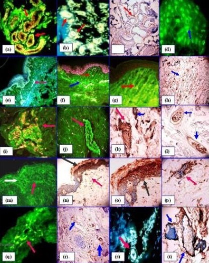
In Figures 1 and 2, we show the most prominent DIF patterns observed, including 1) autoreactivity to 1) sweat glands and nerves; 2) intercellular (ICS) epidermal pemphigus-like staining with IgM antibodies; 3) basement membrane zone (BMZ) findings, 4) antinuclear antibody (ANA) findings, and 5) compartmentalization of some areas of the dermal extracellular matrix, especially when using fibrinogen as an antibody and in areas adjacent to the main immunological process.
Fig. 2.

(a, b, e, f, s DIF), (d, g, j, m monocolor DIF), (c, h, k, l, n, o, p, r and t IHC). a, Positive deposits of FITC-conjugated anti-human-fibrinogen, around the sweat glands (green stain) (red arrows) (200×). b. We utilized egg white at a concentration of 0.05% in PBS and diluted the anti-human-fibrinogen to distinguish between real autoreactivity of the sweat glands, versus the autofluoresence of lipofuscin in these glands. The glands shows real autoreactivity (white staining) (red arrows) (200X). c. The sweat glands were also positive using anti-fibrinogen (red arrow). d. Positive ANAs (speckled pattern) were observed inside several keratinocytes utilizing anti-human total IgG (white arrow). e. Immunoreactivity to vessels (white-bluish stain) using anti-human fibrinogen (fuchsia arrow). The nuclei were counterstained with DAPI (blue stain). f. Positive, wavy deposits of fibrinogen (indicating some compartmentalization of the dermis) in proximity to the areas where the main inflammatory process occurred (red arrow); the blue arrow shows positivity to superficial vessels. In a and f, the nuclei of the cells are also counterstained with TO-PRO®-3/DNA (pink). g. Same as f, but without nuclear counterstaining and at higher magnification. h. The extracellular matrix compartmentalization is visualized by utilizing anti-human fibrinogen (as shown in f and g) (blue arrow). i, j, and k. DIF and correlating IHC, displaying positive nerve reactivity with fibrinogen (in i and j, DIF utilizes FITC-conjugated anti-human fibrinogen) (green) magenta arrows), i, includes nuclei counterstaining with TO-PRO®-3/DNA (pink), k, IHC positivity in the nerve (red arrow) and to vessels (blue arrow) utilizing anti-fibrinogen. l. By IHC, we demonstrate neural staining using anti-PPG.9.5 (blue arrows). m. “Pseudo-pemphigus” pattern, weakly positive ICS was noted between epidermal keratinocytes using FITC-conjugated anti-human IgM-antiserum (green) (white arrow). In addition, some BMZ area deposits of the same immunoglobulin were observed (red arrow). n and o. IHC showing reactivity of the BMZ (red arrow), as well as “pseudo pemphigus” pattern (black arrow) in this case when using anti-human C3c. p. C3c was positive against vessels. q. Immunoreactivity to vessels using anti-human antiserum against FITC-conjugated fibrinogen (blue arrow). r. IHC positive staining against deep vessels using C3c (blue arrow). s Same technique as b, but in this case utilizing anti-human C3c to show positive staining of the sweat glands (red arrow) (white stain). t. Fibrinogen positive staining around the sebaceous glands (blue arrows).
Immunohistochemistry studies (IHC): In our IHC studies, we used a dual endogenous peroxidase blockage system, according to Dako guidelines, with the addition of an Envision dual link. Furthermore, we applied 3, 3 diaminobenzidine and counterstained with hematoxylin. We stained for goat anti-human CD3, CD4, CD8, CD45, IgM, IgG, IgE, IgA, C1q, C3c, C3d, mast cell tryptase (MCT), fibrinogen, and antibody to protein gene product 9.5 (PPG 9.5). We observed positive staining by both DIF and IHC in the sweat glands, sebaceous glands and in dermal blood vessels utilizing various antibodies. The IHC studies displayed correlation with the DIF studies (Figs. 1 and 2). Following the diagnosis of an allergic drug reaction, the patient was treated with oral prednisone tablets; the response to this therapy was characterized by slow regression of the cutaneous lesions.
Discussion
Some of the most challenging diagnoses for clinicians and pathologists are the drug-induced cutaneous dermatoses. Whereas eosinophils are an important histologic sign of a drug-induced eruption, they may also be histologically conspicuous in other skin disorders without a drug etiology[1]. Whether confined to the skin or part of a systemic disease, drug eruptions are characterized by a spectrum of clinical and histologic patterns that include perivascular dermatitis, nodular and diffuse dermatitis, vesiculobullous lesions, pustular dermatosis, sclerodermoid alterations, vasculitis, folliculitis/perifolliculitis and panniculitis[1].
Physicians caring for the patient may or may not be aware that while a single drug can elicit a range of reaction patterns, no single reaction pattern is fully specific for a particular drug[1]. In our case, we presented three different methodologies (H&E, IHC and DIF) that, when combined and correlated with the clinical history, improved the detection and diagnosis of drug-induced-pathology. In contradistinction, less accurate results may result from the use of 1) H& E examination alone, or 2) utilizing classical, monocolor fluorescein isothyocyanate(FITC) fluorescence DIF. Specifically, the use of monocolor DIF may lead to interpretation errors involving non-pathologic, incidental background immunoreactivity.
Although the temporal link between initiation of drug therapy and the onset of the clinical rash is often helpful in establishing the diagnosis, drug reactions may also occur during the course of chronic drug ingestion and/or as a result of drug-drug interactions[2]. Drug eruption pathology may include the presence of overlapping histological reaction patterns, and incongruent clinical and histopathological features[2].
We presented in our case several immunological alterations that may help to improve the diagnostic process, including autoreactivity to sebaceous and sweat glands, vessels, nerves, and pseudo-pemphigus and ANA findings. These alterations include deposition of various immunoreactants including immunoglobulins, complement and fibrinogen. Other well known findings in cutaneous drug eruptions included alterations within the basement membrane layer of the epidermis and around the dermal blood vessels[5]. In these areas, we found positive staining utilizing DIF in the sweat glands, hair follicles and nerve fibers. All of these alterations were present with mild inflammatory infiltrates surrounding the same skin appendices. As previously noted, we detected IgM deposits between epidermal keratinocytes (in a pemphigus-like pattern) and some patchy IgM deposits at the BMZ. Additionally, we observed speckled ANAs within epidermal keratinocytes[3–5].
In this case, the patient displayed a 1:80 ANA on serum examination. The pemphigus-like pattern has been previously documented by DIF in selected cases of other conditions such as toxic epidermal necrolysis, erythema multiforme and skin burns[3–5]. The presence of these pemphigus-like antibodies occurred in patients without clinical pemphigus vulgaris; thus, an enzyme-linked immunosorbent assay (ELISA) examination for desmogleins 1 and 3 were negative. As noted, the pemphigus-like antibodies that we detected in the epidermis and at the BMZ were of IgM isotype, not IgG or IgG4 (the most common subclasses found in pemphigus vulgaris).
In regard to the positive staining observed by DIF on other skin appendices, this phenomenon has been described in drug eruptions by other authors, specifically occurring in some dermal vessels, and at the BMZ[6]. Specifically, other authors have described a series of 34 patients with a diagnosis of erythema multiforme, who displayed pathologic BMZ and vascular fluorescence with both fibrinogen and C3[7,8]. In more severe allergic drug eruptions such as toxic epidermal necrolysis, prior reports have documented positive DIF staining in the sweat ducts and glands[9,10]. In addition to the previously described DIF findings, epidermal and nerve findings by both light and electron microscopy have been previously documented in drug reactions displaying tumefaction, edema and lamellar degeneration[11]. Other authors have also described the presence of a speckled pattern of antinuclear antibodies (ANAs) in reactions to certain drugs such as hydralazines, chlorpromazine, isoniazid and penicillin[7].
Our DIF findings correlated with our IHC and H & E data to demonstrate that, when utilizing simple H&E and monocolor DIF, case findings could be misinterpreted as non-pathologic, background immunofluorescence, when these findings actually represent pathologic alterations. Other authors have also reported diffuse intraepidermal and dermal deposition of immunoreactants on DIF as a clue to the early diagnosis of epidermal necrolysis[12]. Finally fibrin-fibrinogen degradation products have also been previously documented in cutaneous allergic drug reactions[13].
Acknowledgments
Jonathan Jones, HT and Lynn Nabers, HT for their excellent technical assistance at Georgia Dermatopathology Associates.
The work was performed utilizing funding from Georgia Dermatopathology Associates.
References
- 1.Rield MA, Casillas AM. Adverse drug reactions: Types and treatment options. Am Fam Physician. 2003;68:1781–1790. [PubMed] [Google Scholar]
- 2.Ramdial PK, Naidoo DK. Drug-induced cutaneous pathology. J Clin Pathol. 2009;62:493–504. doi: 10.1136/jcp.2008.058289. [DOI] [PubMed] [Google Scholar]
- 3.Cormane RH, Petzoldt D. Immunofluorescence studies on the affinity of pemphigus antibodies to epithelial intercellular substances. Dermatologica. 1970;140:1–8. doi: 10.1159/000252527. [DOI] [PubMed] [Google Scholar]
- 4.Ansel J, Petrozzi JW, Kumar V. Possible drug-induced pemphigus-like antibodies with the clinical manifestation of erythema multiforme. Arch Dermatol. 1983;119:1006–1009. [PubMed] [Google Scholar]
- 5.Gibson LE, van Hale HM, Schroeter AL. Direct immunofluorescence for the study of cutaneous drug eruptions. Acta Derm Venereol. 1986;66:39–44. [PubMed] [Google Scholar]
- 6.Stein KM, Schlappner OL, Heaton CL, Decherd JW. Demonstration of basal cell immunofluorescence in drug-induced toxic epidermal necrolysis. Br J Dermatol. 1972;86:246–252. doi: 10.1111/j.1365-2133.1972.tb02224.x. [DOI] [PubMed] [Google Scholar]
- 7.Cannat A, Seligmann M. Possible induction of antinuclear antibodies by isoniazid. Lancet. 1966;1:185–187. doi: 10.1016/s0140-6736(66)90704-5. [DOI] [PubMed] [Google Scholar]
- 8.Finan MC, Schroeter AL. Cutaneous immunofluorescence study of erythema multiforme: correlation with light microscopic patterns and etiologic agents. J Am Acad Dermatol. 1984;10:497–506. doi: 10.1016/s0190-9622(84)80101-2. [DOI] [PubMed] [Google Scholar]
- 9.Fellner MJ, Prutkin L. Morbilliform eruptions caused by penicillin. A study by electron microscopy and immunologic tests. J Invest Dermatol. 1970;55:390–395. doi: 10.1111/1523-1747.ep12260501. [DOI] [PubMed] [Google Scholar]
- 10.Laplante L, Barcelo R, Carrière S, Legresley LP, Beaudry C. Bullous lesions and necrosis of the sudoriparous glands in acute drug poisoning. Union Med Can. 1971;100:1755–1761. [PubMed] [Google Scholar]
- 11.Ding ZH, Liu WK, Ding JY. Experimental morphological study of injection nerve injuries in rabbits. Zhonghua Hu Li Za Zhi. 1997;32:128–131. [PubMed] [Google Scholar]
- 12.King T, Helm TN, Valenzuela R, Bergfeld WF. Diffuse intraepidermal deposition of immunoreactants on direct immunofluorescence: a clue to the early diagnosis of epidermal necrolysis. Int J Dermatol. 2007;33:634–636. doi: 10.1111/j.1365-4362.1994.tb02922.x. [DOI] [PubMed] [Google Scholar]
- 13.Palma-Carlos AG, de Sá A, Joana-Geraldes M, Ducla-Soares J, Migueis-Clode MH. Fibrin-fibrinogen degradation products in allergic disorders. Allergol Immunopathol (Madr) 1974;2:419–422. [PubMed] [Google Scholar]


