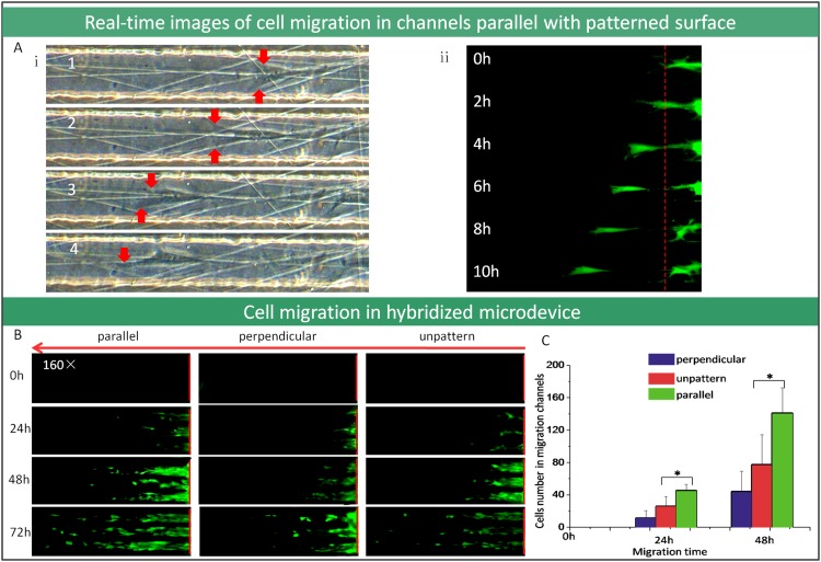
Figure 5.
Cell migration investigation used microdevice by integrating microfluidic channel with nanofiber patterned surface. (a) The real-time images of cell migration in the microchannels which are parallel with the patterned surface (100×). (b) The fluorescent microscope images of cell migration in the microchannels that are parallel or perpendicular with patterned surface (100×). (c) The comparison of migrated cells number in microfluidic channels (*P < 0.05).

