Abstract
Treatment protocols exist for vascular obstruction due to injections with hyaluronic acids. Options for vascular insult due to non-hyaluronic acid products are less defined. The authors report two cases of vascular insult due to calcium hydroxylapatite and discuss treatment options. Patients who have vascular occlusion due to calcium hydroxylapatite require immediate intervention. The authors’ suggested protocol is elucidated and presented as a basis for future discussions and clinical trials.
One of the most significant adverse events associated with injections of soft tissue augmentation products is vascular occlusion. Adverse events associated with vascular occlusion include pain, long-term erythema, neovascularization, epidermal and dermal necrosis, scarring, and pigment changes. While rare, these events are significant for both patient and physician.
Vascular compromise is a function of compression and/or embolization of material into the vasculature. When the material injected is a hyaluronic acid, the compromise may be partially mitigated by use of hyaluronidase. However, when the material is calcium hydroxylapatite, poly L lactic acid, silicone, fat, or methylmethacrylate, there is little mitigation that can be performed. Among injectors of soft tissue augmentation products, this lack of mitigation potential is one of the main reasons that semipermanent products are not used more frequently. Our goal is not to promulgate these as definitive measures, but rather to establish some treatment protocol that may be helpful as well as to provide the basis for future protocols.
The protocol outlined by Glaich et al1 calls for a coherent, sequential treatment for vascular compromise resulting from injections of hyaluronic acids. This protocol elaborates a sequence of events that utilize topical nitroglycerin, hyaluronidase, and other modalities to minimize the damage from impending necrosis. Other authors have also published guidelines for the treatment of impending necrosis following soft tissue augmentation following injections of hyaluronic acid.2,3 Typically, these events most frequently occur in the nasolabial crease where the angular artery is impacted. The glabella is another area that is impacted by vascular events. Early experience with cross-linked bovine collagen (Zyplast) prepared many injectors for this eventuality and many believe that necrosis in this site is linked not only to the nature of Zyplast but also to the proximity of the underlying vessels to the area that the injection needle is placed. The small injection area and bony foundation are likely to be contributing factors for vascular adverse events in this area. Necrosis of the marionette lines with soft tissue augmentation products is also a potential risk with injections into this area.
Illegal injections of hyaluronic acid into the vaginal area has been associated with pulmonary embolism.4 Embolization of material is reported with several soft tissue augmentation products including fat and hyaluronic acid.5 When the embolization involves the retinal artery, loss of vision may result.6,7 Necrosis of the nasal ala has also been reported with injections of soft tissue augmentation products.8 Particulate fillers, such as methylmethacrylate, may also cause embolization, but the rate of this occurrence with these molecules is unknown. Poly L lactic acid is now increasing in popularity. Depending on its reconstitution and time for hydration, it may be more or less of a particulate solute.
A controlled trial of various rescue treatments for vascular injury and compromise is not ethically possible. However, based upon experience with hyaluronic acid fillers and knowledge of rheologic and chemical properties of particulate fillers, it is possible to develop a suggested treatment protocol for vascular compromise with these agents.
Case Series
Case 1. A 40-year-old man presented for a cosmetic evaluation. Examination showed that he had moderate mid-face tissue loss with moderately deep nasolabial creases. He had Fitzpatrick type II skin and had no prior history of filler use. After reviewing the various options including particulate hyaluronic acid fillers and calcium hydroxylapatite (CAHA, Radiesse, Merz Aesthetics, Inc.) it was decided to proceed with injections of CAHA. Each syringe of CAHA was mixed with 0.1cc of 1% lidocaine with 1:100,000 epinephrine. A total of 1.5mL was injected on each side using a serial puncture technique and a 28-guage needle measuring 3/4 of an inch in length. Upon injecting the superior aspect of his right nasolabial crease, a blanching was noted. The blanching extended along the lateral aspect of his nose and up to his inferior eyelid in a distribution that suggested vascular distribution. However, there was no sign of impending necrosis, such as development of a dusky hue, and it was thought that vascular spasm due to the epinephrine was the cause of the blanching.
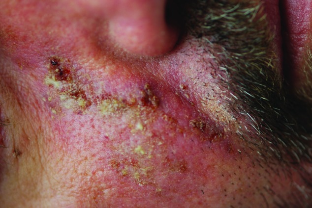
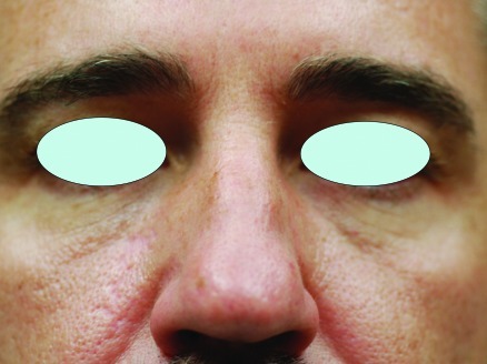
However, the next morning, the patient called the office and complained of pain in the distribution of the angular artery. He was not able to come to the office for evaluation because of travel and a photo sent showed that there was faint erythema, but not any blue discoloration or dusky appearance. Treatment with oral corticosteroids (methylprednisolone) was initiated as well as aspirin. The next day, there was some vesiculation noted on another photograph he had sent. On the third post-procedure day, the patient was seen and there was a superficial erosion noted. Small yellowish papules were also noted at that time (Figure 1). The patient was started on both oral cephalexin and valcyclovir. Cultures taken at that visit were negative for bacterial and viral growth. Over the span of the next few days, the patient was seen at frequent intervals with a superficial slough noted in the medial cheek. No necrosis was noted on any of the distal aspects of the vascular distribution. One month after his injection, the scar was treated with low energy pulsed dye laser and thereafter with a fractionated 1550 erbium laser. Following several visits, the residual scar was minimal (Figure 2).
Figure 1.
Case 1, three days after the injection. Crusted papules are visible in the distribution of the angular artery.
Figure 2.
Case 1, four months after the occlusive incident. Treatments included fractionated erbium laser as well as pulsed dye laser.
Case 2. A 49-year-old man presented for a lower face augmentation with CAHA. He was treated with this product in the past and wanted to enhance his chin and oral commissures. A small amount (0.1cc) was placed in his nasolabial crease in the midpoint of the fold. Unlike the prior case report, the CAHA in this instance was not mixed with lidocaine. Upon injection into the nasolabial crease, there was an immediate blanching in the distribution of the angular artery. The area covered by this blanch was approximately 4.5 x 7.5cm and was triangular in shape.
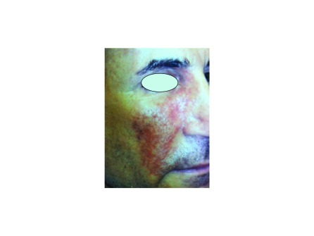
Nitroglycerin paste was immediately applied and the area was massaged. Following these procedures, the size of the blanching was reduced by about 50 percent. Over the span of a few minutes, the blanched areas turned a gray-purple (Figure 3). Approximately 30 minutes after the injection, the patient left the office.
Figure 3.
Case 2 two weeks after the injection. Bruising in the distribution of the angular artery is still prominent. This patient underwent a similar impetigo stage not shown here.
Three hours after the injection, the patient returned for evaluation and nitroglycerin paste was again applied. In addition, 600 units of hyaluronidase was injected as well as 5mL of normal saline. The hyaluronidase was injected with the hope that it would dissolve some native hyaluronic acid, thereby decreasing the pressure on the blood supply. Incision and drainage were performed to attempt to extrude the product, and the upper portion of the occlusion cleared immediately with visible evidence of vascular flow. After consultation with several colleagues, the patient was placed on oral prednisone at a dose of 40mg/day with a gradual taper. In addition, aspirin 81mg/day was added. In an effort to dilate the arterial blood supply, sildenafil (Viagra, Pfizer Inc.) was also added at a dose of 50mg/day. Hyperbaric oxygen therapy was initiated the day after the occlusion.
The patient developed an impetigo and was placed on cephalexin 500mg twice daily as well as mupirocin ointment twice daily. On the 11th day, the patient discontinued all oral medications, but continued the topical mupirocin and sunblock. He continued hyperbaric oxygen for a total of 10 sessions.
Discussion
Based upon the experience with hyaluronic acid occlusion, treatment for particulate fillers that occlude vascular structures should seek to increase blood flow to the affected areas. This may be accomplished by decreasing pressure in the anatomic compartment (using corticosteroids and hyaluronidase), increasing blood flow (with sildenafil or similar drugs, aspirin, and nitroglycerin paste), and increasing the oxygen content to the affected tissues (hyperbaric oxygen). However, unlike hyaluronic acid fillers, there is no simple reversal for CAHA, and injections with hyaluronidase are unlikely to digest the blockage. At the present time, protocols for the treatment of CAHA occlusion are based on relatively small amounts of clinical experience and empiric data rather than by evidence-based clinical trials. Thus, they are presented as suggestions rather than dogma.
One technique that may help to decrease the chance of vascular occlusion when injecting particulate fillers is the use of a cannula instead of a needle. Cannulae are available in two sizes for injection of CAHA. Each has a blunt tip and a port on the side of the cannula. This is in contrast with the needle, which has an opening at the leading edge of the cutting aspect. The design of the latter instrument will tend to introduce material into a vessel should one be encountered during injection while the design of the former will not only tend to push vessels to the side of the leading edge, but also not be as likely to introduce material into the vessel, instead injecting it to the side of it.
As with occlusion from gel-based fillers, it is imperative to minimize the degree of damage caused by vascular occlusion. One way to do this is to dilate the vasculature using 2% nitroglycerin paste applied liberally to the affected area. The authors recommend application of nitroglycerin paste 2 to 3 times daily provided that the patient does not develop symptoms such as headaches or light headedness.
Corticosteroids are indicated to diminish the inflammatory component of the injury, which can further inflame the compartment and lead to more vascular compromise. Oral corticosteroids in doses ranging from 40 to 60mg of prednisone are recommended for the first 2 to 3 days after occlusion. A taper over the first week is then initiated. Alternatively, use of a methylprednisolone dose pack is also reasonable.
Dilation of the blood vessels should be maximized with the use of drugs designed for the treatment of erectile dysfunction.9 These drugs, including sildenafil (Viagra), tadalafil (Cialis, Lilly USA, LLC), and vardenafil (Levitra, Bayer Pharmaceuticals Corporation) are selective inhibitors of cyclic guanosine monophosphate (GMP) specific phosphodiesterase type 5.9 nitric oxide, which is released during normal activities, activates guanylate cyclase, which, in turn, increases cyclic GMP. Increases in GMP cause smooth muscle relaxation, dilation of the vascular wall, and increased blood flow. As with the use of these drugs in any patient, caveats regarding heart disease and other contraindications are still pertinent.
Aspirin is used to block platelet aggregation and has moderate anti-inflammatory properties. If the vascular injury from the particulate filler has not entirely occluded the vessel, aspirin may be able to help blood flow by inhibiting platelet aggregation and blood clotting. Keeping any aspect of the vessel patent will help to increase the viability of any tissue that relies on it for circulatory support. Doses of 81mg/day should be effective in decreasing platelet aggregation, and in the acute setting, aspirin may be placed sublingually.
Hyperbaric oxygen has the potential to deliver oxygen deep into the skin and may help to keep oxygen-dependent tissues viable. Its use in flaps, grafts, and other skin that has potential vascular compromise is controversial. However, if a facility exists that can provide hyperbaric oxygen to a patient with impending necrosis, it may be reasonable to attempt a course of this treatment.
In each of these cases, clinical signs of impetigo appeared after a few days. In both patients, cultures for bacteria as well as for virus were negative. Despite the negative cultures, the patients were placed on cephalosporin antibiotics as soon as the honey-colored crust appeared. It is possible that this crust represented an exudate from a com-promised epidermal barrier. However, in the event that a crust forms following vascular occlusion, it is prudent to use oral antibiotics while the cultures are pending.
Conclusion
Particulate-based fillers are becoming more popular for soft tissue aug-mentation and facial remodeling. As the numbers of patients treated increase, the likely occurrence of adverse events, including vascular obstruction, will also increase. Since there are rational protocols extant for hyaluronic acid based vascular obstruction, it seems reasonable to create a protocol for vascular occlusion with particulate fillers. The authors’ suggested protocol is included in Table 1. The suggestions listed in this article form the basis for a discussion of what optimal treatments should be for vascular occlusion with particulate fillers. The authors look forward to more data as well as discussion on this subject.
TABLE 1.
Suggested treatment for particulate filler vascular compromise
| Nitroglycerin paste 2% | Apply immediately upon suspected necrosis and then for 5 minutes every 1-2 hours |
| Prednisone | 20-40mg each day for 3-5 days |
| Aspirin 325mg | 1 under the tongue immediately and then daily |
| Sildenafil 50mg | 1 per day for 3-5 days |
| Warm compresses | Apply 5-10 minutes every 1-2 hours (avoid burning the skin) |
| Hyperbaric oxygen | Begin treatment daily as soon as possible with continued treatments until the area has improved |
Footnotes
DISCLOSURE: Dr. Beer is an investigator for Merz Aesthetics, Medicis, and Allergan and a consultant for Medicis and Allergan. Dr. Beer is a shareholder of Allergan Corporation. Dr. Downie is a consultant and investigator for Merz, Allergan, and Medicis.
References
- 1.Glaich AS, Cohen JL, Goldberg LH. Injection necrosis of the glabella: protocol for prevention and treatment after use of dermal fillers. Dermatol Surg. 2006;32(2):276–281. doi: 10.1111/j.1524-4725.2006.32052.x. [DOI] [PubMed] [Google Scholar]
- 2.Hirsch RJ, Cohen JL, Carruthers JD. Successful management of an unusual presentation of impending necrosis following a hyaluronic acid injection embolus and a proposed algorithm for management with hyaluronidase. Dermatol Surg. 2007;33(3):357–360. doi: 10.1111/j.1524-4725.2007.33073.x. [DOI] [PubMed] [Google Scholar]
- 3.Dayan SH, Arkins JP, Mathison CC. Management of impending necrosis associated with soft tissue filler injections. J Drugs Dermatol. 2011;10(9):1007–1012. [PubMed] [Google Scholar]
- 4.Park HJ, Jung KH, Kim SY, et al. Hyaluronic acid pulmonary embolism: a critical consequence of an illegal cosmetic vaginal procedure. Thorax. 2010;65(4):360–361. doi: 10.1136/thx.2009.128272. [DOI] [PubMed] [Google Scholar]
- 5.Coronado-Malagón M, Visoso-Palacios P, Arce-Salinas CA. Fat embolism syndrome secondary to injection of large amounts of soft tissue filler in the gluteal area. Aesthet Surg J. 2010;30(3):448–450. doi: 10.1177/1090820X10373381. [DOI] [PubMed] [Google Scholar]
- 6.Peter S, Mennel S. Retinal branch artery occlusion following injection of hyaluronic acid (Restylane) Clin Experiment Ophthalmol. 2006;34(4):363–364. doi: 10.1111/j.1442-9071.2006.01224.x. [DOI] [PubMed] [Google Scholar]
- 7.Park SH, Sun HJ, Choi KS. Sudden unilateral visual loss after autologous fat injection into the nasolabial fold. Clin Ophthalmol. 2008;2(3):679–683. [PMC free article] [PubMed] [Google Scholar]
- 8.Kang MS, Park ES, Shin HS, et al. Skin necrosis of the nasal ala after injection of dermal fillers. Dermatol Surg. 2011;37(3):375–380. doi: 10.1111/j.1524-4725.2011.01891.x. [DOI] [PubMed] [Google Scholar]
- 9. Levitra [package insert]. Wayne, NJ: Bayer Healthcare; 2009.


