Abstract
Background
Chronic lymphocytic leukemia B cells display prolonged survival in vivo, but when cultured in vitro rapidly undergo spontaneous apoptosis. We hypothesize that interactions with endothelial cells in infiltrated tissues and during recirculation may have a pathogenic role in chronic lymphocytic leukemia.
Design and Methods
We evaluated apoptosis of leukemic cells after co-culture on a monolayer of human umbilical vein endothelial cells with addition of fludarabine and antibodies that block adhesion. Then, we compared microarray-based gene expression profiles between leukemic cells at baseline and after co-culture.
Results
We found that the endothelial layer protected leukemic cells from apoptosis inducing a 2-fold mean decrement in apoptotic cells after 2 days of co-culture. Moreover, the endothelial layer decreased the sensitivity of chronic lymphocytic leukemia B cells to fludarabine-induced apoptosis. Physical contact with endothelium mediated by both β1- and β2- integrins is essential for the survival advantage of leukemic cells. In particular, blocking CD106 on endothelial cells or CD18 on leukemic B cells led to the almost complete abrogation of the survival advantage (>70% inhibition of viability). However, a reduction of apoptosis was also measured in leukemic cells cultured in conditioned medium collected after 2 days of co-culture, implying that survival is partially mediated by soluble factors. Overall, the contact with endothelial cells modulated 1,944 genes in chronic lymphocytic leukemia B cells, establishing a peculiar gene expression profile: up-regulation of angiogenesis-related genes, an increase of genes involved in TGFβ and Wnt signaling pathways, secretion of cytokines recruiting stromal cells and macrophages and up-regulation of anti-apoptotic molecules such as Bcl2 and Survivin.
Conclusions
Our study supports the notion that endothelial cells are major players in the chronic lymphocytic leukemia microenvironment. Adhesion to endothelium strongly supports survival, protects from drug-induced apoptosis and extensively modifies the gene expression profile of leukemic cells.
Keywords: chronic lymphocytic leukemia, microenvironment, endothelial cells, cell adhesion
Introduction
Despite an apparent long life in vivo, chronic lymphocytic leukemia (CLL) cells die rapidly in vitro during culture in media supplemented with either autologous or fetal bovine serum.1,2 This observation suggests that the apoptotic resistance is not intrinsic to leukemia B cells but that extrinsic factors are necessary for the prolonged survival of CLL cells.
CLL cells infiltrate bone marrow and lymph node compartments, progressively disrupting the physiological architecture and functionality of tissues and generating hallmark structures called proliferation centers. These pseudo-follicular structures contain pro-lymphocytes and para-immunoblast leukemic cells, are characterized by a higher proportion of Ki-67+ cells as compared to surrounding CLL small lymphocytes and contain a follicular dendritic cell network along with several T cells.3,4 Bidirectional interactions between CLL cells, surrounding non-transformed cells of stromal and immune compartments and extracellular matrix components extend CLL-cell survival, induce genetic instability and protect from the effects of chemotherapeutics. Prolonged survival of CLL cells can be achieved in vitro by co-culture with different accessory cells present in the CLL microenvironment, such as nurse-like cells, mesenchymal marrow stromal cells or follicular dendritic cells.5
Increasing evidence suggests that angiogenesis can play a role in the pathophysiology of CLL. Angiogenesis, i.e. the formation of new blood vessels from pre-existing ones, is a complex process tightly regulated by a dynamic balance between positive and negative regulatory factors.6 Serum or plasma levels of angiogenic factors such as basic fibroblast growth factor (bFGF), vascular endothelial growth factor (VEGF) and angiopoietin 2 (Ang2) were reported to be higher in CLL patients than in normal controls.7–10 Moreover, high serum or plasma concentrations of VEGF and Ang2 define a subset of CLL patients with a poor clinical outcome.8,10,11 CLL cells induce increased angiogenesis in vitro, which is mediated by both leukemia-derived VEGF and Ang2.12 In addition, CLL-infiltrated bone marrow and lymph nodes contain abnormal vascular elements that are related to disease stage and are predictors of poor clinical outcome.7,13–15
CLL lymphocytes reside in tissues enriched with new microvessels and interact with activated endothelium during recirculation from peripheral blood to the marrow and lymph node compartments.16 Contradictory results were reported about the role of endothelial cells (EC) in CLL survival. Long et al. reported that apoptosis of CLL cells can be prevented by contact with EC hybrids EA.hy926.17 In contrast, Moreno et al. reported that the ECV-304 endothelial cell line inhibits apoptosis of CLL cells mainly through soluble factors, in particular interleukin-6 dimers.18 Elevated levels of the anti-apoptotic proteins Bcl-2, Mcl-1 and Bcl-XL, increased expression of CD38 and CD49d and NF-κB activation were reported in CLL cells co-cultured with EC.19 Likewise, Badoux et al. found that CLL cells attached to an adherent EC layer and were protected from undergoing spontaneous apoptosis through cell-cell contact.16 Conversely, a lack of survival advantage after co-culture with EC was reported in another study.20
Here, we co-cultured CLL cells on EC layers investigating the role of endothelial contact in the survival of leukemic cells. To highlight cellular pathways and molecular networks involved in this crosstalk, we analyzed gene expression changes induced in CLL cells as a result of co-culture with EC. Dissecting the complex array of interactions and studying their relative importance in induction of survival of CLL cells is necessary for future work on new therapeutic targets.
Design and Methods
Patients and samples
After obtaining informed consent in accordance with the Declaration of Helsinki with a protocol approved by the Institutional Review Board, blood samples were collected from 34 untreated CLL patients fulfilling standard clinical, morphological and immunophenotypic criteria21 at the Hematology Division of Modena Hospital. Peripheral blood mononuclear cells, taken at the time of diagnosis, were isolated by density gradient centrifugation (Ficoll, Pharmacia LKB Biotechnology, Piscataway, NY, USA). To enrich for CLL cells, the peripheral blood mononuclear cells were incubated with CD19Microbeads (Miltenyi Biotech, Auburn, CA, USA), obtaining a purity >99% as assessed by flow cytometry using phycoerythrin (PE)-conjugated CD19 (Miltenyi Biotech).
Cell culture conditions
Purified CD19+ CLL cells were suspended at a final concentration of 1×10⁶/mL in AIM-V medium (Invitrogen, Carlsbad, CA, USA) and then plated in 24-well plates alone (CLL only) or onto endothelial layers (CLL HC) formed by human umbilical vein endothelial cells (HUVEC, Cascade Biologics, Invitrogen, Portland, OR, USA). Further details on the culture conditions, sample collection and treatments are provided in the Online Supplementary Design and Methods. The in vitro angiogenesis assay (Chemicon, Millipore, Billerica, MA, USA) was performed as previously described.12
Flow cytometry and confocal microscopy
Apoptotic cell death was analyzed by flow cytometry using annexin V-FITC and propidium iodide (PI)-PE staining (eBioscience, San Diego, CA, USA). Tie2-expressing CLL cells were evaluated using PE-conjugated Tie2 (R&D System). For confocal microscopy studies, HUVEC were visualized by staining with a mouse anti-human CD31 Alexa 647-conjugated (AbD Serotec, Oxford, UK). Tie-2 was visualized using a goat polyclonal antibody (R&D Systems) followed by Dylight 488-conjugated bovine anti-goat IgG (Jackson Immunoresearch, West Grove, PA, USA). Nuclei were counter-stained with DAPI (Invitrogen, Milan, Italy). Further details are provided in the Online Supplementary Design and Methods.
Genome-wide expression profiling
Large-scale gene expression profiling (GEP) was performed on total RNA extracted from purified CD19+ cells (RNeasy Midi kit Plus, QIAGEN, Valencia, CA, USA) isolated from nine individual CLL patients. Three experimental subsets were inspected: (i) freshly isolated cells (CLL baseline), (ii) CLL cells cultured in medium alone for 48 h (CLL only) and (iii) CLL cells co-cultured for 48 h on a HUVEC layer (CLL HC). Cy3-labeled RNA were hybridized on a 4×44K Whole Human Genome Microarray (Agilent). Further details are provided in the Online Supplementary Design and Methods. The data discussed in this publication have been deposited in the NCBI Gene Expression Omnibus (GEO, http://www.ncbi.nlm.nih.gov/geo/) and are accessible through GEO Series accession number GSE28787.
Enzyme-linked immunosorbent assay
Ang2, Ang1, CCL2 (MCP-1), endoglin (CD105), VEGFC and THBS-1 protein levels were determined in conditioned media by using solid-phase enzyme-linked immunosorbant assays (Quantikine ELISA for Ang2, Ang1, CD105, VEGFC and THBS-1, R&D Systems; eBioscience for CCL2). Each sample was tested in duplicate.
Immunohistochemistry
Lymph node biopsies were incubated for 30 min with the primary monoclonal mouse antibody anti-endothelin1 (Calbiochem, Merck, Darmstadt, Germany, dilution 1:250) or with the primary monoclonal antibody mouse anti–human Ang2 (MAB0983, R&D Systems, dilution 1:50). Further details are provided in the Online Supplementary Design and Methods.
Results
Endothelial cells protect chronic lymphocytic leukemia cells from spontaneous apoptosis
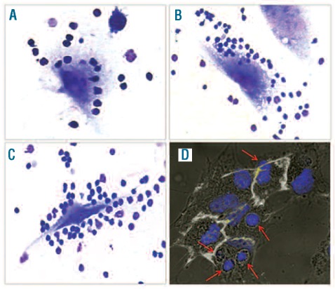
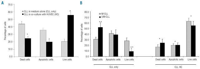
Purified CLL cells at a concentration of 1×10⁶/mL were cultured in direct contact with HUVEC at 70% confluence (HC condition) or in medium alone (CLL only condition) in 24-well plates (Figure 1). CLL apoptotic death was determined at 48 h in 34 CLL using annexin V-PI staining by flow cytometry. The percentage of apoptotic cells was substantially reduced in the co-culture condition compared to in the medium alone (Figure 2). The mean percentage of apoptotic cells was 35.7±2.5% for CLL in medium alone and 19.9±1.5% for CLL cells co-cultured with HUVEC (P<0.001). Likewise, the percentage of live CLL cells in the HC condition (55.7±3.3%) was higher than that in the CLL only condition (20.4±3.2%) (P<0.0001) (Figure 2A). When patients were divided into two subsets according to IGHV mutational status, spontaneous apoptosis appeared to be increased in unmutated IGHV CLL (n=16, UM-CLL) compared to in mutated ones (n=17, M-CLL) (live cells, 9.0%±5.8% in UM-CLL versus 28.0%±5.5% in M-CLL, P=0.04). However, when CLL cells were co-cultured on HUVEC, the survival of M-CLL and UM-CLL was similar, implying a 2.2-fold increase in relative viability in M-CLL compared with a 6.1-fold increase in UM-CLL (Figure 2B).
Figure 1.
Intimate contact between CLL and EC in vitro. CLL cells from representative patients were co-cultured for 48 h on HUVEC, and non-adherent cells were carefully removed before May-Grunwald Giemsa staining (A–C). Specimens were photographed at 400X magnification. Confocal immunofluorescence analysis is shown in panel (D) HUVEC were stained using anti-CD31 monoclonal antibody (white), while nuclei were visualized using DAPI (blue). Slides were analyzed using a TCSSP5 Leica confocal microscope at 63x magnification. Confocal planes are shown after merging.
Figure 2.
Enhanced survival of CLL cells in contact with EC. (A) Purified CLL cells were co-cultured on an EC layer (HC condition) or in medium alone (CLL only). Histograms represent percentages of apoptotic cells (annexin+/propidium iodide−), dead cells (propidium iodide+) and live cells (annexin−/propidium iodide−) measured by flow cytometry at 48 h in 34 CLL cultured together with EC or alone.*P<0.0001 in HC compared to CLL only (Wilcoxon’s non-parametric test). (B) CLL patients were divided into M-CLL (n=17) or UM-CLL (n=16) subsets according to immunoglobulin mutational status. Of note, UM-CLL appeared more prone to spontaneous apoptosis compared to M-CLL, but more sensitive to EC-derived survival signals. *P<0.05 in HC compared to CLL only, **P<0.05 in M-CLL compared to UM-CLL subsets (Mann-Whitney non-parametric test). Data are presented as mean ± SEM.
Physical contact with endothelial cells through β1- and β2- integrins is essential for protection from spontaneous apoptosis
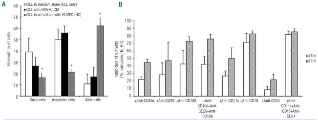
CLL cells were cultured in the presence of conditioned medium collected from HUVEC cultured alone near confluence (70–80%) (HUVEC CM) and apoptosis was evaluated at 48 h by flow cytometry. HUVEC CM did not exert a protective effect on CLL cells (apoptotic cells 50.2±9.1% in CLL only versus 56.1±5.5% CLL with HUVEC CM) (Figure 3A). When a microporous membrane was inserted between the HUVEC layer and CLL cells to prevent physical contact, the protective effect of endothelial cells was completely lost (data not shown). These data indicate that a direct cellular interaction is necessary to mediate CLL survival. To investigate the mechanism of adhesion between CLL cells and HUVEC, neutralizing monoclonal antibodies were added to co-cultures to antagonize β1- and β2- integrin pathways. As shown in Figure 3B, blocking CD106 on endothelial cells or CD18 on CLL cells led to almost complete abrogation of apoptosis protection (>70% inhibition of viability compared to the HC condition). Anti-CD49d, anti-CD29 and anti-CD11a induced moderate inhibition of survival (nearly 50% inhibition). There was a slight alteration of the HC system with blocking of CD54 (22% inhibition). With simultaneous blocking of CD49d, CD29 and CD106 or CD11a, CD18 and CD54, 76% and 85% inhibition was observed (Figure 3B).
Figure 3.
Blocking adhesion of CLL cells to endothelial cells abrogates the survival advantage. (A) Purified CLL cells (n=5) were cultured in the presence of conditioned medium collected from HUVEC near confluence (HUVEC CM). Following 2 days of culture, leukemic cells were harvested and labeled with annexin V and propidium iodide to assess cell apoptosis. *P<0.05 in HC compared to CLL only (Wilcoxon’s test). (B) CLL or HUVEC were pre-incubated for 1 h with blocking antibodies before starting the co-culture condition. Then, leukemic cells (n=6) were cultured for 48–72 h either in medium alone or on a HUVEC layer in the presence or absence of 10 μg/mL of the indicated monoclonal antibodies. Data are presented as mean ± SEM. Histograms represent the percentage of viability inhibition determined by blocking antibodies relative to the viability observed in the HC condition with irrelevant isotypic control antibody, added.
Endothelial cell-induced survival of chronic lymphocytic leukemia cells is partially mediated by soluble factors
We found that intimate contact with HUVEC cells is essential to rescue CLL cells from spontaneous apoptosis. However, we investigated whether the interaction between endothelial and CLL cells determined secretion of soluble factors able to promote and maintain CLL survival. We, therefore, collected media from CLL co-cultured on HUVEC for 48 h (HC CM) and then added them to CLL cultures of five patients. A significant reduction of dead cells was measured in CLL cells cultured with the addition of HC CM (41.2±3.4%) compared to cells in unconditioned medium or in medium obtained from CLL cells cultured alone in suspension (52.7±2.3%) (P<0.05). In addition, CLL cells were co-cultured in close contact with the HUVEC layer for 48 h and then leukemic cells were collected and cultured alone in the same HC medium for another 2 days (Online Supplementary Figure S1). We found that CLL cells, always maintained in medium alone for 96 h, had a level of 15.4±13.7% of live cells, whereas CLL cells stimulated by contact with HUVEC and then cultured for 48 h alone showed 57.8±12.2% of live cells, comparable to the survival of CLL cells in the permanent co-culture condition (58.2±14.7%) (Online Supplementary Figure S1). Overall, these observations suggest that soluble factors present in the co-culture conditioned medium were contributing to the observed inhibition of apoptosis in CLL cells.
Endothelial cells protect chronic lymphocytic leukemia cells from drug-induced apoptosis
We next investigated whether HUVEC could protect CLL cells from drug-induced apoptosis, testing cell viability in 12 CLL cultured with fludarabine 10 μM in medium alone or in combination with confluent HUVEC. The endothelial cell layer decreased the in vitro sensitivity of CLL cells to fludarabine-induced apoptotic cell death (Figure 4A–B). The mean viability of CLL cells treated with 10 μM fludarabine in the absence of HUVEC was 57.3% (±4.1%) after 24 h, 19.8% (±4.4%) after 48 h, 3.8% (±1.3%) after 72 h and 1.0% (±0.5%) after 96 h. In the presence of the HUVEC layer, the mean viability of CLL cells treated with 10 μM fludarabine was 63.7% (±7.5%) after 24 h, 37.8% (±9.1%) after 48 h, 14.3% (±3.2%) after 72 h and 7.6% (±3.9%) after 96 h (P<0.05, at 48, 72, and 96 h) (Figure 4A).
Figure 4.
Endothelial cells inhibit drug-induced CLL apoptosis. (A) Histograms represent the percentage of viable CLL cells in the presence (HC) or absence (CLL only) of a HUVEC layer and/or 10 μM fludarabine (+FLU) at the time points indicated on the horizontal axis. Data are presented as mean ± SEM for 12 CLL. HUVEC provided a significant level of protection from fludarabine-induced apoptosis. *P<0.05 in HC compared to CLL only, **P<0.05 in HC (HC+FLU) compared to CLL only (CLL only + FLU) both with addition of fludarabine (Wilcoxon’s test). (B) Time-course experiments for two representative CLL patients.
Endothelial cells induce a peculiar gene expression profile in chronic lymphocytic leukemia cells
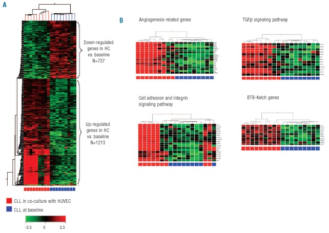
Given the ability of EC to protect CLL cells from apoptosis, we investigated the mechanisms underlying the EC/CLL cell crosstalk by comparative GEP of CLL cultured in medium alone or in contact with EC. From nine patients, three set of samples were collected: (i) CLL cells from peripheral blood (CLL at baseline); (ii) CLL cells cultured for 48 h in medium alone (CLL only); and (iii) CLL cells cultured for 48 h in contact with the HUVEC layer (CLL HC). The whole-genome GEP of all CLL samples was inspected by microarrays. We compared GEP between CLL cells cultured in contact with the EC layer and CLL at baseline to unravel the transcriptional modifications induced by EC. Overall 1,944 genes were found to be modulated by co-culture [fold change (FC)≥2, P<0.05]: 1,217 genes were up-regulated in the HC condition and 727 genes were down-regulated (Figure 5A). Differentially expressed genes were analyzed with the GeneSpring Gene Ontology browser tool to identify the Gene Ontology categories most represented in gene-lists (Online Supplementary Table S1). CLL cells in co-culture with EC up-regulated a series of genes involved in angiogenesis and regulation of EC function. Moreover, genes involved in cell motion and migration, in reorganization of the actin cytoskeleton as well as in response to external stimuli, to stress, and to hypoxia were significantly modulated in HC. The most represented cellular pathways included the TGFβ and Wnt signaling pathways, inflammation mediated by chemokine and cytokine signaling pathways and the integrin signaling pathway as well as a set of genes coding for BTB-Kelch proteins (Figure 5B). We next compared our GEP to those previously found in co-culture of CLL cells with nurse-like cells and the bone marrow-derived stromal cell line HS-5.22,23 We found that different microenvironmental elements determined distinct gene expression signatures in CLL cells with only few genes being modulated in a similar way by different cell types (Online Supplementary Figure S2).
Figure 5.
Contact with endothelial cells modulates the CLL gene expression profile. (A) Heat map depicting differentially expressed genes between CLL cells at baseline and CLL cells in co-culture for 48 h. (B) Sets of differentially expressed genes involved in angiogenesis, cell adhesion and the TGFβ signaling pathway and BTB-kelch genes are indicated. The data are represented in a grid format in which each column represents a case and each row a single gene. Normalized intensity signals are depicted in the pseudo color scale as indicated.
Contact with endothelial cells increases the pro-angiogenic and anti-apoptotic profile of chronic lymphocytic leukemia cells
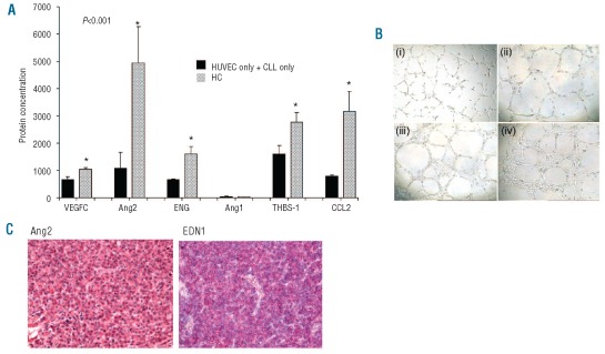
Deeper analyses of GEP data were performed focusing on two categories of differentially expressed genes: (i) those coding for soluble factors involved in angiogenesis and/or regulating microenvironment components, and (ii) those coding for proteins involved in apoptosis regulation (Online Supplementary Table S2). We found that CLL cells in culture with EC showed a 22.6-fold increase of the C-C motif chemokine CCL2, capable to inducing pro-angiogenic stimuli on EC, recruiting tumor-associated monocytes (TAM) and suppressing cytotoxic lymphocytes (P=0.0032). In addition, a 6.5-fold increase of PDGFC expression, a chemoattractant for mesenchymal cells, was induced in CLL cells by EC co-culture (P=0.0051). Other soluble factors up-regulated by EC/CLL contact compared to baseline were VEGFC (FC=9.4, P=0.0061), ANGTL4 (FC=8.6, P=0.015), ENG (FC=4.0, P=0.025), EDN1 (FC=9.2, P=0.0061), AMOTL2 (FC=4.3, P=0.019) and THBS1 (FC=45.1, P=0.0004) as well as the metalloproteases MMP2 (FC=8.3, P=0.02) and MMP14 (FC=3.0, P=0.039). All these transcriptional modifications were not detected in the GEP of CLL cells cultured alone. In order to confirm the modulation of vascular cytokines in CLL cells cultured in the presence of the EC layer, we measured the secreted levels in conditioned media collected after 48 h-culture (Figure 6A). We found that HUVEC cultured in medium alone secreted VEGFC (643.3±71.7 pg/mL), Ang2 (1054.9±570.0 pg/mL), ENG (910.0±10.0 pg/mL), THBS-1 (1602.0±310.0 ng/mL) and CCL2 (772.5±60.0 pg/mL) but small levels of Ang1 (24.5±3.1 pg/mL). When cultured alone, CLL cells secreted low levels of VEGFC (33.4±27.9 pg/mL), Ang2 (31.4±8.6 pg/mL), Ang1 (24.5±3.5 pg/mL) and CCL2 (11.1±1.7 pg/mL) and no detectable levels of THBS-1 or ENG. The CLL/EC co-culture condition increased the secretion of VEGFC (1043.2±57.7 pg/mL), Ang2 (4935.7±1325.1 pg/mL), ENG (1603.1±273.7 pg/mL), THBS-1 (2774.8±357.4 ng/mL) and CCL2 (3162.1±721.2 pg/mL) whereas it maintained low Ang1 secretion (32.7±3.7 pg/mL). In addition, CLL cells (n=3) were cultured for 48 h on the HUVEC layer and then collected and cultured for a further 48 h in fresh medium. We found that media conditioned by CLL cells previously stimulated by EC contact compared to media conditioned by unstimulated CLL cells were enriched in THBS-1 (from 0 to 5 ng/mL), Ang2 (from 27 to 59 pg/mL), ENG (from 0 to 370 pg/mL), VEGFC (from 89 to 897 pg/mL) and CCL2 (from 11 to 3188 pg/mL) but not Ang1 (from 27 to 21 pg/mL). Furthermore, conditioned media collected after 48 h of CLL/EC co-culture significantly increased the tube formation on a matrigel angiogenesis assay with complex mesh-like structures just developed at 6 h, compared to control medium in which isolated cells or cells just starting to elongate and migrate to align themselves were visible at 6 hours (Figure 6B).
Figure 6.
Increase of angiogenesis-related proteins in CLL cells by EC contact. (A) Purified CLL cells were co-cultured on a HUVEC layer near confluence for 48 h at which time the condition medium (CM) was harvested for ELISA determination of VEGFC, Ang2, ENG, Ang1, THBS-1 and CCL2. Histograms represent mean protein concentration ± SEM for eight separate experiments. HUVEC only + CLL only columns represent the sum of protein levels obtained from CM of HUVEC and CLL cells cultured in medium alone for 48 h, whereas HC columns represent protein levels in CM obtained from CLL co-culture on a HUVEC layer. Note the increase in secretion of several angiogenesis-related proteins when CLL cells were co-cultured on HUVEC compared to secretion obtained from both separated cellular elements. Values presented are in pg/mL for VEGFC, Ang2, ENG, Ang1 and CCL2 and in ng/mL for THBS-1. *P<0.05 for HC compared to HUVEC and CLL cells separately (Mann-Whitney test). (B) HUVEC cells were plated onto Matrigel (ECMatrix) and incubated with (i) M200 medium alone (control sample) or (ii) M200 supplemented with CLL only-derived CM [1vol:1vol] or (iii–iv) M200 supplemented with CLL HC-derived CM [1vol:1vol] for 6 h. The increased effect of CLL HC-derived supernatants on angiogenesis is visible. Original magnification, 40X. (C) Immunohistochemical evaluation of CLL lymph nodes (n=5) stained with antibodies against angiopoietin-2 (Ang2) and endothelin-1 (EDN1) showing CLL cells positive for the two pro-angiogenic factors up-regulated by EC contact. Original magnification, 400X.
The Ang2 tyrosine kinase receptor Tie2 mRNA was found to be increased in CLL cells in co-culture (FC=10.7, P=0.017) and not modified in the CLL only condition. We confirmed the GEP data by analyzing Tie2 expression on the surface of CLL cells (n=8) co-cultured on the EC layer. The percentage of Tie2+CLL cells was increased by 2-fold and 4.3-fold at 48 h and 72 h, respectively. The peripheral CLL population comprised 1.7±0.3% (range, 0.8–3.7%) Tie2+ cells at baseline and 7.0±2.0% (range, 3.4–21.1%) at 72 h in the HC condition (Online Supplementary Figure S3). Expression of low levels of surface Tie2 by CLL cells was also visualized by confocal microscopy, after plating purified leukemic cells on a HUVEC monolayer for 72 h (Online Supplementary Figure S3).
Given the ability of a HUVEC layer to provide marked protection against spontaneous and drug-induced apoptosis, we evaluated the transcriptional modulation of genes involved in apoptotic cellular pathways (Online Supplementary Figure S4). CLL cells cultured for 48 h in the presence of an endothelial layer maintained or increased the expression levels of anti-apoptotic factors such as Bcl-2, Bcl2A1, BIRC3/c-IAP2 and BIRC5/Survivin compared to CLL cells at baseline. In contrast, a marked decrease of anti-apoptotic factors was detected in CLL cells cultured alone. In addition, CLL cells in culture alone showed increased expression of pro-apoptotic factors such as BAX, BCL2L10, BCL2L13 and BCL2L15, and decreased expression of several poly(ADP-ribose) polymerases (PARP) compared to cells cultured in the HC condition. Significant correlations were found between Bcl-2 expression levels and viability at 48 h as assessed by flow-cytometry (dead cells, r=−0.615, P=0.007; live cells, r=0.8, P<0.0001; apoptotic cells, r=−0.708, P=0.001) (Online Supplementary Figure S4).
Discussion
In this study we investigated the interaction between endothelial and CLL cells in an in vitro co-culture system. Although an in vitro model is never going to be able to recapitulate the in vivo microenvironment completely, it may be useful for dissecting specific cellular signals ascribed to different elements directly interacting with leukemic cells in vivo. Our study was devised in the light of several observations. First, although CLL cells exhibit characteristics consistent with prolonged cell survival in vivo, when cultured in vitro these cells often undergo spontaneous apoptosis.1 This suggests that extrinsic signals from microenvironmental elements surrounding leukemic cells in vivo are essential to support prolonged CLL survival. Second, several pieces of evidence indicate that increased angiogenesis in infiltrated bone marrow and lymph nodes may be involved in the pathogenesis and progression of CLL.8,12 Moreover, increased serum and plasma levels of several angiogenic factors in CLL patients have been shown to correlate with adverse prognostic factors, fludarabine-resistance and reduced survival.8,9,11
The increased vascularization may provide infiltrated tissues with oxygen and metabolites, as well as with signals and conditions involved in regulating leukemic cell homing and recirculation. We hypothesize that the presence of activated EC in bone marrow and lymph nodes as well as the direct interaction with EC during recirculation from peripheral blood to tissues may also contribute to CLL cell survival and may be involved in drug resistance, thus facilitating disease progression and relapse after conventional therapy.
Thus, we co-cultured CLL cells on a layer of adherent HUVEC allowing the establishment of functional interactions through intimate contact between cellular elements. We found that EC rescued CLL from spontaneous apoptosis and that the endothelial protection was mediated by integrins and soluble factors. Despite the individual variability of apoptotic rate, all CLL cells co-cultured in direct contact with endothelial layers showed a strong survival advantage which was relatively stable for several days after an initial decrease in the first 24–48 h. In agreement with the results of a recent study, we found that the survival advantage was completely abrogated by separating the cells with a microporous membrane.16 Moreover, conditioned medium collected from HUVEC near confluence failed to recapitulate the survival improvement observed in the direct co-culture system. These results indicate that the intimate contact between leukemic cells and supporting endothelial elements is necessary to protect CLL cells from spontaneous apoptosis. Blocking monoclonal antibodies against the α4β1/VCAM-1 and αLβ2/ICAM axes demonstrated that both signals were involved in the interaction between CLL and endothelium. Disrupting the integrin-mediated contact significantly decreased the survival advantage in the co-culture system. Approximately 70% to 80% inhibition of survival could be obtained with anti-CD106 or anti-CD18.
Surprisingly, however, we observed that when CLL cells were removed from the EC layer, the leukemic cells maintained a survival advantage compared to CLL cultured in suspension. In addition, conditioned medium collected from 2-day CLL/EC co-culture significantly inhibited spontaneous apoptosis of CLL cells. Overall, our data demonstrated that physical contact between CLL cells and the endothelium is essential to trigger the cellular secretion of several soluble factors mediating additional survival hits on leukemic cells. Furthermore, in agreement with recently reported results,24 we observed that spontaneous apoptosis in vitro was higher in IGHV-unmutated CLL cells than in IGHV-mutated ones. When CLL cells were co-cultured on the endothelial layer, CLL survival was comparable in the two prognostic subsets. These observations suggest that IGHV-unmutated CLL clones may be more dependent on the interaction with the microenvironment to reach the long-lived phenotype. Indeed, we found that EC had a greater effect on the survival of IGHV-unmutated cells than on the IGHV-mutated ones.
Growing evidence suggests that CLL cells are protected from conventional drugs in tissue microenvironments, with facilitation of drug-resistant residual disease, ultimately paving the way to clonal evolution and relapse.25 Interestingly, we found that EC protected CLL cells from fludarabine-induced apoptosis, suggesting that EC in intimate contact with CLL cells in vivo may contribute to establishing a protective niche hiding residual CLL cells from conventional drugs.
To explore the mechanisms underlying the enhanced survival consequent to co-culture with EC, we performed GEP of CLL transcriptome changes that occur as a consequence of the CLL-EC interaction, finding a wide spectrum of peculiar gene expression changes. We found that interaction between endothelium and CLL cells affected the levels of several anti-apoptotic factors including Bcl-2 and BIRC5/Survivin. When circulating CLL cells were cultured in medium alone, their Bcl-2 expression decreased rapidly. EC contact was able to maintain elevated levels of Bcl-2 comparable to those of CLL cells in vivo and to increase the expression of Survivin significantly. This suggests that physical contact with endothelium may trigger signals able to preserve the anti-apoptotic phenotype of CLL cells and to deliver additional survival hits. Moreover, the endothelium contact increased the pro-angiogenic phenotype of leukemic cells by causing dramatic alterations in the secreted levels of several angiogenesis-related factors. This finding is consistent with the reported ability of stromal cells to activate CLL cells to secrete VEGF and bFGF in co-culture assays.26,27 This imbalance in the production of angiogenic regulators may cause a microenvironmental-driven angiogenic switch in CLL cells. In particular, we found that intimate contact with endothelium determined the up-regulation of endothelin-1 (EDN1), endoglin (ENG), angiomotin-like protein 2 (AMOTL2), angiopoietin-related protein 4 (ANGPTL4), Ang2, VEGFC, and thrombospondin 1 (THBS-1) in CLL cells. EDN1 modulates various stages of neovascularization, including EC proliferation, migration, invasion, protease production and tube formation as well as stimulating the production of VEGF by increasing hypoxia-inducible factor 1α and matrix metalloproteases. Moreover, EDN1 was found to be involved in cell survival and proliferation in many solid tumors through a rapid induction of c-FOS, c-JUN and c-MYC and also by triggering anti-apoptotic signaling through Bcl2-dependent and phosphatidyl-inositolol 3-kinase Akt pathways.28 We also found that CLL cells increased the secretion of ENG involved in the activation and survival of EC. ENG (CD105) is a disulfite-linked homodimeric transmembrane glycoprotein, component of the receptor complex of TGF-β. ENG is mainly expressed by activated vascular EC but also by several neoplastic cells, including solid tumors such as meningiomas, ovary and breast carcinomas and hematologic malignancies such as refractory anemia with excess of blasts, blast crisis of chronic myelogenous leukemia, the most immature subtypes of acute leukemias and also CLL.29,30 Other soluble factors up-regulated by CLL cells in contact with endothelium were AMOTL2, involved in EC migration and tube formation, and two angiopoietins (Ang2 and ANPTL4) capable of inducing destabilization of pre-existing vessels and neovascularization as well as migration and an immunosuppressive phenotype of monocytes.31,32 Of note Tie2, the Ang2 tyrosine kinase receptor, was expressed in about 2% of circulating CLL cells, with a value increasing to nearly 7% in the HC co-culture condition. Further studies are necessary to increase the understanding of the role of the Tie2+ CLL cell subset. Another angiogenesis-related factor secreted by CLL cells in contact with EC was a member of the VEGF family, VEGFC. VEGFC has been characterized as a lymphangiogenic and angiogenic growth factor and shown to signal through the receptors VEGFR-3 and VEGFR-2. Apart from its effect on angiogenesis, VEGFC signaling was reported to induce proliferation, promote cell survival and protect leukemic cells from chemotherapy-induced apoptosis by induction of Bcl-2 in a subset of acute leukemias.33 In contrast with the reported decrease of THBS-1 in CLL cells adhering to stromal cells, we found that CLL cells, when co-cultured on an endothelial layer, up-regulated THBS-1. Classically considered an anti-angiogenic factor,34 THBS-1 can also be considered a stimulator of tumor progression and angiogenesis by inducing proteolytic degradation of the extracellular matrix, cell migration and invasion through regulation of the plasminogen/plasmin system, up-regulation of MMP9 and activation of TGF-β1.35 Of interest, CLL cells were reported to secrete THBS-1 and to express the thrombospondin receptor CD36.36 Overall, our finding that the interaction between CLL cells and endothelium leads to an increase in several pro-angiogenic cytokines involved in survival, proliferation and migration of EC, and vessel sprouting indicates that a kind of circularly sustained angiogenic milieu may be present in CLL-infiltrated tissues in order to maintain increased microvessel density and to improve growth conditions for the CLL clone.
Another indication from the microarray experiments was that EC induced expression changes of factors able to orchestrate the cellular composition and function of several microenvironmental elements including stromal fibroblasts and macrophages such as Ang2, PDGFC and CCL2. In particular, the C-C motif chemokine CCL2 is one of the key chemokines regulating migration and infiltration of monocytes/macrophages via activation of the CCR2 receptor. In addition, CCL2 can directly mediate tumor angiogenesis and augments the Th2 response.37 Interestingly, about half of CLL clones were reported to express CCR2 receptor.38 Very recently, CCL2 protein was detected in supernatants obtained from CLL co-culture with the bone marrow-derived stromal cell line HS-5, implying a chemotactic activity on monocytes.23
Among the up-regulated entities, we also found several genes involved in cell adhesion and integrin pathways as well as a set of genes containing the BTB-Kelch domains. BTB-kelch proteins seem to be involved in a variety of biological processes, such as stabilization/remodeling of the cytoskeleton and cell migration. Of note, we observed in all CLL co-cultured on an EC layer that, compared to the baseline condition, there was up-regulation of KLHL6, which is implicated in B-cell receptor signaling and germinal center formation and mutated in about 1.8% of cases of CLL.39,40
In conclusion, we demonstrated that CLL cells interact with endothelium through α4β1/VCAM-1 and αLβ2/ICAM axes and cell adhesion is necessary to trigger the survival signals on leukemic cells. CLL/EC physical contact profoundly modifies the transcriptional profile of leukemic cells and leads to the establishment of a complex network of soluble factors that seem to provide additional survival advantages. Molecular mechanisms triggered by CLL cell-EC crosstalk are complex and partially dissected by this study. However, our study supports the line of evidence indicating that EC are major players in CLL-infiltrated microenvironments being able to create a vicious cycle of interactions that strongly support leukemic cell survival, thus protecting CLL cells from drug-induced apoptosis and profoundly modifying CLL gene expression profile.
Footnotes
Funding: this work was supported by grants from the Associazione Italiana per la Ricerca sul Cancro (AIRC IG10621-2010), Milan, Italy; Associazione Italiana contro le Leucemie (AIL), Modena, Italy; Programma Ricerca Regione-Università 2007–2009, Emilia Romagna, Italy; and Programmi di Ricerca di Interesse Nazionale (PRIN) 2008, Ministero dell’Università e della Ricerca (MIUR), Rome, Italy.
The online version of this article has a Supplementary Appendix.
Authorship and Disclosures
The information provided by the authors about contributions from persons listed as authors and in acknowledgments is available with the full text of this paper at www.haematologica.org.
Financial and other disclosures provided by the authors using the ICMJE (www.icmje.org) Uniform Format for Disclosure of Competing Interests are also available at www.haematologica.org.
References
- 1.Collins RJ, Verschuer LA, Harmon BV, Prentice RL, Pope JH, Kerr JF. Spontaneous programmed death (apoptosis) of B-chronic lymphocytic leukaemia cells following their culture in vitro. Br J Haematol. 1989;71(3):343–50. doi: 10.1111/j.1365-2141.1989.tb04290.x. [DOI] [PubMed] [Google Scholar]
- 2.McConkey DJ, Aguilar-Santelises M, Hartzell P, Eriksson I, Mellstedt H, Orrenius S, et al. Induction of DNA fragmentation in chronic B-lymphocytic leukemia cells. J Immunol. 1991;146(3):1072–6. [PubMed] [Google Scholar]
- 3.Schmid C, Isaacson PG. Proliferation centres in B-cell malignant lymphoma, lymphocytic (B-CLL): an immunophenotypic study. Histopathology. 1994;24(5):445–51. doi: 10.1111/j.1365-2559.1994.tb00553.x. [DOI] [PubMed] [Google Scholar]
- 4.Lampert IA, Wotherspoon A, Van Noorden S, Hasserjian RP. High expression of CD23 in the proliferation centers of chronic lymphocytic leukemia in lymph nodes and spleen. Hum Pathol. 1999;30(6):648–54. doi: 10.1016/s0046-8177(99)90089-8. [DOI] [PubMed] [Google Scholar]
- 5.Burger JA. Nurture versus nature: the microenvironment in chronic lymphocytic leukemia. Hematology Am Soc Hematol Educ Program. 2011;2011:96–103. doi: 10.1182/asheducation-2011.1.96. [DOI] [PubMed] [Google Scholar]
- 6.Yancopoulos GD, Davis S, Gale NW, Rudge JS, Wiegand SJ, Holash J. Vascular-specific growth factors and blood vessel formation. Nature. 2000;407(6801):242–8. doi: 10.1038/35025215. [DOI] [PubMed] [Google Scholar]
- 7.Peterson L, Kini AR. Angiogenesis is increased in B-cell chronic lymphocytic leukemia. Blood. 2001;97(8):2529. doi: 10.1182/blood.v97.8.2529. [DOI] [PubMed] [Google Scholar]
- 8.Molica S, Vacca A, Ribatti D, Cuneo A, Cavazzini F, Levato D, et al. Prognostic value of enhanced bone marrow angiogenesis in early B-cell chronic lymphocytic leukemia. Blood. 2002;100(9):3344–51. doi: 10.1182/blood-2002-01-0084. [DOI] [PubMed] [Google Scholar]
- 9.McCabe D, Bacon L, O’Regan K, Condron C, O’Donnell JR, Murphy PT. CD38 expression on B-cell chronic lymphocytic leukemic cells is strongly correlated with vascular endothelial growth factor expression. Leukemia. 2004;18(3):649–50. doi: 10.1038/sj.leu.2403282. [DOI] [PubMed] [Google Scholar]
- 10.Huttmann A, Klein-Hitpass L, Thomale J, Deenen R, Carpinteiro A, Nuckel H, et al. Gene expression signatures separate B-cell chronic lymphocytic leukaemia prognostic subgroups defined by ZAP-70 and CD38 expression status. Leukemia. 2006;20(10):1774–82. doi: 10.1038/sj.leu.2404363. [DOI] [PubMed] [Google Scholar]
- 11.Maffei R, Martinelli S, Santachiara R, Rossi D, Guarnotta C, Sozzi E, et al. Angiopoietin-2 plasma dosage predicts time to first treatment and overall survival in chronic lymphocytic leukemia. Blood. 2010;116(4):584–92. doi: 10.1182/blood-2009-11-252494. [DOI] [PubMed] [Google Scholar]
- 12.Maffei R, Martinelli S, Castelli I, Santachiara R, Zucchini P, Fontana M, et al. Increased angiogenesis induced by chronic lymphocytic leukemia B cells is mediated by leukemia-derived Ang2 and VEGF. Leuk Res. 2010;34(3):312–21. doi: 10.1016/j.leukres.2009.06.023. [DOI] [PubMed] [Google Scholar]
- 13.Kurtova AV, Balakrishnan K, Chen R, Ding W, Schnabl S, Quiroga MP, et al. Diverse marrow stromal cells protect CLL cells from spontaneous and drug-induced apoptosis: development of a reliable and reproducible system to assess stromal cell adhesion-mediated drug resistance. Blood. 2009;114(20):4441–50. doi: 10.1182/blood-2009-07-233718. [DOI] [PMC free article] [PubMed] [Google Scholar]
- 14.Martinelli S, Maffei R, Castelli I, Santachiara R, Zucchini P, Fontana M, et al. Increased expression of angiopoietin-2 characterizes early B-cell chronic lymphocytic leukemia with poor prognosis. Leuk Res. 2008;32(4):593–7. doi: 10.1016/j.leukres.2007.09.002. [DOI] [PubMed] [Google Scholar]
- 15.Shanafelt TD, Kay NE. The clinical and biologic importance of neovascularization and angiogenic signaling pathways in chronic lymphocytic leukemia. Semin Oncol. 2006;33(2):174–85. doi: 10.1053/j.seminoncol.2006.01.008. [DOI] [PubMed] [Google Scholar]
- 16.Badoux X, Bueso-Ramos C, Harris D, Li P, Liu Z, Burger J, et al. Cross-talk between chronic lymphocytic leukemia cells and bone marrow endothelial cells: role of signal transducer and activator of transcription 3. Hum Pathol. 2011;2(12):1989–2000. doi: 10.1016/j.humpath.2011.02.027. [DOI] [PMC free article] [PubMed] [Google Scholar]
- 17.Long BW, Witte PL, Abraham GN, Gregory SA, Plate JM. Apoptosis and interleukin 7 gene expression in chronic B-lymphocytic leukemia cells. Proc Natl Acad Sci USA. 1995;92(5):1416–20. doi: 10.1073/pnas.92.5.1416. [DOI] [PMC free article] [PubMed] [Google Scholar]
- 18.Moreno A, Villar ML, Camara C, Luque R, Cespon C, Gonzalez-Porque P, et al. Interleukin-6 dimers produced by endothelial cells inhibit apoptosis of B-chronic lymphocytic leukemia cells. Blood. 2001;97(1):242–9. doi: 10.1182/blood.v97.1.242. [DOI] [PubMed] [Google Scholar]
- 19.Buggins AG, Pepper C, Patten PE, Hewamana S, Gohil S, Moorhead J, et al. Interaction with vascular endothelium enhances survival in primary chronic lymphocytic leukemia cells via NF-kappaB activation and de novo gene transcription. Cancer Res. 2010;70(19):7523–33. doi: 10.1158/0008-5472.CAN-10-1634. [DOI] [PubMed] [Google Scholar]
- 20.Giannoni P, Scaglione S, Quarto R, Narcisi R, Parodi M, Balleari E, et al. An interaction between hepatocyte growth factor and its receptor (c-MET) prolongs the survival of chronic lymphocytic leukemic cells through STAT3 phosphorylation: a potential role of mesenchymal cells in the disease. Haematologica. 2011;96(7):1015–23. doi: 10.3324/haematol.2010.029736. [DOI] [PMC free article] [PubMed] [Google Scholar]
- 21.Hallek M, Cheson BD, Catovsky D, Caligaris-Cappio F, Dighiero G, Dohner H, et al. Guidelines for the diagnosis and treatment of chronic lymphocytic leukemia: a report from the International Workshop on Chronic Lymphocytic Leukemia updating the National Cancer Institute-Working Group 1996 guidelines. Blood. 2008;111(12):5446–56. doi: 10.1182/blood-2007-06-093906. [DOI] [PMC free article] [PubMed] [Google Scholar]
- 22.Burger JA, Quiroga MP, Hartmann E, Burkle A, Wierda WG, Keating MJ, et al. High-level expression of the T-cell chemokines CCL3 and CCL4 by chronic lymphocytic leukemia B cells in nurselike cell cocultures and after BCR stimulation. Blood. 2009;113(13):3050–8. doi: 10.1182/blood-2008-07-170415. [DOI] [PMC free article] [PubMed] [Google Scholar]
- 23.Schulz A, Toedt G, Zenz T, Stilgenbauer S, Lichter P, Seiffert M. Inflammatory cytokines and signaling pathways are associated with survival of primary chronic lymphocytic leukemia cells in vitro: a dominant role of CCL2. Haematologica. 2011;96(3):408–16. doi: 10.3324/haematol.2010.031377. [DOI] [PMC free article] [PubMed] [Google Scholar]
- 24.Coscia M, Pantaleoni F, Riganti C, Vitale C, Rigoni M, Peola S, et al. IGHV unmutated CLL B cells are more prone to spontaneous apoptosis and subject to environmental pro-survival signals than mutated CLL B cells. Leukemia. 2011;25(5):828–37. doi: 10.1038/leu.2011.12. [DOI] [PubMed] [Google Scholar]
- 25.Meads MB, Hazlehurst LA, Dalton WS. The bone marrow microenvironment as a tumor sanctuary and contributor to drug resistance. Clin Cancer Res. 2008;14(9):2519–26. doi: 10.1158/1078-0432.CCR-07-2223. [DOI] [PubMed] [Google Scholar]
- 26.Kay NE, Shanafelt TD, Strege AK, Lee YK, Bone ND, Raza A. Bone biopsy derived marrow stromal elements rescue chronic lymphocytic leukemia B-cells from spontaneous and drug induced cell death and facilitates an “angiogenic switch”. Leuk Res. 2007;31(7):899–906. doi: 10.1016/j.leukres.2006.11.024. [DOI] [PMC free article] [PubMed] [Google Scholar]
- 27.Edelmann J, Klein-Hitpass L, Carpinteiro A, Fuhrer A, Sellmann L, Stilgenbauer S, et al. Bone marrow fibroblasts induce expression of PI3K/NF-kappaB pathway genes and a pro-angiogenic phenotype in CLL cells. Leuk Res. 2008;32(10):1565–72. doi: 10.1016/j.leukres.2008.03.003. [DOI] [PubMed] [Google Scholar]
- 28.Nelson J, Bagnato A, Battistini B, Nisen P. The endothelin axis: emerging role in cancer. Nat Rev Cancer. 2003;3(2):110–6. doi: 10.1038/nrc990. [DOI] [PubMed] [Google Scholar]
- 29.Dallas NA, Samuel S, Xia L, Fan F, Gray MJ, Lim SJ, et al. Endoglin (CD105): a marker of tumor vasculature and potential target for therapy. Clin Cancer Res. 2008;14(7):1931–7. doi: 10.1158/1078-0432.CCR-07-4478. [DOI] [PubMed] [Google Scholar]
- 30.Kay NE, Bone ND, Tschumper RC, Howell KH, Geyer SM, Dewald GW, et al. B-CLL cells are capable of synthesis and secretion of both pro- and anti-angiogenic molecules. Leukemia. 2002;16(5):911–9. doi: 10.1038/sj.leu.2402467. [DOI] [PubMed] [Google Scholar]
- 31.Coffelt SB, Tal AO, Scholz A, De Palma M, Patel S, Urbich C, et al. Angiopoietin-2 regulates gene expression in TIE2-expressing monocytes and augments their inherent proangiogenic functions. Cancer Res. 2010;70(13):5270–80. doi: 10.1158/0008-5472.CAN-10-0012. [DOI] [PubMed] [Google Scholar]
- 32.Coffelt SB, Chen YY, Muthana M, Welford AF, Tal AO, Scholz A, et al. Angiopoietin 2 stimulates TIE2-expressing monocytes to suppress T cell activation and to promote regulatory T cell expansion. J Immunol. 2011;186(7):4183–90. doi: 10.4049/jimmunol.1002802. [DOI] [PubMed] [Google Scholar]
- 33.Dias S, Choy M, Alitalo K, Rafii S. Vascular endothelial growth factor (VEGF)-C signaling through FLT-4 (VEGFR-3) mediates leukemic cell proliferation, survival, and resistance to chemotherapy. Blood. 2002;99(6):2179–84. doi: 10.1182/blood.v99.6.2179. [DOI] [PubMed] [Google Scholar]
- 34.Margosio B, Marchetti D, Vergani V, Giavazzi R, Rusnati M, Presta M, et al. Thrombospondin 1 as a scavenger for matrix-associated fibroblast growth factor 2. Blood. 2003;102(13):4399–406. doi: 10.1182/blood-2003-03-0893. [DOI] [PubMed] [Google Scholar]
- 35.Sargiannidou I, Zhou J, Tuszynski GP. The role of thrombospondin-1 in tumor progression. Exp Biol Med (Maywood) 2001;226(8):726–33. doi: 10.1177/153537020222600803. [DOI] [PubMed] [Google Scholar]
- 36.Rutella S, Rumi C, Puggioni P, Barberi T, Di Mario A, Larocca LM, et al. Expression of thrombospondin receptor (CD36) in B-cell chronic lymphocytic leukemia as an indicator of tumor cell dissemination. Haematologica. 1999;84(5):419–24. [PubMed] [Google Scholar]
- 37.Zhang J, Patel L, Pienta KJ. CC chemokine ligand 2 (CCL2) promotes prostate cancer tumorigenesis and metastasis. Cytokine Growth Factor Rev. 2010;21(1):41–8. doi: 10.1016/j.cytogfr.2009.11.009. [DOI] [PMC free article] [PubMed] [Google Scholar]
- 38.Trentin L, Cabrelle A, Facco M, Carollo D, Miorin M, Tosoni A, et al. Homeostatic chemokines drive migration of malignant B cells in patients with non-Hodgkin lymphomas. Blood. 2004;104(2):502–8. doi: 10.1182/blood-2003-09-3103. [DOI] [PubMed] [Google Scholar]
- 39.Kroll J, Shi X, Caprioli A, Liu HH, Waskow C, Lin KM, et al. The BTB-kelch protein KLHL6 is involved in B-lymphocyte antigen receptor signaling and germinal center formation. Mol Cell Biol. 2005;25(19):8531–40. doi: 10.1128/MCB.25.19.8531-8540.2005. [DOI] [PMC free article] [PubMed] [Google Scholar]
- 40.Puente XS, Pinyol M, Quesada V, Conde L, Ordonez GR, Villamor N, et al. Whole-genome sequencing identifies recurrent mutations in chronic lymphocytic leukaemia. Nature. 2011;475(7354):101–5. doi: 10.1038/nature10113. [DOI] [PMC free article] [PubMed] [Google Scholar]


