Abstract
An outbreak of myocarditis occurred among adults in Illinois in 2003. Diagnostic testing of myocardial tissues from 3 patients and comprehensive tests for enterovirus and adenovirus of other specimens from patients were inconclusive. Appropriate specimen collection from patients with idiopathic cardiomyopathy and further enhancement of diagnostic techniques are needed.
Keywords: myocarditis, cardiomyopathy, viral, outbreak, biopsy, polymerase chain reaction, surveillance, enzyme immunoassay, immunohistochemistry, Illinois, dispatch
Acute myocarditis is characterized by inflammatory infiltrates of the myocardium. Disease has been attributed to multiple infectious and noninfectious causes, but viruses, particularly the enteroviruses group B coxsackievirus and echoviruses, are believed to be the most common agents of infection in the United States (1). An infectious cause of myocarditis is usually suspected when unexplained heart failure or arrhythmia occurs in a person with a systemic febrile illness or upper respiratory tract infection. Acute myocarditis is typically sporadic, although clusters have been reported during outbreaks of viral disease (2,3). Most cases are idiopathic without a known cause (1). Myocardial biopsy specimens used for pathologic examination, the conventional standard for diagnosis (4,5), have been considered difficult to collect in nonfatal cases. Viruses are infrequently cultured from tissue specimens, although viral nucleic acid identification by polymerase chain reaction (PCR) assays on myocardium has recently enhanced viral detection (6–8). Viral serologic tests and PCR assays of blood, stool, urine, and nasopharyngeal specimens are adjunctive techniques for diagnosing myocarditis that have not been validated.
On March 21, 2003, the Kane County Health Department was notified about 6 cases of presumptive myocarditis and 1 case of pericarditis that occurred in patients hospitalized in Kane County, Illinois, within a 2-week period from February 26 to March 10. Five case-patients were <50 years of age, 1 of whom died within 24 hours of hospitalization. Five of the 6 case-patients were hospitalized at hospital A. Illinois Department of Public Health (IDPH) and Kane County Health Department initiated an investigation to identify additional cases and determine the cause of illness.
The Study
On March 22, IDPH distributed a notice describing the cluster of myocarditis cases to local health departments and healthcare providers in Illinois and requested urgent reporting of similar cases. At hospital A, where most of the initial cases were diagnosed, active surveillance was instituted for patients with a clinical syndrome consistent with myocarditis or pericarditis or an upper respiratory tract illness with profound fatigue or disproportionate shortness of breath of >2 weeks' duration. For patients with suspected cases, a testing protocol was implemented, which included a 2-dimensional echocardiogram; electrocardiogram; chest radiograph; measure of serum cardiac enzymes; complete blood count; nasopharyngeal, stool, and urine samples for enterovirus assays; and acute- and convalescent-phase serologic testing for enterovirus.
A review of all records for patients with discharge diagnoses of myocarditis or cardiomyopathy at all 5 hospitals in Kane County from October 1, 2002, through March 31, 2003, was conducted to find unreported cases of myocarditis. Persons with ischemic, alcoholic, postpartum, or chronic cardiomyopathy were excluded. To determine the background number of myocarditis cases for all patients <50 years of age in Kane County, a database search of medical records during the preceding 2-year period (October 1, 2000 to September 30, 2002) at all 5 hospitals was performed by principal International Classification of Diseases, 9th revision (ICD-9) discharge diagnosis codes (Appendix).
A case of myocarditis was defined as 1) a person with myocarditis diagnosed by electrocardiogram, echocardiogram, or cardiac catheterization, which indicates the presence of unexplained arrhythmia or decreased ejection fraction without apparent cause or 2) myocardial inflammatory infiltrates on tissue pathologic examination by using the Dallas criteria (9) or 3) viral isolation or nucleic acid identification in myocardial tissue specimens in persons living in northern Illinois from October 1, 2002, through May 30, 2003.
Medical records of patients were reviewed, and physicians who treated case-patients were interviewed when available. Information was collected about patient demographics; antecedent illness; underlying medical condition; exposure to toxins, pets, or ill persons; recent travel; and smallpox vaccination history.
The results of echocardiograms and routine specialized laboratory tests, including enterovirus complement-fixation serologic screening, conducted by physicians who evaluated patients at hospitals, were recorded. Nasopharyngeal, urine, and stool specimens from patients were cultured for enterovirus at the IDPH laboratory. Any available serum and myocardial tissue specimens from patients were tested at the California Department of Health Services Viral and Rickettsial Disease Laboratory by using real-time PCR nucleic acid amplification (Amersham Eclipse, Piscataway, NJ, USA) and immunoglobulin M (IgM) enzyme immunoassay for detecting enterovirus and adenovirus (10,11).
Pathology reports on autopsy specimens from patients with fatal cases and myocardial biopsy specimens from patients with nonfatal cases were reviewed. Formalin-fixed, paraffin-embedded tissue from the autopsy of 1 available patients was submitted to the Centers for Disease Control and Prevention (CDC) Unexplained Deaths and Critical Illnesses (UNEX) Laboratory for Gram and calcium staining, enteroviral 5´ noncoding region gene PCR assay, and immunohistochemical staining to detect enterovirus, cytomegalovirus, influenza A, influenza B, and hantavirus.
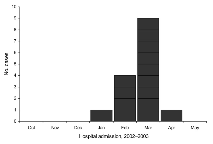
Sixteen cases, 1 of which (that of patient 8) was recognized through retrospective medical record review, were identified. All patients were hospitalized and admitted between January 28 through April 7 (Figure 1), and 13 patients (81%) were adults <50 years of age. Six (38%) of the 16 patients were hospitalized at hospital A during January through March. For comparison, the number of diagnoses of myocarditis in patients <50 years of age (16 patients) from October 1, 2000, to September 30, 2002, was <1 per month.
Figure 1.
Reported myocarditis case-patients by month of hospital admission, northern Illinois, 2003. (N = 15 because the exact date of admission to hospital was unknown for 1 patient).
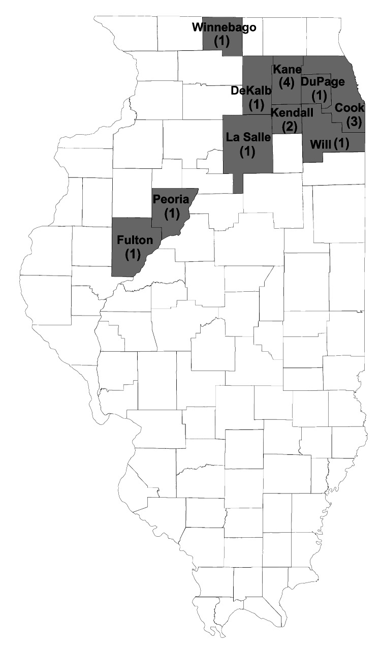
The median age for patients was 38 years (range 20–70 years). Among the 16 case-patients, 4 (25%) were residents of Kane County, 8 (50%) were from 5 counties bordering Kane County, and 4 (25%) were from 4 other counties in northern Illinois (Figure 2).
Figure 2.
County of residence of reported myocarditis case-patients (N = 16), northern Illinois, 2003.
Thirteen case-patients (81%) had an acute, viral-like illness within 1 month before onset of myocarditis. Two female patients, 26 and 39 years of age, had ventricular fibrillation that required an automatic implantable cardioverter defibrillator (AICD) and recovered. There were 2 deaths (Table 1).
Table 1. Demographic and clinical features of reported myocarditis patients, northern Illinois, 2003.
|
|
Cardiac test results |
||||||
|---|---|---|---|---|---|---|---|
| Patient and county of residence | Age/Sex | Date of hospital admission | Illness prodrome | Echocardiogram ejection fraction (abnormal <45%) | Cardiac catheterization | Endomyocardial biopsy | Other |
| 1, Kane | 31 F | 3/8 | Cough, shortness of breath, malaise for 3–5 d, diarrhea for 2 d | Decreased | Normal coronary arteries | Autopsy: lymphocytic infiltration of the myocardium | – |
| 2, La Salle | 47 M | 3/10 | None | 15%–20% | None | None | EKG*: new onset atrial fibrillation |
| 3, Kendall | 70 M | 3/4 | Upper respiratory tract infection for 2 wk | 20%–25% | None | None | – |
| 4, Kane |
45 F |
3/10 |
Fever, shortness of breath, obtundation for 1 d |
30% |
None |
None |
– |
| 5, DeKalb | 26 F | 3/4 | Viral bronchitis 1 mo before admission | None | None | None | EKG: ventricular fibrillation arrest |
| 6, Kendall | 32 M | 1/28 | Upper respiratory tract infection and diarrhea for 10 d | 20% | Normal coronary arteries | None | – |
| 7, Kane | 42 M | 03/25 | Cough for 2 wk | 20%–25% | None | None | EKG: new onset atrial fibrillation |
| 8, Kane |
45 F |
3/6 |
Viral illness 3 mo before, increasing palpitations for 3 mo |
30% |
Normal coronary arteries |
None |
– |
| 9, Will | 33 M | 3/19 | Upper respiratory tract infection for 5 d, shortness of breath for 2 d | Dilated cardiomyopathy | Normal coronary arteries | Lympohocytic and eosinophilic infiltration | – |
| 10, Fulton | 56 M | 2/8 | Upper respiratory tract infection 1 mo before, fever for 1 d | 20% | None | None | – |
| 11, Peoria | 38 M | 2/9 | Upper respiratory tract infection for 1 wk | 20%–25% | Normal coronary arteries | None | – |
| 12, Cook |
28 M |
Unknown |
Fevers for 2 wk |
20% |
None |
None |
– |
| 13, Cook | 60 M | 3/20 | Fever, cough, shortness of breath for 6 d | Decreased with global hypokinesis | None | None | – |
| 14, Cook† | 34 F | 2/28 | Unknown | Decreased, pericardial effusion | None | None | – |
| 15, DuPage | 39 F | 2/28 | Upper respiratory tract infection symptoms for 1 wk | 20%–25% | None | None | EKG: ventricular fibrillation arrest |
| 16, Winnebago | 20 M | 4/6 | Weight loss for 6 wk, vomiting and hemoptysis for 2 wk | None | None | Acute dilated cardiomyopathy | EKG: asystolic arrest |
*EKG, electrocardiogram. †Patient 14 had a diagnosis of myopericarditis.
No common exposures could be identified among the patients. None of the patients had recently been vaccinated for smallpox.
Information on acute serologic testing for group B coxsackievirus performed at hospitals was known for 5 patients. Two patients (patients 11 and 14) had elevated antibody titers to group B coxsackievirus. Patient 14 had a convalescent-phase serum specimen collected for group B coxsackievirus antibody testing that had a 2-fold greater titer than the acute-phase sample. Acute serologic testing for echovirus was performed for 2 patients; results were positive for patient 14 and negative for patient 13. Patient 14 also had an elevated acute-phase influenza B antibody titer but a negative convalescent-phase antibody titer. Patient 12 had no change in acute- and convalescent-phase–positive titers for group B coxsackievirus (Table 2).
Table 2. Laboratory features of reported myocarditis case-patients, northern Illinois, 2003*.
| Patient and county of residence | Local test results |
California laboratory tests |
Outcome | |||
|---|---|---|---|---|---|---|
| Group B coxsackie virus serology (reference range <1:8) | Other | Specimens | Date collected | Results | ||
| 1, Kane | None | Blood cultures: negative; pericardial fluid culture: negative | Serum, myocardial tissue | 3/8, 3/9 | Negative | Died |
| 2, La Salle | None | – | Serum | 4/2 | Negative | Recovered |
| 3, Kendall | None | – | Serum | 4/1 | Negative | Recovered |
| 4, Kane |
None |
Legionella urinary antigen: negative; sputum, blood, CSF, urine, stool viral cultures: negative |
Serum |
3/24 |
Negative |
Recovered |
| 5, DeKalb | None | – | Serum | 4/2 | Negative | AICD, recovered |
| 6, Kendall | None | – | Serum | 4/1 | Negative | Recovered |
| 7, Kane | Acute: negative | Influenza A and B: negative; blood, throat, urine, stool viral culture: negative | Serum | 3/25 | Negative | Recovered |
| 8, Kane |
None |
– |
Serum |
4/1 |
Negative |
Recovered |
| 9, Will | None | – | Myocardial tissue | 3/03 | Negative | Recovered |
| 10, Fulton | None | – | None | – | – | Recovered |
| 11, Peoria | Acute: positive 1:80; convalescent: positive 1:80 | Mycoplasma IgM serology: negative (reference range < 0.77 U/L); mycoplasma IgG serology: positive 1.47 U/L (reference range < 0.77 U/L); EBV, CMV serology: negative | Serum | 4/3 | Negative | Recovered |
| 12, Cook |
None |
Nasopharyngeal, stool culture: negative |
None |
– |
– |
Recovered |
| 13, Cook | Acute: negative | Echovirus serology: negative Influenza A and B, RSV rapid tests: negative; Legionella urinary antigen: negative | Serum | 4/12 | Negative | Recovered |
| 14, Cook† | Acute: positive 1:320; convalescent: 1:640 | Blood culture: negative; mycoplasma IgM serology: 0.11 negative (reference range < 0.77 U/L); mycoplasma IgG serology: negative 0.07 U/L (reference range < 0.77 U/L); acute echovirus type 11 serology: 1:320 (reference range >1:10); acute influenza A serology: negative (reference range >1:8; acute influenza B serology:positive 1:32; convalescent influenza B serology: negative (reference range <1:8); endotracheal viral culture: negative | None | – | – | Recovered |
| 15, DuPage | Acute: negative | CMV, EBV serology: negative | None | – | – | AICD, recovered |
| 16, Winnebago | None | – | Myocardial tissue | 4/7 | Negative | Died |
*CSF, cerebrospinal fluid; EBV, Epstein-Barr virus; CMV, cytomegalovirus; RSV, respiratory syncytial virus; AICD, automatic interventricular cardiac defibrillator. †Patient 14 had myopericarditis.
IDPH laboratory cultured nasopharyngeal (n = 5), urine (n = 6), stool (n = 6), and myocardial tissue (n = 1) specimens from 9 patients for enterovirus viral isolation. All cultures were negative. Among specimens (serum samples from 11 patients and myocardial tissue from 2 patients) tested for enterovirus and adenovirus by PCR and enzyme immunoassay , all were negative (Table 2).
For the 2 patients with fatal cases, the primary autopsy diagnosis was acute myocarditis. Autopsy tissue specimens from the 1 case-patient submitted to CDC were negative for viral agents (patient 1).
Conclusions
An outbreak of myocarditis of unknown cause occurred among adults in Kane County (population 400,000) and adjacent areas during winter and early spring 2003. Surveillance for myocarditis cases was initiated throughout Illinois in March and April, although clustering of cases was only evident in and limited to Kane County and surrounding communities. The reporting of myocarditis cases from other counties likely reflected baseline rates of idiopathic myocarditis in those populations that only came to the attention of public health officials through enhanced surveillance.
No common exposures were identified among case-patients. The outbreak occurred within the same period that adverse events of myopericarditis were being reported after smallpox vaccinations among military and healthcare personnel in the United States, including Illinois (12); however, no patients in this outbreak had recently been vaccinated against smallpox. Most illnesses were preceded by a prodrome that suggested the outbreak was viral in origin. Substantial illness and death occurred in these reported cases. All reported patients were hospitalized, 2 required AICD devices, and 2 deaths occurred, a reminder of the severe sequelae associated with this illness.
Despite extensive laboratory testing on submitted specimens, no specific agent was identified. Cross-reactivity of group B coxsackievirus serology with several agents was apparent from initial laboratory tests performed at the hospitals. These results were insufficient to support a specific cause of illness. Tissue specimens from only 3 of the 16 patients were available for testing, which was a major laboratory limitation in the investigation, particularly for detecting viral nucleic acid by PCR assays. The inability to implicate a responsible agent is a common outcome of myocarditis outbreak investigations (1,13).
A better understanding of myocarditis through enhanced diagnostic and therapeutic strategies, increased awareness of possible clusters of illness, and rapid reporting of clusters to public health departments will help improve prevention of future outbreaks. Recent biopsy-based studies suggest that a proportion of life-threatening myocarditis or idiopathic cardiomyopathy in otherwise healthy adults may arise from enteroviral and cytomegalovirus infections (14,15). Research is needed to assess the effect of potential antiviral treatment on illness and death in this patient population. In addition to encouraging appropriate viral testing of acute- and convalescent-phase serologic specimens, further study is required to examine the usefulness of endomyocardial tissue collection for advanced molecular analyses in patients with unexplained cardiomyopathy.
Acknowledgments
We thank Julu Bhatnagar, Marc Fischer, Andrea Winquist, and Carol Glaser for their contributions to this study and acknowledge the activities of CDC's UNEX Project and CDC's Division of Viral and Rickettsial Diseases Infectious Disease Pathology Laboratory.
We note with sadness that one of our coauthors, Douglas Passaro, died suddenly on April 18, 2005. Dr Passaro was a talented and outstanding researcher, who initiated a nationally recognized program in the 1990s to investigate infectious causes of unexplained deaths; ironically, his death also remains unexplained.
Biography
Dr Huhn completed an infectious diseases fellowship at Rush University Medical Center, Chicago, Illinois, in June 2005. From 2002 to 2004, he was an Epidemic Intelligence Service officer with CDC. His research interests include emerging infectious diseases, tropical medicine, infection control, and HIV.
Appendix
Selected Codes for Myocarditis and Pericarditis from the International Classification of Diseases, 9th Revision, Clinical Modification (ICD-9-CM).
Myocarditis: 422.90, 422.92, 422.93, 422.99
Other primary cardiomyopathies: 42.4
Footnotes
Suggested citation for this article: Huhn GD, Gross C, Schnurr D, Preas C, Yagi S, Reagan S, et al. Myocarditis outbreak among adults, Illinois, 2003. Emerg Infect Dis [serial on the Internet]. 2005 Oct [date cited]. http://dx.doi.org/10.3201/eid1110.041152
References
- 1.Savoia MC, Oxman MN. Myocarditis and pericarditis. In: Mandell GL, Bennett JE, Dolin R, editors. Mandell, Douglas, and Bennett's principles and practice of infectious diseases. 5th ed. Philadelphia: Churchhill Livingstone; 2000. p. 925–41. [Google Scholar]
- 2.Woodruff JF. Viral myocarditis: a review. Am J Pathol. 1980;101:425–84. [PMC free article] [PubMed] [Google Scholar]
- 3.Helin M, Sarola J, Lapinleimu K. Cardiac manifestations during a Coxsackie B5 epidemic. BMJ. 1968;3:97. 10.1136/bmj.3.5610.97 [DOI] [PMC free article] [PubMed] [Google Scholar]
- 4.Billingham ME. The safety and utility of endomyocardial biopsy in infants, children, and adolescents. J Am Coll Cardiol. 1990;15:443–5. 10.1016/S0735-1097(10)80075-0 [DOI] [PubMed] [Google Scholar]
- 5.Fowles RE, Mason JW. Endomyocardial biopsy. Ann Intern Med. 1982;97:885–94. [DOI] [PubMed] [Google Scholar]
- 6.Katsuragi M, Yutani C, Mukai, T. Arai Y, Imakita M, Ishibashi-Ueda H, et al. Detection of enteroviral genome and its significance in cardiomyopathy. Cardiology. 1993;83:4–13. 10.1159/000175941 [DOI] [PubMed] [Google Scholar]
- 7.Severini GM, Mestroni L, Falaschi A, Camerini F, Giacca M. Nested polymerase chain reaction for high-sensitivity detection of enteroviral RNA in biological samples. J Clin Microbiol. 1993;31:1345–9. [DOI] [PMC free article] [PubMed] [Google Scholar]
- 8.Tracy S, Wiegand V, McManus B, Gauntt C, Pallansch M, Beck M, et al. Molecular approaches to enteroviral diagnosis in idiopathic cardiomyopathy and myocarditis. J Am Coll Cardiol. 1990;15:1688–94. 10.1016/0735-1097(90)92846-T [DOI] [PubMed] [Google Scholar]
- 9.Aretz HT. Myocarditis: the Dallas criteria. Hum Pathol. 1987;18:619–24. 10.1016/S0046-8177(87)80363-5 [DOI] [PubMed] [Google Scholar]
- 10.Rotbart HA, Sawyer MH, Fast S, Lewinski C, Murphy N, Keyser EF, et al. Diagnosis of enteroviral meningitis by using PCR with a colorimetric microwell detection assay. J Clin Microbiol. 1994;32:2590–2. [DOI] [PMC free article] [PubMed] [Google Scholar]
- 11.Schnurr D, Yagi S, Devlin R. IgA and IgM ELISA for the study of an echovirus 30 outbreak in California [abstract S23]. In: Programs and abstracts of the 10th Annual Clinical Virology Symposium. Clearwater (FL): Pan American Society for Clinical Virology; 1994. [Google Scholar]
- 12.Centers for Disease Control and Prevention. Cardiac adverse events following smallpox vaccination—United States, 2003. MMWR Morb Mortal Wkly Rep. 2003;52:248–50. [PubMed] [Google Scholar]
- 13.Mounts AW, Amr S, Jamshidi R, Groves C, Dwyer D, Guarner J, et al. A cluster of fulminant myocarditis cases in children, Baltimore, Maryland, 1997. Pediatr Cardiol. 2001;22:34–9. 10.1007/s002460010148 [DOI] [PubMed] [Google Scholar]
- 14.Kuhl U, Pauschinger M, Noutsias M, Seeberg B, Bock T, Lassner D, et al. High prevalence of viral genomes and multiple viral infections in the myocardium of adults with "idiopathic" left ventricular dysfunction. Circulation. 2005;111:887–93. 10.1161/01.CIR.0000155616.07901.35 [DOI] [PubMed] [Google Scholar]
- 15.Kyto V, Vuorinen T, Saukko P, Lautenschlager I, Lignitz E, Saraste A, et al. Cytomegalovirus infection of the heart is common in patients with fatal myocarditis. Clin Infect Dis. 2005;40:683–8. 10.1086/427804 [DOI] [PubMed] [Google Scholar]


