Fig. 4.
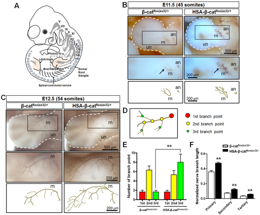
Augmented extension and branching of brachial plexuses in HSA-β-catflox(ex3)/+ embryos. (A) Schematic lateral view of mouse embryo. Roman numerals indicate cranial nerves. C, cervical; T, thoracic. (B) Embryos at 45-somite stage stained whole-mount with anti-NF antibody, which was visualized with diaminobenzidine (DAB). Arrows indicate brachial plexus. Areas in rectangles in top panels are enlarged in middle panels. Lower panels show camera lucida drawing of axons. an, axillary nerve; rn, radial nerve; un, ulnar nerve. (C) Embryos at 54-somite stage stained as described in B. Areas in rectangles in top panels are enlarged in middle panels. Lower panels show camera lucida drawing of axons. (D) Schematic of axon branches. Color matches data shown in E. (E) Increased numbers of rn tertiary branch points in HSA-β-catflox(ex3)/+ embryos shown in C. **P<0.01, n=3, one-way ANOVA. (F) Increased length of rn branches in HSA-β-catflox(ex3)/+ embryos shown in C. Shown are ratios of rn branch length over limb bud length. **P<0.01, n=3, one-way ANOVA. Error bars indicate s.e.m.

