SUMMARY
Evarcha culicivora is an East African jumping spider that feeds indirectly on vertebrate blood by choosing blood-fed female Anopheles mosquitoes as prey. Previous studies have shown that this predator can identify its preferred prey even when restricted to using only visual cues. Here, we used lures and virtual mosquitoes to investigate the optical cues underlying this predator's prey-choice behaviour. We made lures by dissecting and then reconstructing dead mosquitoes, combining the head plus thorax with different abdomens. Depending on the experiment, lures were either moving or motionless. Findings from the lure experiments suggested that, for E. culicivora, seeing a blood-fed female mosquito's abdomen on a lure was a necessary, but not sufficient, cue by which preferred prey was identified, as cues from the abdomen needed to be paired with cues from the head and thorax of a mosquito. Conversely, when abdomens were not visible or were identical, spiders based their decisions on the appearance of the head plus thorax of mosquitoes, choosing prey with female characteristics. Findings from a subsequent experiment using animated 3D virtual mosquitoes suggest that it is specifically the mosquito's antennae that influence E. culicivora's prey-choice decisions. Our results show that E. culicivora uses a complex process for prey classification.
KEY WORDS: Anopheles gambiae, Evarcha culicivora, Salticidae, Culex quinquefasciatus, vision, prey choice, animation, decision making
INTRODUCTION
A fundamental task for a predator that relies on vision is to determine whether objects that come into view are prey or non-prey, but the extent to which predators further classify prey can vary considerably. At one end of the continuum, we find predators that, by relying primarily on a few key prey features [‘sign stimuli’ (Tinbergen, 1951)], make rapid decisions and do minimal classifying of prey into particular types. Of particular note are the remarkably similar prey identification algorithms used by neurologically diverse animals, including amphibians (Ingle, 1983; Ewert, 2004), cuttlefish (Darmaillacq et al., 2004) and mantises (Prete et al., 2011). In some of these examples, such as frogs and toads, predators rapidly capture prey with a ballistic flick of the tongue after swift, efficient classification based on seeing an object of a specific size range moving in a specific orientation (Barlow, 1953; Lettvin et al., 1959; Ewert, 1997; Ewert, 2004). Many jumping spiders (Salticidae) may, like a toad (Ewert, 1997) or a mantis (Prete et al., 2011), rapidly decide on the basis of a few key features whether an object is prey or non-prey, followed by a swift prey-capture sequence (Drees, 1952; Forster, 1982). However, it is also among the salticids that some of the most distinctive examples of predators at the other end of the continuum are found.
Several salticid species adopt different prey-specific capture behaviour for different kinds of prey, express distinctive preferences for particular prey types and generally adopt a slower, more deliberate style of predation (Harland and Jackson, 2000; Nelson and Jackson, 2011). For example, araneophagic salticids are species that show pronounced preferences for spiders as prey and classify spiders into numerous different categories.
Salticids have eight eyes, with the terms ‘principal’ and ‘secondary’ eyes being used to denote their anatomical and functional distinctions (Homann, 1928; Land, 1985). The secondary eyes, spaced laterally around the salticid's carapace, are especially effective at detecting moving objects in the periphery and triggering orientation behaviour that enables fixation of the principal eyes on the object (Land, 1972; Zurek et al., 2010). However, the eyes for which salticids are best known are the large forward-facing principal eyes. These eyes enable salticids to see with exceptionally high spatial acuity (Land, 1969; Williams and McIntyre, 1980) and it has been shown that, for araneophagic and other salticids that do considerable classifying of prey into distinctive categories, identification of prey type can be achieved even when they are restricted to using vision alone.
The most precise expression of preference for a particular prey type known for a salticid, and possibly for any predator, comes from Evarcha culicivora (Wesolowska and Jackson, 2003). This East African salticid is unique in that feeds indirectly on vertebrate blood by choosing as prey female mosquitoes (particularly Anopheles) that have recently fed on blood (Jackson et al., 2005; Jackson and Nelson, 2012; Nelson and Jackson, 2012). Previous findings show that, even when restricted to using vision alone, E. culicivora can distinguish between blood-fed female mosquitoes and similar prey that are not carrying blood, such as male mosquitoes, female mosquitoes that have not fed on blood, and various similar-sized non-mosquito prey (Jackson et al., 2005; Nelson and Jackson, 2006; Nelson and Jackson, 2012). These experiments are often carried out using motionless dead prey in a life-like posture (lures), and here we also used lures, although we additionally ran experiments using moving lures made from dead prey and using 3D animation, as moving stimuli are more effective at maintaining the interest of the spider.
As little is known about the mechanisms underlying prey classification by predators that make fine distinctions between prey categories, we investigated the optical cues by which E. culicivora distinguishes between male and female mosquitoes and between mosquitoes that are and are not carrying blood. Our hypothesis was that this predator attends especially to the appearance of the mosquito's antennae and the abdomen. The rationale for considering antennae is that a male mosquito's antennae are plumose in appearance, while the antennae of female mosquitoes appear comparatively naked (Clements, 1999). The rationale for considering the abdomen is that the female mosquito's abdomen takes on a distinctive rounded shape after a blood meal.
MATERIALS AND METHODS
General
In our experiments, we used two of the mosquito species on which E. culicivora preys in nature (Wesolowska and Jackson, 2003), Culex quiquefasciatus and Anopheles gambiae (henceforth simply Culex and Anopheles). All spiders were from laboratory cultures (2nd and 3rd generation) initiated from specimens collected in Mbita Point, in Western Kenya. All test spiders were unmated adults that had matured 2–3 weeks before testing (4.5–5.5 mm), or in some cases juveniles (body length, 1.5 mm). Standard rearing, maintenance and testing methods were as in earlier studies (for details, see Jackson et al., 2005; Nelson and Jackson, 2006). Each salticid was allowed to feed to satiation three times per week on blood-fed female mosquitoes (Anopheles and Culex) from laboratory culture, and on chironomids and chaoborids collected as needed from the field (see Jackson et al., 2005). All mosquitoes had continuous access to sugar (6% glucose solution), but some female mosquitoes of both species had a human-blood meal 4–5 h before being used for making the lures used in experiments. The shorter expressions ‘blood-fed’ and ‘sugar-fed’ are used for female mosquitoes that did or did not receive a blood meal before use.
Testing was carried out between 08:00 h and 12:00 h in a laboratory lit by a 200 W incandescent lamp positioned 400 mm overhead with additional ambient lighting from fluorescent ceiling lamps (laboratory photoperiod 12 h:12 h L:D, lights on at 07:00 h). A 7 day pre-test fast ensured that the test spiders would be motivated to feed during testing. For each test, individuals were randomly chosen from the stock culture and no test spider was used more than once. Results were analysed using binomial tests (Ho 50:50).
Motionless-lure tests
During each test, adult test spiders had simultaneous access to two lure types. Anopheles and Culex mosquitoes used to make lures were immobilized with CO2 and then placed in 80% ethanol for 4 h, after which each was removed and dissected into two parts (abdomen and head+thorax). The head+thorax and the abdomen of different mosquitoes were then glued together to make a reconstructed mosquito (i.e. the ‘lure’). The reconstructed mosquito was then mounted in a life-like posture on the centre of one side of a thin disc-shaped piece of cork (diameter 7 mm) and sprayed with a transparent plastic adhesive for preservation. The body lengths of all reconstructed mosquitoes were 4.5 mm (accurate to the nearest 0.5 mm). When referring to the head+thorax, our interest was in the mosquito's antennae, but transferring antennae alone between lures was excessively difficult. This meant that we could not rule out the possibility that appearance features in addition to the antennae mattered to the test spider. However, when using virtual mosquitoes (see below), the appearance of heads and thoraces remained constant when we altered solely the appearance of antennae.
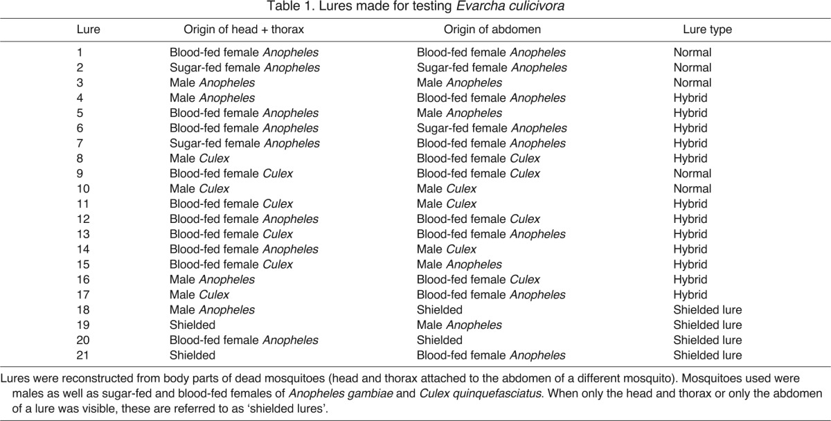
‘Normal’ lures (Table 1) were made by combining an abdomen and head+thorax of the same type (i.e. both from blood-fed female, both from sugar-fed female, or both from male), but with the proviso that the abdomen and head+thorax always came from different individuals (sham controls). ‘Hybrid’ lures were made by combining an abdomen of one type of mosquito with a head+thorax of another type of mosquito.
Table 1.
Lures made for testing Evarcha culicivora
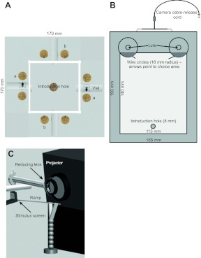
A square transparent glass box served as a testing arena (Fig. 1A). Four vials were fitted into holes spaced around the four sides of the box, with a lure on each side of each vial, such that eight lures were space around the box. The box was mounted on a wooden platform surrounded by a white wooden frame serving as a background against which E. culicivora saw the lures. Each lure sat on the platform and faced directly toward the side of the box. Two lure types were present during each test. One type was placed on one pair of opposing sides (positions ‘a’) and the other type was placed on the other opposing sides (positions ‘b’) (for details, see Jackson et al., 2005). Which of the two lure types was placed in positions ‘a’ was decided at random.
Fig. 1.
Equipment used for prey-choice testing. (A) Testing arena for motionless-lure tests. The apparatus consisted of a glass arena (square box, 100×100 mm, walls 35 mm high), with a removable glass lid. Holes in the box connected with four ‘choice’ vials flanked by lures. Lures in position ‘a’ are different from lures in position ‘b’. (B) Testing apparatus for moving-lure tests. A 35 mm deep rectangular glass box (pale grey) with a glass lid sat on top of a wooden stand (grey). Moving lures were controlled through a camera release-cord. The ‘choice area’ is the dark grey semicircular area within wire circles. (C) Apparatus for virtual-prey testing. Images pass from a data projector lens through a second lens (for reducing image size) onto a stimulus screen positioned in front of the higher end of the ramp.
When a test spider entered any one of the four vials and remained inside for 30 s, the lure type beside the vial was recorded as its ‘choice’. After introducing the test spider into the centre of the arena and then plugging the hole in the lid with a rubber stopper, tests lasted 30 min or until the test spider made a choice. Between tests, the box, the stopper and all vials were washed with 80% ethanol followed by distilled water, and then dried.
Moving-lure tests
The testing arena (Fig. 1B) was a glass box with a removable glass lid that was centred on top of a 150 mm high stand. The arena was surrounded a white frame serving as a background against which E. culicivora saw the lures. We introduced spiders into the arena through a hole in the floor. This hole, situated with its closer side 10 mm from one end of the box, was plugged with a removable rubber bung. At the opposite end of the arena, there was a ‘left lure hole’ and a ‘right lure hole’ (diameter of each, 5 mm). Two types of lures (position randomised) were placed outside the arena so that spiders could only see them through the arena's glass walls. During tests, a lure was centred on top of the right hole and another lure was centred on the top of the left hole. The lure was placed such that it faced directly toward the side of the arena. The lure stayed in place because the diameter of the hole in the stand was less than the diameter of the cork disc holding the lure.
Lures were moved by connecting a metal prong, which was attached to a camera cable-release cord, to the underside of each of the two cork discs. Pressing the cable-release moved each lure 5 mm up from the floor of the arena and then, by releasing the cable, each lure was lowered back to the floor. As soon as the test spider entered the arena, the cable-release was pressed once every 30 s and then released immediately, causing the lure/s to move up once and down once for each press.
Two circles made from thin copper wire were situated on the platform. A lure hole was at the centre of each circle and a part of each wire circle extended under the arena, remaining visible because the arena was made of glass. The part of the circle under the arena was the ‘choice area’ (Fig. 1B). Our operational definition of a choice was seeing the test spider fixate its gaze on a lure and then, while retaining fixation, enter the choice area. ‘Fixate’ refers to the corneal lenses of the salticid's large forward-facing principal eyes being held oriented toward a lure. There were rare instances (<5%) of the 15 min test period ending with the test spider outside the choice area but with its gaze fixated on a lure. In these instances, we extended the test period until the test spider either made its choice or turned away.
Some lures (see Table 1) were shielded with a 10 mm high black paper card glued upright on the cork disc such that only a head+thorax or only an abdomen of Anopheles was visible to the test spider (anterior end of head+thorax facing out from the card; posterior end of abdomen facing out).
Virtual-prey tests
Our basic methods for working with virtual mosquitoes were as described previously (Nelson and Jackson, 2006) except for modifications of the methods required for varying the antennae that we combined with virtual-mosquito bodies. Using 3D Studio Max, virtual mosquitoes were drawn based on images of blood-fed Anopheles derived from microscopy (see Nelson and Jackson, 2006).
Each mosquito antenna was created by ‘surfacing’ a transparent virtual box with a photograph of either a male or female antenna. Each box was then modified (using ‘bend’ and ‘twist’) to give a 3D appearance to the antennae. During tests, spiders were presented with side-on views of two virtual mosquitoes (placed side by side). Each virtual mosquito had an enlarged abdomen and was presented in ‘greyscale’. The two virtual mosquitoes differed only in the appearance of their antennae. Whether a particular virtual prey was on the left or right was determined at random. Movement that we added for animation was based on frame-by-frame analysis of digital video footage of Anopheles females that were grooming.
A 10 s mosquito-animation movie file was set to loop continuously on a computer. Rendered movies (AVI format) were forward-projected onto a glass screen using a Telex P400 LCD data projector (800×600 pixels; frame rate of animation files 25 frames s–1). The screen (fine-ground matte unmarked type D Nikon F3 focusing screen, 39 mm wide×30 mm high) was situated ∼150 mm from the projector lens, in front of which there was a ramp (stainless steel, 15 mm wide×150 mm long) (Fig. 1C). The distance between the screen and the top end of the ramp was 2 mm when testing juvenile spiders and 5 mm when testing adults. These screen–ramp distances ensured that spiders that attacked virtual prey had to leap rather than simply walk onto the screen. The projector was angled down by 10 deg and the screen sat in front of the top end of a stainless steel ramp, angling up by 25 deg. With this configuration, spiders walked up the ramp without entering the light path from the projector.
Adult spiders were first taken into a transparent PVC tube (10 mm long, 8 mm i.d.) and the two ends were plugged with corks. The tube was then positioned along the midline of the ramp, oriented in the same direction as the ramp, with the closer end 50 mm from the top of the inclined ramp. We removed the cork on the upward-facing end, and testing began when the spider walked out of the tube and onto the ramp.
Preliminary trials revealed that this method was problematic when using small juveniles because at a distance of 50 mm from the screen they often seemed not to notice the virtual mosquitoes and, when closer to the screen, the tube cast a shadow on the screen. Our solution was first to entice a juvenile onto the tip of a soft paintbrush and then to touch the ramp with the tip of the brush 10 mm from the ramp's upper end (juvenile on top of brush). Testing began when the spider walked slowly off the brush onto the ramp.
Tests lasted 15 min. However, if the test spider had begun stalking, testing continued until the end of the stalking bout (this never exceeded 18 min). Stalking is a readily identifiable behaviour characterised by the salticid slowly stepping toward the prey, with its body lowered close to the substrate and its palps waving, all the while visually fixated on the prey. In successful tests, the spider stalked to the end of the ramp and then either stayed quiescent and fixated on one of the virtual mosquitoes for 30 s or else leapt and landed on one of the virtual mosquitoes. There were no instances of leaping and landing on the screen at any location other than on one of the projected virtual mosquitoes.
RESULTS
Motionless-lure tests
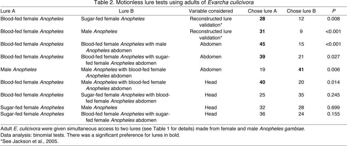
As in earlier work with intact lures (Jackson et al., 2005), test spiders chose blood-fed females significantly more often than they chose sugar-fed females or males when reconstructed normal lures were used (Table 2). On this basis, we concluded that the effectiveness of the lures we made using our reconstruction methods were comparable to intact lures.
Table 2.
Motionless lure tests using adults of Evarcha culicivora
Regardless of origin of the head+thorax, test spiders chose reconstructed lures that had a blood-fed female abdomen significantly more often than lures that had a male abdomen or a sugar-fed female abdomen (Table 2). When lures were made with the abdomens of sugar-fed mosquitoes (male or female), no significant tendency to choose one instead of the other was evident when the alternatives were lures with male or with female heads+thoraces. There was no significant difference in the number of spiders that chose normal sugar-fed females and the number that chose normal males, nor was there a significant difference in the number that chose normal sugar-fed females and the blood-fed female head+thorax on a sugar-fed female abdomen or those that chose normal blood-fed females instead of a sugar-fed female head+thorax on a blood-fed female abdomen. However, when the lures were made with the abdomens of blood-fed mosquitoes, E. culicivora chose normal blood-fed females significantly more often than they chose a male head+thorax on a blood-fed female abdomen (Table 2). These results imply that, although cues from the mosquito's head+thorax are also salient, they influenced the spider's choice only when the appropriate cues from the abdomen were present (i.e. for E. culicivora, seeing a blood-fed female abdomen seems to be a necessary prey-choice cue).
Moving-lure tests
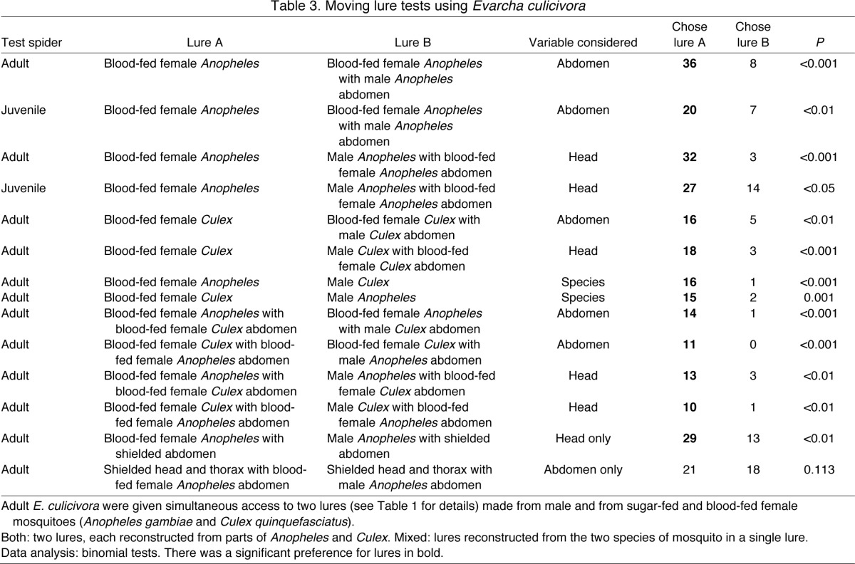
Test spiders chose normal blood-fed females significantly more often than a blood-fed female head+thorax on a male abdomen or a male head+thorax on a blood-fed female abdomen, regardless of whether the mosquito was reconstructed entirely from Anopheles, entirely from Culex or from parts of the two mosquito species (Table 3). However, when the abdomens of lures were identical or shielded, test spiders chose the head+thorax of females more often than the head+thorax of males. When only abdomens were visible there was no difference between the number of spiders that chose the blood-fed female abdomen or the male abdomen (Table 3).
Table 3.
Moving lure tests using Evarcha culicivora
Virtual-prey tests
Unlike in lure tests, the stimuli did not differ in appearance (orientation) depending on the position of the spider. Evarcha culicivora juveniles attended to cues from the antennae of virtual prey that were otherwise identical, choosing virtual prey with female antennae (N=17) significantly more often than virtual prey with male antennae (P=0.009, N=22). The same trend held when we tested E. culicivora adults, as 75% (N=15) chose the virtual prey with female antennae when the alternative was virtual prey with male antennae (P=0.04, N=20).
DISCUSSION
Using lures and virtual prey, we investigated how the appearance of mosquitoes influences E. culicivora's prey-choice behaviour. These methods removed potentially confounding variables related to odour, sound or substrate vibration, and movement was standardised. Lures provided biologically realistic features, but at the cost of being unable to alter the appearance of a mosquito's antennae while keeping all other features of mosquito appearance constant, as was possible with virtual mosquitoes. As hypothesised, our findings show that the appearance of the mosquito's abdomen and the appearance of its antennae are two especially salient prey-choice cues for E. culicivora. Accepting this hypothesis implies that these small predators, using eyes that are minute by vertebrate standards, can base decisions on remarkably fine visual detail. Although the capacity to detect fine detail may be largely explained by the exceptional spatial acuity achieved by the principal eyes of salticids (Land, 1969; Williams and McIntyre, 1980), questions remain concerning how the necessary processing of visual information is achieved with the number of receptors present in these small eyes.
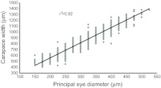
The number of receptors in the principal eyes of E. culicivora has not been determined directly, but we use what we know about other salticids to derive estimates. Phiddipus johnsoni is the only species for which we know the diameter of the principal eye corneal lens [∼600 μm for adults, based on previous data (Jackson, 1978)] and also have a reliable calculation of the number of receptors in the principal eye [∼1184 receptors in each eye for adults (Land, 1969)]. Assuming that receptor diameter and spacing remain more or less constant through development and that variation among salticid species is moderate, we can crudely calculate the number of receptors in each principal eye retina of the E. culicivora we used. The diameter of the principal eyes of adult E. culicivora is about 500 μm (Fig. 2) and, on this basis, we estimate that these spiders had about 1000 receptors per retina. Juveniles, with a principal eye diameter of only 150–200 μm, would only have about 300 receptors per retina with which to discriminate between mosquitoes that differed only in the appearance of the abdomen or the antennae.
Fig. 2.
Linear regression of the size of corneal lenses of the principal eyes of Evarcha culicivora in relation to carapace width. Eyes were measured using an ocular micrometer to the nearest 25 μm (N=977 spiders).
The strength of the expression of E. culicivora's preference for female mosquitoes varies with the spider's size and prior feeding condition, with animals becoming less discerning over time (Nelson and Jackson, 2012). When sated, adults choose sugar-fed female Anopheles over males, so the preference for females is present independent of the blood meal, although after a 7 day fast this expression of preference is lost (Nelson and Jackson, 2012), as also seen in the present study. Except when nutritionally stressed, predators are expected to select more profitable prey (Stephens and Krebs, 1986). Both active foraging spiders and sedentary web-building spiders have been shown to optimise nutrient composition (Greenstone, 1979; Mayntz et al., 2005), and preference by E. culicivora for blood-carrying mosquitoes suggests that indirect blood meals may be more profitable for E. culicivora than meals from other potential prey. However, why E. culicivora should select females that have not fed on blood in preference to males, and why they should have mechanisms that enable them to make this distinction (attending to the antennae), is unclear and unlikely to be amenable to simplistic explanation.
The engorged abdomens of female mosquitoes may be, albeit transiently, an easily detected visual indicator of the presence of blood, yet the finer distinction based on the appearance of antennae was only made when the abdomens were engorged or not visible. Perhaps whether the mosquito is male or female does not matter when the cue for blood (engorged abdomen) is absent. This is not an entirely satisfactory explanation because the presence of blood necessarily implies that the mosquito is female, yet E. culicivora attends to cues from the head when both lures are engorged or when lures had shielded abdomens. However, mosquitoes may be engorged because they are gravid, have fed on blood or, to a lesser extent, because they have fed on water or nectar (Amerasinghe and Amerasinghe, 1999). It is possible that, for E. culicivora, it is important to ascertain that engorgement is due to the presence of blood and not something else. While using cues from the antennae is not an especially accurate predictor of blood, seeing male antennae would be sufficient to inform the spider of its absence. Another clue may be the colour or brightness of the distended abdomen. Here, colour and shape co-varied when using lures, while with virtual mosquitoes neither colour nor abdomen shape varied. Consequently, it remains to be seen whether E. culicivora can distinguish between blood-fed females (which take on a red hue in the abdomen) and other engorged mosquitoes. This is the basis for an ongoing study concerned with the effect of colour and brightness in E. culicivora's prey-choice behaviour.
While research on the neural basis for salticid behaviour is in its infancy, we do have some evidence for homologous processing compared with other taxa. For example, the orientation response of Servaea vestita is very selective to stimuli of certain sizes, with very small stimuli essentially ignored (despite being detected; D. B. Zurek and X.J.N., submitted). Furthermore, these responses are relatively velocity invariant, as found in the tectal neurons of frogs (see Ewert, 2004), although at ‘optimal’ sizes (between 2 and 4 deg), there does appear to be an optimal ‘not too fast’ speed (Zurek et al., 2010). This suggests that salticids, which have limited neural capacity in their minute brains, adopt mechanisms similar to those that have been found in several other groups, including toads and mantises (Ewert, 2004; Kral and Prete, 2004). The basic premise behind these mechanisms is that predators are able to recognise and respond with predatory behaviour to a particular class of objects – potential prey items. Of course, prey will not be viewed from the same perspective every time (for example, a spider might be on the underside of a horizontal leaf viewing a mosquito on the topside of a leaf angled at 70 deg), and an explicit representation of prey based on a neural ‘image’ or ‘photographic representation’ for each angle, distance and prey type is unlikely given the constraints imposed by the small nervous systems. Consequently, category-specific, spatiotemporal features shared by various prey-like stimuli are likely to be used to create a representation of prey. This implicit recognition is achieved through simultaneous processing of spatio-temporal features of objects that fit a limited combination of biologically relevant parameters, including movement (Edwards and Jackson, 1994; Ewert, 2004; Kral and Prete, 2004). Therefore, for neurally constrained predators in particular, objects that elicit appetitive behaviour will be defined by their inclusion within a perceptual envelope that includes a variety of images sharing a subset of certain key stimulus characteristics. The particular weighting of the key characteristics is modifiable by experience (Edwards and Jackson, 1994; VanderSal and Hebets, 2007), hunger level (Nelson and Jackson, 2012), and the presence of other characteristics (in this case cues that are directly related to obtaining prey that have fed on blood), among others.
Blood meals play an important role in the sexual behaviour of E. culicivora. By feeding on blood-fed mosquitoes, these spiders acquire an odour that makes them attractive to the opposite sex (Cross et al., 2009). The evidence presented here provides support for the hypothesis that the presence of blood in prey is a primary factor in the fine-tuning of E. culicivora's prey-choice behaviour, as completely visible lures with engorged abdomens were preferred to any alternative. Our findings suggest that whether the head and thorax is from a male or a female only becomes relevant to E. culicivora when the spider fails to see an abdomen that comes from a blood-fed female. Additionally, E. culicivora's prey preference behaviour is innate (Nelson et al., 2005), suggesting that there may be some sort of unlearnt template (search image), or perceptual envelope, for what the spider is seeking in its prey. One hypothesis is that when a spider encounters stimuli providing unreliable (‘noisy’) cues, such as male antennae corresponding with the abdomen of a female mosquito or, somewhat more realistically, a mosquito with an obscured abdomen, E. culicivora ‘defaults’ to further assessment, perhaps based on longer inspection of the stimulus, serving as the best proxy to finding blood – namely, the appearance of a female's antennae.
As mentioned, several other factors influence E. culicivora's complex prey-choice decisions. For example, this spider makes predatory decisions based on prey size relative to itself, with smaller juveniles choosing smaller prey (Jackson et al., 2005), and also makes predatory decisions based on hunger level (Nelson and Jackson, 2012). When sated, adult E. culicivora chooses Anopheles mosquitoes in preference to Culex, and chooses sugar-fed female Anopheles over male Anopheles, but these underlying preferences are not revealed when spiders are hungry. Juvenile spiders have a preference for Anopheles that is stronger than that of adults (Nelson and Jackson, 2012), possibly because the tilted resting posture of Anopheles makes them easier for a small spider to attack and hold on to than Culex, which rest with the body parallel to the substrate (Nelson et al., 2005). Consistent with this, E. culicivora also distinguishes between Anopheles and Culex mosquitoes based on their resting posture (Nelson and Jackson, 2006).
It seems that E. culicivora uses a decision network for positive identification of its preferred prey. Although it appears that the first ‘filter’ of the perceptual envelope includes looking for evidence of a blood meal based largely on cues from the mosquito's abdomen, it is clear that E. culicivora is not simply using a hierarchical ‘identification key’ when choosing its prey. Depending on what information the spider received from looking at the abdomen, it may use resting posture as a cue to identify whether the mosquito is Anopheles (Nelson and Jackson, 2006) or, as shown here, it may look for cues based on the antennae in order to distinguish mosquito sex. In addition to hunger level, each of these steps is contingent upon other factors such as prey size, and in the case of looking at the antennae, possibly based on cues from the abdomen itself, making this spider an unusually discerning predator.
ACKNOWLEDGEMENTS
We are grateful to Godfrey Otieno Sune, Stephen Alluoch, Silas Ouko Orima and Jan McKenzie for technical assistance and to two anonymous referees for excellent suggestions.
FOOTNOTES
FUNDING
This research was supported by grants to R.R.J. from the National Geographic Society [8676-09, 6705-00] and the US National Institutes of Health [R01-AI077722]. X.J.N. was supported by a doctoral scholarship from the University of Canterbury and a William and Ina Cartwright scholarship. Deposited in PMC for release after 12 months.
REFERENCES
- Amerasinghe P. H., Amerasinghe F. P. (1999). Multiple host feeding in field populations of Anopheles culicifacies and An. subpictus in Sri Lanka. Med. Vet. Entomol. 13, 124-131 [DOI] [PubMed] [Google Scholar]
- Barlow H. B. (1953). Summation and inhibition in the frog’s retina. J. Physiol. 119, 69-88 [DOI] [PMC free article] [PubMed] [Google Scholar]
- Clements A. N. (1999). The Biology of Mosquitoes. Wallingford, UK: CABI Publishing; [Google Scholar]
- Cross F. R., Jackson R. R., Pollard S. D. (2009). How blood-derived odor influences mate-choice decisions by a mosquito-eating predator. Proc. Natl. Acad. Sci. USA 106, 19416-19419 [DOI] [PMC free article] [PubMed] [Google Scholar]
- Darmaillacq A. S., Chichery R., Poirier R., Dickel L. (2004). Effect of early feeding experience on subsequent prey preference by cuttlefish, Sepia officinalis. Dev. Psychobiol. 45, 239-244 [DOI] [PubMed] [Google Scholar]
- Drees O. (1952). Untersuchungen über die angeborenen Verhaltensweisen bei Springspinnen (Salticidae). Z. Tierpsychol. 9, 169-207 [Google Scholar]
- Edwards G. B., Jackson R. R. (1994). The role of experience in the development of predatory behaviour in Phidippus regius, a jumping spider (Araneae, Salticidae) from Florida. N. Z. J. Zool. 21, 269-277 [Google Scholar]
- Ewert J. P. (1997). Neural correlates of key stimulus and releasing mechanism: a case study and two concepts. Trends Neurosci. 20, 332-339 [DOI] [PubMed] [Google Scholar]
- Ewert J. P. (2004). Motion perception shapes the visual world of amphibians. In Complex Worlds from Simpler Nervous Systems (ed. Prete F. R.), pp, 117-160 Cambridge, MA: MIT Press; [Google Scholar]
- Forster L. M. (1982). Vision and prey catching strategies in jumping spiders. Am. Sci. 70, 165-175 [Google Scholar]
- Greenstone M. H. (1979). Spider feeding behavior optimizes dietary essential amino acid composition. Nature 282, 501-503 [Google Scholar]
- Harland D. P., Jackson R. R. (2000). ‘Eight-legged cats’ and how they see: a review of recent research on jumping spiders (Araneae: Salticidae). Cimbebasia 16, 231-240 [Google Scholar]
- Homann H. (1928). Beiträge zur Physiologie der Spinnenaugen. I. Untersuchungsmethoden II. Das sehvermögen der Salticiden. Z. vergl. Physiol. 7, 201-268 [Google Scholar]
- Ingle D. J. (1983). Brain mechanisms of visual localization by frogs and toads In Advances in Vertebrate Neuroethology (ed. Ewert J. P., Capranica R. R., Ingle D. J.), pp. 177-226 New York: Plenum; [Google Scholar]
- Jackson R. R. (1978). Life history of Phidippus johnsoni (Araneae, Salticidae). J. Arachnol. 6, 1-29 [Google Scholar]
- Jackson R. R., Nelson X. J. (2012). Evarcha culicivora chooses blood-fed Anopheles mosquitoes but other East African jumping spiders do not. Med. Vet. Entomol. 26, 233-235 [DOI] [PubMed] [Google Scholar]
- Jackson R. R., Nelson X. J., Sune G. O. (2005). A spider that feeds indirectly on vertebrate blood by choosing female mosquitoes as prey. Proc. Natl. Acad. Sci. USA 102, 15155-15160 [DOI] [PMC free article] [PubMed] [Google Scholar]
- Kral K., Prete F. R. (2004). In the mind of a hunter: the visual world of the praying mantis. In Complex Worlds from Simpler Nervous Systems (ed. Prete F. R.), pp, 75-116 Cambridge, MA: MIT Press; [Google Scholar]
- Land M. F. (1969). Structure of the retinae of the principal eyes of jumping spiders (Salticidae: Dendryphantinae) in relation to visual optics. J. Exp. Biol. 51, 443-470 [DOI] [PubMed] [Google Scholar]
- Land M. F. (1972). Stepping movements made by jumping spiders during turns mediated by the lateral eyes. J. Exp. Biol. 57, 15-40 [DOI] [PubMed] [Google Scholar]
- Land M. F. (1985). Fields of view of the eyes of primitive jumping spiders. J. Exp. Biol. 119, 381-384 [Google Scholar]
- Lettvin J. Y., Maturana H. R., McCulloch W. S., Pitts W. H. (1959). What the frog’s eye tells the frog’s brain. Proc. Inst. Radio Engr. 47, 1940-1951 [Google Scholar]
- Mayntz D., Raubenheimer D., Salmon M., Toft S., Simpson S. J. (2005). Nutrient-specific foraging in invertebrate predators. Science 307, 111-113 [DOI] [PubMed] [Google Scholar]
- Nelson X. J., Jackson R. R. (2006). A predator from East Africa that chooses malaria vectors as preferred prey. PLoS ONE 1, e132 doi:10.1371/journal.pone.0000132 [DOI] [PMC free article] [PubMed] [Google Scholar]
- Nelson X. J., Jackson R. R. (2011). Flexibility in the foraging strategies of spiders. In Spider Behaviour: Flexibility and Versatility (ed. Herberstein M. E.), pp. 31-56 New York: Cambridge University Press; [Google Scholar]
- Nelson X. J., Jackson R. R. (2012). Fine-tuning of vision-based prey-choice decisions by a predator that targets malaria vectors J. Arachnol. 40, 23-33 [Google Scholar]
- Nelson X. J., Jackson R. R., Sune G. O. (2005). Use of Anopheles-specific prey-capture behavior by the small juveniles of Evarcha culicivora, a mosquito-eating jumping spider. J. Arachnol. 33, 541-548 [Google Scholar]
- Prete F. R., Komito J. L., Domínguez S., Svenson G., López Y. L., Guillen A., Bogdanivich N. (2011). Visual stimuli that elicit appetitive behaviors in three morphologically distinct species of praying mantis, J. Comp. Physiol. A, 197, 877-894 [DOI] [PubMed] [Google Scholar]
- Stephens D. W., Krebs J. R. (1986). Foraging Theory. Princeton, NJ: Princeton University Press; [Google Scholar]
- Tinbergen N. (1951). The Study of Instinct. Oxford: Clarendon Press; [Google Scholar]
- VanderSal N. D., Hebets E. A. (2007). Cross-modal effects on learning: a seismic stimulus improves color discrimination learning in a jumping spider. J. Exp. Biol. 210, 3689-3695 [DOI] [PubMed] [Google Scholar]
- Wesolowska W., Jackson R. R. (2003). Evarcha culicivora sp. nov., a mosquito-eating jumping spider from East Africa (Araneae: Salticidae). Ann. Zool. 53, 335-338 [Google Scholar]
- Williams D. S., McIntyre P. (1980). The principal eyes of a jumping spider have a telephoto component. Nature 288, 578-580 [Google Scholar]
- Zurek D. B., Taylor A. J., Evans C. S., Nelson X. J. (2010). The role of the anterior lateral eyes in the vision-based behaviour of jumping spiders. J. Exp. Biol. 213, 2372-2378 [DOI] [PubMed] [Google Scholar]


