Abstract
Perineural cysts are believed to be asymptomatic; however, they rarely cause symptoms related to nerve root compression. Cervical symptomatic perineural cysts are in fact exceedingly rare. There are no reported cervical perineural cysts in the literature that present like cubital tunnel syndrome. A patient with motor weakness of the abductor and adductor muscles of the fingers of the left hand and hypoesthesia in the hypothenar region of the left hand presented at our clinic. A neurological examination, and neuroradiological and electrophysiological evaluations supported the finding that the patient’s clinical condition was caused by a perineural cyst located around the C8 neural root. The neurological symptoms of the patient markedly improved after medical treatment. We reported the first cervical perineural cyst as presenting like cubital tunnel syndrome patient in the literature. The visualization of perineural cyst may need extra magnetic resonance imaging (MRI) sections in order to view the nerve root through the neural foramen or extraforaminal area. These lesions are benign, and the appropriate treatment is curative.
Keywords: Perineural cyst, Tarlov cyst, Cubital tunnel syndrome, Radiculopathy
Introduction
Perineural cysts, also known as Tarlov cysts, are largely incidental findings in imaging studies. The majority are clinically insignificant. The standard visualization used for these cystic lesions is magnetic resonance imaging (MRI). The cysts rarely associate with radiculopathy especially in the lumbar and sacral levels, headache, and cauda equina syndrome. Symptomatic cervical perineural cysts are extremely rare in the literature [1–7].
Cubital tunnel syndrome is one of the most common entrapment neuropathies for the upper limb. Yet there is still considerable difficulty in pinpointing the location of the pathologic compression of the nerve and thus choosing the correct treatment. Radiological and electrodiagnostic studies allow for the correct localization of compression on the nerve.
In this paper, we report on the first symptomatic cervical perineural cyst in the literature, presenting like cubital tunnel syndrome and treated with medical and physical therapy.
Case report
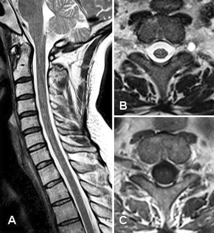
A 50-year-old man presented with a one-week history of pain, and numbness in the left hand. He expressed that the onset of symptoms had literally happened overnight. Neurological examination on admission revealed paresis of finger abductors and adductors and decreased superficial sensation in the hypothenar region on the left hand with normal sensation in the distal left arm. The remaining muscle strength was normal. Deep tendon reflexes were normal and symmetrical at the bilateral biceps, brachioradial, and triceps tendons. There was no evidence of upper motor neuron involvement found in the physical examination. An electromyography (EMG) test was given to the patient because the possible leading diagnosis was cubital tunnel syndrome. After a normal EMG of the intrinsic hand muscles and a normal nerve conduction study with inching at the level of the cubital tunnel, an MRI of the brain and cervical spine was ordered. It revealed a cystic lesion that filled the entire circumference of the left C8 neural root right after exiting the neural foramen. The lesion was hyperintense on the T2-weighted image, but it did not enhance after gadolinium (Fig. 1). Steroid medication was started, and physical therapy given to the patient.
Fig. 1.
T2-weighted sagittal image shows normal spinal cord and spinal anatomy (a), and T2-weighted axial image (b) shows homogeneous high signal intensity cystic area on the C8 nerve root. This cyst does not show contrast enhancement (c)
A subsequent EMG and somatosensorial evoked potential (SEP) tests in the fifth week revealed denervation of the left first and fourth dorsal interosseous muscles and normal bilateral ulnar nerve F latencies, bilateral normal median, and tibial nerve SEP results. The ulnar nerve SEP was normal on the right, but it could not be recorded on the left. These findings indicated a left C8 nerve root lesion that affected only the fibers of the ulnar division. On a follow-up examination 2 months later, the patient’s muscle strength had returned to normal. Because the patient rejected a control cervical MRI, we could not visualize the cyst. One year after therapy, we called the patient and learned everything was normal.
Discussion
Cubital tunnel syndrome is mostly caused by the compression of the ulnar nerve at the elbow. External trauma and deformity are common causes of ulnar nerve entrapment in cubital tunnel. In our case, the patient’s symptoms are related to the perineural cyst around the C8 nerve root.
A differential diagnosis of cubital tunnel syndrome includes C8–T1 cervical radiculopathy, thoracic outlet syndrome, lower brachial plexopathy, syringomyelia, and motor neuron disease. In C8–T1 radiculopathy, ulnar sensory potentials in C8 are intact and there are no focal conduction abnormalities across the elbow segment. A cervical MRI may show the reason for the radiculopathy. In the thoracic outlet syndrome and lower brachial plexopathy, sensory symptoms involve not only the fourth and fifth fingers, but also the medial forearm where weakness involves both the hypothenar and the thenar muscles, and electro diagnostic studies show normal conduction and a lesion in the lower trunk of the brachial plexus. Dissociated sensory loss is characteristic in syringomyelia and often associated long tract findings are found in the legs. Electrodiagnosis shows normal ulnar sensory potentials, and an MRI is an effective diagnostic tool. Motor neuron disease is characterized by weakness and wasting of the intrinsic hand muscles. The thenar and hypothenar muscles are also affected. Sensory disturbances are not found, while present fasciculation will indicate widespread nature of the disease.
Perineural cysts are pathological formations located in the space between the peri- and endoneurium of the spinal posterior nerve root sheath at the dorsal root ganglion [1]. The distinctive feature of perineural cysts versus other cystic spinal lesions is the presence of spinal nerve root fibers within the cyst wall, or the cyst cavity itself [1, 8, 9].
Perineural cysts are believed to be asymptomatic but they rarely cause symptoms related to nerve root compression. Symptomatic cases found in the literature are mostly located at the lumbar and sacral levels of the spine. If symptomatic, they have been reported to cause sacral radiculopathy, hip, leg, foot, or perineal pain, paresthesias, bowel or bladder dysfunction, neck pain, numbness in the arm, and mid-back pain [2, 3, 5–7, 10, 11]. Symptoms can be exacerbated by standing, coughing, or other Valsalva maneuvers because elevated subarachnoid pressure will drive the cerebrospinal fluid (CSF) from the spinal subarachnoid space through small ball valve–like communication into the perineurial cyst cavity [12]. Cubital tunnel-like syndrome was the presenting style in our case, but not reported in the literature. An MRI is particularly useful in studying perineural cysts that have CSF-like characteristics, which produces a low signal on T1-weighted images and a high signal on T2-weighted images. An MRI can also delineate the exact relationship of the cyst to the thecal sac, as well as the total volume of fluid lying within the cyst. It may also demonstrate bone and pedicle erosion, spinal canal widening, and neural foramina enlargement [1, 13]. In our case, a lesion exhibited similar MRI features to those described above. This case emphasizes that during the standard cervical MRI procedure, additional images that visualize the entire nerve within the foramen and distal to foramen must be included and evaluated. This inclusion is especially important if no other clear reason for the symptoms can be found.
Perineural cysts can additionally cause objective neurophysiological abnormalities, including decreased sural nerve action potentials and sensory nerve conduction velocities. These cysts cause mostly sensory disturbances because of their location at the dorsal root ganglion [1]. Denervation findings were seen in EMG 5 weeks after the onset of symptoms.
In conclusion, perineural cysts are benign lesions, but direct nerve root compression by such cyst may cause rare clinical conditions as in our case. Paraspinal areas distal to neural foramen are important and must be visualized on an MRI with additional axial and sagittal sections especially in a patient with standard normal cervical MRI.
Conflict of interest
None of the authors has any potential conflict of interest.
References
- 1.Acosta FL, Jr, Quinones-Hinojosa A, Schmidt MH, Weinstein PR. Diagnosis and management of sacral tarlov cysts. Case report and review of the literature. Neurosurg Focus. 2003;15(2):E15. doi: 10.3171/foc.2003.15.2.15. [DOI] [PubMed] [Google Scholar]
- 2.Dimitroulias AP, Stenner RC, Cavanagh PM, Madhavan P, Webb PJ. Multiple bilateral sacral perineural cysts unusually distal to the exit foramina. Br J Neurosurg. 2007;21(5):521–522. doi: 10.1080/02688690701436670. [DOI] [PubMed] [Google Scholar]
- 3.Langdown AJ, Grundy JR, Birch NC. The clinical relevance of tarlov cysts. J Spinal Disord Tech. 2005;18(1):29–33. doi: 10.1097/01.bsd.0000133495.78245.71. [DOI] [PubMed] [Google Scholar]
- 4.Mitra R, Kirpalani D, Wedemeyer M. Conservative management of perineural cysts. Spine. 2008;33(16):E565–E568. doi: 10.1097/BRS.0b013e31817e2cc9. [DOI] [PubMed] [Google Scholar]
- 5.Takatori M, Hirose M, Hosokawa T. Perineural cyst as a rare cause of l5 radiculopathy. Anesth Analg. 2008;106(3):1022–1023. doi: 10.1213/ANE.0b013e3181632583. [DOI] [PubMed] [Google Scholar]
- 6.Edeiken J, Zervas NT, Clearfield R. Cervical perineural or extradural cysts mimicking bone erosion of neurofibromata. Clin Orthop Relat Res. 1966;44:187–190. [PubMed] [Google Scholar]
- 7.McEvoy SD, DiLuna ML, Baird AH, Duncan CC. Symptomatic thoracic tarlov perineural cyst. Pediatr Neurosurg. 2009;45(4):321–323. doi: 10.1159/000235751. [DOI] [PubMed] [Google Scholar]
- 8.Goyal RN, Russell NA, Benoit BG, Belanger JM. Intraspinal cysts: a classification and literature review. Spine. 1987;12(3):209–213. doi: 10.1097/00007632-198704000-00003. [DOI] [PubMed] [Google Scholar]
- 9.Nabors MW, Pait TG, Byrd EB, Karim NO, Davis DO, Kobrine AI, Rizzoli HV. Updated assessment and current classification of spinal meningeal cysts. J Neurosurg. 1988;68(3):366–377. doi: 10.3171/jns.1988.68.3.0366. [DOI] [PubMed] [Google Scholar]
- 10.North RB, Kidd DH, Wang H. Occult, bilateral anterior sacral and intrasacral meningeal and perineurial cysts: case report and review of the literature. Neurosurgery. 1990;27(6):981–986. doi: 10.1227/00006123-199012000-00020. [DOI] [PubMed] [Google Scholar]
- 11.Jain SK, Chopra S, Bagaria H, Mathur PP. Sacral perineural cyst presenting as chronic perineal pain: a case report. Neurol India. 2002;50(4):514–515. [PubMed] [Google Scholar]
- 12.Ju CI, Shin H, Kim S, Kim H. Sacral perineural cyst accompanying disc herniation. J Korean Neurosurg Soc. 2009;45(3):185–187. doi: 10.3340/jkns.2009.45.3.185. [DOI] [PMC free article] [PubMed] [Google Scholar]
- 13.Rodziewicz GS, Kaufman B, Spetzler RF. Diagnosis of sacral perineural cysts by nuclear magnetic resonance. Surg Neurol. 1984;22(1):50–52. doi: 10.1016/0090-3019(84)90228-3. [DOI] [PubMed] [Google Scholar]


