Abstract
INTRODUCTION
The aim of this study was to develop a standardised management plan for boys with abnormal appearance of meatus at circumcision for balanitis xerotica obliterans (BXO).
METHODS
Between 1995 and 2008, 107 boys underwent circumcision for BXO (confirmed on histology). Of these, 23 had abnormal appearance of the meatus at operation; their case notes were reviewed for age, presenting symptoms, management, outcome and follow up.
RESULTS
The age range at operation was 3–15 years (mean: 9 years). Patients commonly presented with phimosis and balanitis. Seven patients had an additional procedure at circumcision: six had meatotomy, one had meatal dilatation. Thirteen were treated with topical steroid cream post-operatively. Eight of these (62%) subsequently required meatotomy. Three patients were observed and did not require further intervention. Meatotomy was required in 9 patients, 6–29 months after circumcision (mean: 11 months). Two patients required dilatation, including one with a previous intraoperative meatotomy, who required multiple dilatations.
CONCLUSIONS
We propose the following standardised management plan: 1. With clinical evidence of BXO at circumcision, prepuce should be sent for histology. 2. If BXO is confirmed but the meatus appears normal, patients should be seen once post-operatively to give information about meatal stenosis. 3. When the meatus appears scarred with a narrowed lumen at operation, a meatotomy should be performed, with follow up for at least two years. 4. If the lumen is scarred but adequate, patients should be followed up in clinic for the same period for possible development of stenosis. 5. Topical steroid cream can be considered for voiding discomfort without decreased urine stream.
Keywords: Balanitis xerotica obliterans, Meatal stenosis, Children, Management, Circumcision
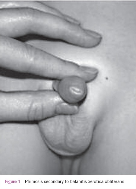
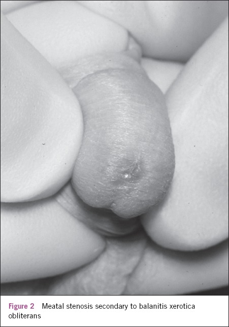
Balanitis xerotica obliterans (BXO) has long been recognised as a cause of pathological phimosis in children (Fig 1).1,2 It is an inflammatory process is of unknown aetiology, affecting the prepuce, glans and urethral meatus. The natural history of the disease in adults is well documented; progression of the disease can cause meatal stenosis and urethral stricture.3 There are few available data regarding such complications in boys. Therefore, when presented with abnormal appearances of the meatus at circumcision for BXO (Fig 2), the optimal management plan is uncertain. Subsequent treatment and follow up can be variable, even within an institution. Our aim was to develop a standardised management plan for such patients within our unit.
Figure 1.

Phimosis secondary to balanitis xerotica obliterans
Figure 2.

Meatal stenosis secondary to balanitis xerotica obliterans
Methods
We carried out a retrospective review of clinical and histological findings from a series of circumcisions performed under the care of a single consultant between 1995 and 2008. A total of 107 boys underwent circumcision for phimosis with a clinical diagnosis of BXO that was subsequently confirmed on histology. Of these, 23 (21%) had an abnormal appearance of the meatus at operation. Their case notes were reviewed for age at operation, presenting symptoms, intraoperative management, post-operative management, subsequent outcome and follow up.
Results
Patients' ages ranged from 3 to 15 years (mean: 9 years). Patients commonly presented with phimosis and recurrent balanitis. Other symptoms included poor urinary stream (n=4), urinary retention (n=4), paraphimosis (n=1) and hydronephrosis (n=1). Sixteen patients (70%) had a meatus that was scarred but with an adequate lumen. Seven patients (30%) had a meatus with a narrowed lumen and a further procedure was performed at the time of circumcision (Table 1). Of these, six had a meatotomy and one had dilatation of the meatus.
Table 1.
Outcome of patients with histologically confirmed balanitis xerotica obliterans and abnormal meatus at circumcision
| Total number of patients | Follow up only | Steroids only | Late meatotomy | Late dilatation | |
|---|---|---|---|---|---|
| Meatus scarred but adequate | 16 (70%) | 3 | 4 | 8 | 1 |
| Meatus scarred and lumen narrowed | 7 (30%) | 4 | 1 | 1 | 1 (multiple, self) |
After the operation, three patients were simply observed following circumcision and did not require any further intervention. Thirteen patients (56%) were treated with topical steroid cream in the post-operative period (including two who had early meatotomy/dilatation). Of these patients, eight (62%) subsequently required meatotomy, including the patient who had an intraoperative dilatation. Late meatotomy was necessary in a total of 9 patients, between 6 and 29 months after circumcision (mean: 11 months).
Two patients required meatal dilatation, including one with a previous intraoperative meatotomy. This patient required multiple (self) dilatations and had a prolonged follow-up period of 13 years. Another patient was lost to follow up. After excluding these two patients, the mean follow-up period was 14 months (range: 1.5–42 months).
Discussion
BXO has long been recognised as a cause of secondary phimosis in boys.1,2 More recently, there have been several studies that have found the incidence of BXO in circumcised boys to be higher than previously thought.4,5 It has also been suggested that BXO may be underestimated on clinical examination alone.6 In the literature, BXO in adults has been shown to be associated with a higher incidence of meatal stenosis and urethral strictures.3 Our data highlight the fact that BXO in children can also progress to involve the meatus.
Although meatal stenosis in boys with BXO has been described previously,7 we were surprised to discover that it was in a high proportion of cases (21%). It is difficult to establish the true incidence of meatal involvement as there is little available literature on the subject. In a large series of 1,178 childhood circumcisions, Kiss et al found that of 471 boys with BXO only 2% had meatal involvement.4 In two smaller studies, meatal involvement was found in 4 of 37 patients (11%)6 and 20 of 41 patients (49%)8 respectively.
It has been suggested that circumcision is potentially curative for BXO, even when extending onto the glans.2,4 The majority of our patients underwent circumcision at the time of surgery only, as it was hoped that removal of the foreskin would halt disease progression and allow any meatal involvement to resolve. However, when meatal stenosis was demonstrated at the time of circumcision, we felt it was appropriate to perform a further intervention (meatotomy/dilatation) to prevent obstruction to the urinary stream. The importance of follow up is clearly demonstrated by our study: a large proportion of patients (47%) required further intervention at an average of 11 months post-operatively (9 meatotomies, 2 dilatations). Without follow up, these complications may have been missed or diagnosed late, allowing further progression of the disease.
Regular application of a topical anti-inflammatory has been recommended as treatment after circumcision to prevent progression/recurrence.6,9 Over half our patients received topical steroid cream. Unfortunately, 62% of these patients required a meatotomy at a later date. It is possible that patients who were prescribed steroid cream had ‘worse’ appearances at circumcision than those who were not prescribed steroids and were therefore at greater risk of developing meatal stenosis. We would be reluctant to conclude that the use of topical steroids was ineffective from the results of our small study population, particularly as there is some evidence in the literature to suggest it has some benefits in the treatment of BXO albeit in the pre-operative period.
A randomised, placebo controlled study of children with BXO by Kiss et al showed some clinical improvement in histologically early and intermediate stage BXO when a topical steroid treatment was applied pre-operatively.10 They also suggested that it may inhibit further worsening in late stage BXO. Topical steroids may therefore be a useful adjunct in the post-operative management of more advanced BXO.
Conclusions
Our data demonstrate that BXO in children can progress to involve the meatus in a high proportion of cases. They also highlight the importance of following up such patients as in the months following circumcision nearly half developed complications that necessitated further surgical intervention. Taking this into account, we propose the following standardised management plan:
With clinical evidence of BXO at circumcision, the prepuce should be sent for histology.
If BXO is confirmed but the meatus appears normal, patients should be seen once post-operatively at approximately three months, to give information about meatal stenosis, and then discharged if voiding is normal.
When the meatus appears scarred with a narrowed lumen at operation, a meatotomy should be performed, with follow up for at least two years. In the first year follow-up visits should be at three-monthly intervals. If there are no concerns, follow-up appointments can then be every six months.
If the lumen is scarred but adequate, patients should be followed up in clinic at the same intervals and for the same period as above for possible development of stenosis.
Topical steroid cream can be considered for voiding discomfort without decreased urine stream. Patients should be alerted to further intervention in situations with worsening symptoms.
References
- 1.Chalmers RJ, Burton PA, Bennett RF, et al. Lichen sclerosus et atrophicus. A common and distinctive cause of phimosis in boys. Arch Dermatol. 1984;120:1,025–1,027. doi: 10.1001/archderm.120.8.1025. [DOI] [PubMed] [Google Scholar]
- 2.Meuli M, Briner J, Hanimann B, Sacher P. Lichen sclerosus et atrophicus causing phimosis in boys: a prospective study with 5-year followup after complete circumcision. J Urol. 1994;152:987–989. doi: 10.1016/s0022-5347(17)32638-1. [DOI] [PubMed] [Google Scholar]
- 3.Das S, Tunuguntla HS. Balanitis xerotica obliterans – a review. World J Urol. 2000;18:382–387. doi: 10.1007/PL00007083. [DOI] [PubMed] [Google Scholar]
- 4.Kiss A, Király L, Kutasy B, Merksz M. High incidence of balanitis xerotica obliterans in boys with phimosis: prospective 10-year study. Pediatr Dermatol. 2005;22:305–308. doi: 10.1111/j.1525-1470.2005.22404.x. [DOI] [PubMed] [Google Scholar]
- 5.Yardley IE, Cosgrove C, Lambert AW. Paediatric preputial pathology: are we circumcising enough? Ann R Coll Surg Engl. 2007;89:62–65. doi: 10.1308/003588407X160828. [DOI] [PMC free article] [PubMed] [Google Scholar]
- 6.Bochove-Overgaauw DM, Gelders W, De Vylder AM. Routine biopsies in pediatric circumcision: (non) sense? J Pediatr Urol. 2009;5:178–180. doi: 10.1016/j.jpurol.2008.11.008. [DOI] [PubMed] [Google Scholar]
- 7.Baron M, Heloury Y, Stalder JF, et al. Acquired phimosis, or preputial sclero-atrophic lichen in children. J Chir (Paris) 1991;128:368–371. [PubMed] [Google Scholar]
- 8.Gargollo PC, Kozakewich HP, Bauer SB, et al. Balanitis xerotica obliterans in boys. J Urol. 2005;174:1,409–1,412. doi: 10.1097/01.ju.0000173126.63094.b3. [DOI] [PubMed] [Google Scholar]
- 9.Ebert AK, Vogt T, Rösch WH. Topical therapy of balanitis xerotica obliterans in childhood. Long-term clinical results and an overview. Urologe A. 2007;46:1,682–1,686. doi: 10.1007/s00120-007-1577-1. [DOI] [PubMed] [Google Scholar]
- 10.Kiss A, Csontai A, Pirót L, et al. The response of balanitis xerotica obliterans to local steroid application compared with placebo in children. J Urol. 2001;165:219–220. doi: 10.1097/00005392-200101000-00062. [DOI] [PubMed] [Google Scholar]


