Abstract
Longitudinally extensive transverse myelitis (LETM) is usually associated with neuromyelitis optica and other autoimmune and inflammatory disorders but this is the first report linking it with dengue fever. Dengue infection can cause a variety of neurological complications which may result in poor recovery and long-term disability. The authors report here a patient who developed LETM in the para-infectious stage of dengue fever. The patient had a complicated clinical course resulting in severe paraparesis and urinary retention. Treatment with immunoglobulins and antiviral agents supported by a spell of early intensive rehabilitation programme produced excellent results in terms of recovery.
Background
Dengue is the most important arboviral infection of man with estimated 100 million cases per year and 2.5 billion people at risk. Dengue viruses now affect almost every country between the tropics of Capricorn and Cancer. The expansion of this flavivirus infection has been linked to resurgence of mosquito vector Aedes aegypti, to overcrowding, and increasing travel.1 2
Neurological sequelae of dengue infection are well recognised but are fortunately rare in uncomplicated dengue infection. Neurological complications are generally considered to be associated with poor or delayed recovery, and includes a variety of conditions like mono and polyneuropathies, Guillaine–Barre syndrome or one of its variant, and encephalitis but there have been very few reports of spinal cord involvement.2 When transverse myelitis extends across to involve more than three vertebral segments and shows hyper intensity on the sagittal MRI T2 scan then it is termed as longitudinally extensive transverse myelitis (LETM).3
This report is about a young man with dengue infection which was complicated by extensive myelitis, but in the end achieving good recovery.
Case presentation
A 43-year-old man presented to the emergency department with clinical features of dengue fever such as body rash, generalised myalgia and high-grade fever started 3 days before his admission. Dengue infection was later confirmed on serology with positive dengue IgM and RNA. He was fallen ill on an overseas trip in an area known to be endemic for dengue fever. His clinical parameters were stable and therefore, he was discharged home after 2 days of admission; however, only to be readmitted a day after the discharge. By then, he had developed urinary retention and bilateral leg weakness with continued high grade temperature. There was no report of headaches, neck stiffness, visual blurring or altered consciousness.
On initial assessment, the main features were flaccid paraparesis with power of one in all muscle groups on Medical Research Council grading, absent deep tendon reflexes in legs and equivocal plantar reflexes, bilaterally. On American spinal injury association impairment scale, he was classed at ASIA ‘B’ with sensory level at T4. Examination of arms did not reveal any abnormality, apart from some generalised fatigue due to acute illness. He had a catheter put in for urinary retention but had intact perianal sensation and sphincter function.
Investigations
Dengue virus RNA and IgM were again found to be positive on serology; however dengue virus was not isolated from cerebrospinal fluid (CSF) sample, checked a few days later. CSF analysis was largely unremarkable and showed white blood cells 5, no red blood cells, proteins 0.39 g/l and glucose 3.9 mmol/l. Oligoclonal bands and viral cultures including of human simplex virus and dengue were also negative. Blood tests showed haemoglobin of 16.6 g/dl, white blood cell’s 9.9×10⁹/l, platelet 245×109/l and C reactive protein 0.9 mg/l. A list of relevant blood tests and CSF lab results is given in table 1.
Table 1.
Lab results
| Blood tests | |
| Dengue IgM | Positive |
| Dengue virus RNA | Positive |
| HIV | Non-reactive |
| anti DsDNA | Negative |
| VZV IgG | Positive |
| VZV DNA | Negative |
| EBV Ab | Negative |
| ANA | Negative |
| CMV | Negative |
| Syphilis | Negative |
| Mycoplasma | Negative |
| CSF analysis | |
| WBC | 5/mm3 |
| Total proteins | 0.39 g/l |
| Glucose | 3.9 mmol/l |
| CNS culture | No organisms |
| No bacterial growth | |
| Polymorph 1+ | |
| Oligoclonal bands | Negative |
| Tetraplex PCR (CMV DNA, VZV DNA, HSV DNA and Toxoplasma gondii DNA) | Negative |
| Neurotropic viruses CFT (HSV Ab, Measles Ab, Mumps Ab) | Negative |
| Fungi | Negative |
| AFB | Negative |
ANA, antinuclear antibody; AFB, acid-fast bacilli; CFT, complement fixation test; CMV, cytomegalovirus; CNS, central nervous system; CSF, cerebrospinal fluid; EBV Ab, Epstein–Barr virus antibodies; HSV, human simplex virus; IgM, immunoglobulin M; WBC, white blood cell; VZV, varicella zoster virus.
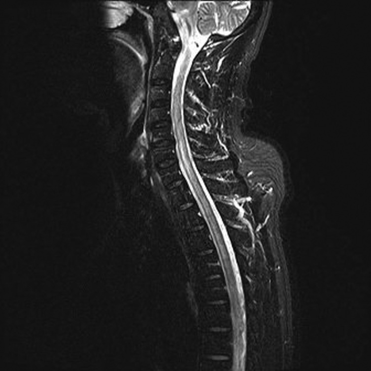
A spinal MRI showed patchy areas of T2 prolongation in the cervical cord from C2 down to C7 and a diffusely scattered T2 hyper intensity within the thoracic cord extending up to T9 vertebral level as shown in figure 1. MRI brain was however deemed normal.
Figure 1.
T2-weighted MRI scan (sagittal view) showing hyperintensity in the cervical and thoracic cord.
Treatment
On the day of admission, intravenous immunoglobulin was administered in a dose of 0.4 g/kg for 5 days, followed by intravenous penicillin, azithromycin and acyclovir for 2 weeks, to cover all possible infective causes of myelitis. However, steroids were not given due to the fact that the patient was in viraemic stage and that his condition had already started to improve.
Outcome and follow-up
After the first week in acute care, he spent about 5 weeks in the rehabilitation ward. His swallowing which was found to be transiently weak in the first week of admission, improved with dysphagia programme. MRI brain revealed high signal in the ventral pons on the T2-weighted scan. The pontine changes were thought to be the aftermath of dengue viraemia.
The first 3 weeks of admission showed little functional improvement. However, from the 4th week onwards, he started to have dramatic recovery. By the end of the 4th week, he started to manage his transfers independently which progressed further to walking with aids and eventually independent mobilisation in week 6. The sensory deficits improved to normal and the strength in his legs rose to grade four out of five. On discharge from the hospital, his American spinal injury association impairment scale had improved to ASIA ‘D’ with neurological level at L1.
He remained continent of bowels but had little improvement with his bladder function. He was discharged home with a urinary catheter and planned to have urodynamic studies before further interventions could be considered.
Discussion
Dengue fever is generally considered a benign clinical entity when comparing with its clinically complicated counterparts such as dengue haemorrhagic fever and dengue shock syndrome. As far as we know, there are only four patients reported in the literature as having had transverse myelitis following dengue infection but dengue has not known to have caused LETM before.4 5
LETM, regardless of its cause, often results in catastrophic consequences and severe disability. The clinical presentation in our patient and the lab results performed, did not support other possible diagnosis like neuromyelitis optica as a triggering factor, which is present in about 50 per cent of LETM cases.3 6–8 On the basis of dengue positive serology and development of spinal cord related symptoms with appearance of MRI features at about the same time, suggest dengue as the most likely cause of LETM in this case.
The other interesting aspect was the quick and near complete recovery the patient achieved without the administration of steroids. This patient improved within his 6 weeks of hospital admission (combined neurology and rehabilitation admission) from completely paraplegic and dysphagic in the 1st week of admission to being independently mobile without aids and having regained full swallowing function by the time of discharge. The recovery could be attributed to factors like timely treatment with antiviral and antimicrobial agents but intravenous immunoglobulins may have played a significant part in limiting the immune mediated neurological damage. Early rehabilitation intervention may have made a significant contribution in getting him back on his feet so quickly.
There is increasing evidence of dengue viral neurotropism causing neurological manifestation such as encephalopathy, in some patients.9–13 In our patient, on MRI, high signal in the pontine area coincided well with transient dysphagia, thought to be due to direct insult caused by dengue viraemia.
This report has highlighted the fact that the dengue fever, which usually takes an uncomplicated course, may cause severe neurological dysfunction and that the recovery is sometimes unpredictably fast, contrary to the general belief. However, more studies are required to substantiate these assumptions.
Learning points.
-
▶
Patients diagnosed with LETM should be investigated for dengue infection especially when there is a travel history to dengue endemic areas or in cases where the cause of LETM could not be easily explained.
-
▶
Neurological complications of dengue infection are not always associated with poor recovery.
-
▶
Early neurological rehabilitation may improve the patient outcome even in complicated presentation.
Footnotes
Competing interests None.
Patient consent Obtained.
References
- 1.Solomon T, Dung NM, Vaughn DW, et al. Neurological manifestations of dengue infection. Lancet 2000;355:1053–9 [DOI] [PubMed] [Google Scholar]
- 2.Halstead SB. Dengue. Lancet 2007;370:1644–52 [DOI] [PubMed] [Google Scholar]
- 3.Kitley J, Leite M, George J, et al. The differential diagnosis of longitudinally extensive transverse myelitis. Mult Scler 2012;18:271–85 [DOI] [PubMed] [Google Scholar]
- 4.Chanthamat N, Sathirapanya P. Acute transverse myelitis associated with dengue viral infection. J Spinal Cord Med 2010;33:425–7 [DOI] [PMC free article] [PubMed] [Google Scholar]
- 5.Seet RC, Lim EC, Wilder-Smith EP. Acute transverse myelitis following dengue virus infection. J Clin Virol 2006;35:310–2 [DOI] [PubMed] [Google Scholar]
- 6.Trebst C, Raab P, Voss EV, et al. Longitudinal extensive transverse myelitis–it’s not all neuromyelitis optica. Nat Rev Neurol 2011;7:688–98 [DOI] [PubMed] [Google Scholar]
- 7.Campana A, Buonuomo PS, Insalaco A, et al. Longitudinal myelitis in systemic lupus erythematosus: a paediatric case. Rheumatol Int. Published Online First: 27 July 2011. doi:10.1007/s00296-011-2061-1 [DOI] [PubMed] [Google Scholar]
- 8.Espinosa G, Mendizábal A, Mínguez S, et al. Transverse myelitis affecting more than 4 spinal segments associated with systemic lupus erythematosus: clinical, immunological, and radiological characteristics of 22 patients. Semin Arthritis Rheum 2010;39:246–56 [DOI] [PubMed] [Google Scholar]
- 9.Varatharaj A. Encephalitis in the clinical spectrum of dengue infection. Neurol India 2010;58:585–91 [DOI] [PubMed] [Google Scholar]
- 10.Strobel M, Lamaury I, Contamin B, et al. [Dengue fever with neurologic expression. Three cases in adults]. Ann Med Interne (Paris) 1999;150:79–82 [PubMed] [Google Scholar]
- 11.Kunishige M, Mitsui T, Tan BH, et al. Preferential gray matter involvement in dengue myelitis. Neurology 2004;63:1980–1 [DOI] [PubMed] [Google Scholar]
- 12.Renganathan A, Ng WK, Chong TT. Transverse myelitis in association with dengue infection. Neurol J Southeast Asia 1996;1:61–3 [Google Scholar]
- 13.Patey O, Ollivaud L, Breuil J, et al. Unusual neurologic manifestations occurring during dengue fever infection. Am J Trop Med Hyg 1993;48:793–802 [DOI] [PubMed] [Google Scholar]


