Abstract
The inside of our gut is inhabited with enormous number of commensal bacteria. The mucosal surface of the gastrointestinal tract is continuously exposed to them and occasionally to pathogens. The gut-associated lymphoid tissue (GALT) play a key role for induction of the mucosal immune response to these microbes1, 2. To initiate the mucosal immune response, the mucosal antigens must be transported from the gut lumen across the epithelial barrier into organized lymphoid follicles such as Peyer's patches. This antigen transcytosis is mediated by specialized epithelial M cells3, 4. M cells are atypical epithelial cells that actively phagocytose macromolecules and microbes. Unlike dendritic cells (DCs) and macrophages, which target antigens to lysosomes for degradation, M cells mainly transcytose the internalized antigens. This vigorous macromolecular transcytosis through M cells delivers antigen to the underlying organized lymphoid follicles and is believed to be essential for initiating antigen-specific mucosal immune responses. However, the molecular mechanisms promoting this antigen uptake by M cells are largely unknown. We have previously reported that glycoprotein 2 (Gp2), specifically expressed on the apical plasma membrane of M cells among enterocytes, serves as a transcytotic receptor for a subset of commensal and pathogenic enterobacteria, including Escherichia coli and Salmonella enterica serovar Typhimurium (S. Typhimurium), by recognizing FimH, a component of type I pili on the bacterial outer membrane 5. Here, we present a method for the application of a mouse Peyer's patch intestinal loop assay to evaluate bacterial uptake by M cells. This method is an improved version of the mouse intestinal loop assay previously described 6, 7. The improved points are as follows: 1. Isoflurane was used as an anesthetic agent. 2. Approximately 1 cm ligated intestinal loop including Peyer's patch was set up. 3. Bacteria taken up by M cells were fluorescently labeled by fluorescence labeling reagent or by overexpressing fluorescent protein such as green fluorescent protein (GFP). 4. M cells in the follicle-associated epithelium covering Peyer's patch were detected by whole-mount immunostainig with anti Gp2 antibody. 5. Fluorescent bacterial transcytosis by M cells were observed by confocal microscopic analysis. The mouse Peyer's patch intestinal loop assay could supply the answer what kind of commensal or pathogenic bacteria transcytosed by M cells, and may lead us to understand the molecular mechanism of how to stimulate mucosal immune system through M cells.
Keywords: Neuroscience, Issue 58, M cell, Peyer's patch, bacteria, immunosurveillance, confocal microscopy, Glycoprotein 2
Protocol
1. Preparation of bacterial cells
Streak glycerol stocked fluorescent bacteria (such as GFP expressing S. Typhimurium) on LB an agar plate containing 100 μg/ml of ampicillin.
Culture a single colony from LB agar overnight in 2 ml of new LB medium.
Add 0.5 ml of bacterial culture to 4.5 ml of new LB medium and incubate until optical density of 1.0 at 600 nm is reached.
Harvest bacterial cells by centrifugation (3,000 x g, 5 min, 4°C).
Discard the supernatant and wash twice with 5.0 ml of sterile phosphate buffer saline (PBS).
Resuspend bacterial pellet with 5 ml of PBS, and use 50 μl of the suspension containing approximately 107 colony forming unit (CFU) as the inoculum.
In case of using fluorescently labeled bacteria, the bacterial cells were labeled by fluorescence labeling reagent according to standard protocol.
2. Anesthesia
Fill a small plastic box (10 x 10 x 5 cm) with 5% (v/v) vaporized isoflurane mixed with air (flow rate: 200 ml/min).
Anesthetize eight- to sixteen-weak-old male or female mice in the box.
Move the mice to an autopsy table after anesthesia.
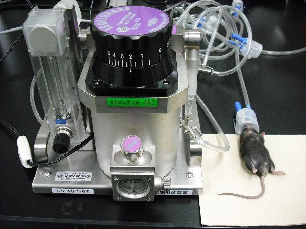
Continuously anesthetize the mice by 2% (v/v) vaporized isoflurane mixed with air (flow rate: 200 ml/min) (Figure 1). This is a terminal procedure.
3. Ligated Peyer's patch loop assay
Incise 1cm of the abdominal skin and then cut the abdominal peritoneum of an anesthetized mouse and take out the small intestine containing the Peyer's patch.
Ligate the intestine with sewing yarn, taking care to avoid blood vessels. Note: only bind one side of the intestine, and leave the other side loose.
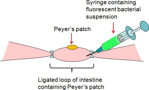
Inject 50 μl of bacterial suspension or PBS (control) with a syringe into ligated Peyer's patch loop on the loose side of the intestine (Figure 2).
Bind loose side, and close the mouse's abdomen with a clip.
After 1 h, remove the ligated Peyer's patch loop, and euthanize the mouse by cervical dislocation.
Excise Peyer's patch from ligated Peyer's patch loop.
Flashing the apical side of Peyer's patch by 1ml of PBS with using syringe attached to a needle to remove excess mucosal fluid and bacteria. Flashing the Peyer's patch an additional two times.
4. Whole mount staining and confocal microscopic analysis of Peyer's patch
Fix Peyer's patch in BD Cytofix/Cytoperm solution on ice for 1 hr.
Wash Peyer's patch three times with 1ml of BD Perm/Wash buffer for 5 min, and then block with 1 ml of blocking buffer containing 0.1% (w/v) saponin, 0.2% (w/v) BSA in PBS for 30 min on ice.
Add 200-fold diluted anti-mouse GP2 monoclonal antibody (5 μg/ml) to the specimen to detect M cells.
Incubate for 2 hrs at room temperature or overnight at 4°C.
Wash three times with 1 ml of cold PBS, then add 200-fold diluted anti-rat IgG antibody conjugated with DyLight549 (20 μg/ml)to the specimen.
Incubate sample on ice for 2 hrs.
Wash three times with 1 ml of cold PBS, then add 50-fold diluted Alexa 633 conjugated Phalloidin to the specimen to detect F-actin.
Incubate on ice for 2 hrs.
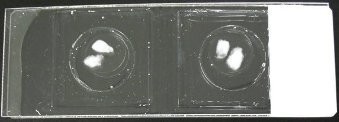
Wash lightly with 1 ml of cold PBS. Then place three to four pieces of circular cover glass on a slide, and embed the specimens in a 30% solution of glycerol in PBS on the slide (Figure 3).
Observe specimens with a DM-IRE2 confocal laser scanning microscope or a DeltaVision Restoration deconvolution microscope.
5. Representative Results:
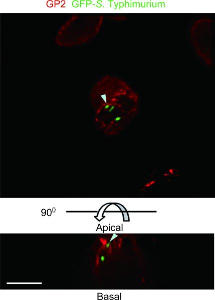
The specimen example of a mouse ligated Peyer's patch loop assay with GFP-S. Typhimurium was observed with a DeltaVision Restoration deconvolution microscope (Figure 4). GFP-S. Typhimurium is transcytosed by GP2+M cells. A mouse Peyer's patch intestinal loop assay can identify the kind of commensal or pathogenic bacteria that can be transcytosed by M cells.
 Figure 1. Anesthesia of mice under vaporized isoflurane condition. Vaporized isoflurane was supplied by an anesthesia-dedicated apparatus (left side).
 Figure 2. Summary of ligated Peyer's patch intestinal loop assay. The fluorescent bacterial suspension was injected by syringe into ligated Peyer's patch loop on the loose side of the intestine.
 Figure 3. Mount of the specimens. The specimens were embedded in a 30% solution of glycerol in PBS on a special slide in which a perforated plastic plate (1mm thick) was bound by adhesive agent on a glass slide.
 Figure 4. Example of a mouse ligated Peyer's patch loop assay with GFP-S. Typhimurium. GP2, which is reported as M cell specific marker5, was stained with anti-mouse GP2 antibody (Red).The specimen was observed with a DeltaVision Restoration deconvolution microscope. GFP-expressing S. Typhimurium was co-localized with GP2 on the apical plasma membrane of M cell and in the subapical cytoplasmic vesicles (arrowheads). Top, apical view. Bottom and side, lateral views. Scale bar: 10 μm.
Discussion
The incubation time of ligated Peyer's patch with bacterial suspension is usually for 1 hr to observe the bacterial incorporation into M cells. In case of 4 hrs incubation, the bacteria are often detected in the T cell zone of Peyer's patches. As the vaporized isoflurane inhalant anesthesia could keep mice stable, the incubation time of ligated Peyer's patch with bacterial suspension is extendable to observe the bacteria which move into T-cell zone in Peyer's patch. The important point of this experiment is to take care not to scratch the blood vessel when ligating the intestine. In the case of not to use GFP-expressed bacteria, we could alternatively use fluorescently labeled bacteria by commercial fluorescence labeling reagent. If the reagent possibly inhibits the bacterial, which you want to use, attachment to the molecule expressed on M cell, you should use the specific primary antibody, if available, to detect the incorporated bacteria.
The ligated loop assay method was previously described6, 7. We improve some points of this method as follows: 1. Isoflurane is used as an anesthetic agent for keeping the mice steady. 2. Approximately 1 cm ligated intestinal loop including Peyer's patch is set up. 3. Bacteria taken up by M cells are fluorescently labeled by fluorescence labeling reagent or by overexpressing fluorescent protein such as GFP. 4. M cells in the follicle-associated epithelium covering Peyer's patch are detected by whole-mount immunostainig with anti Gp2 antibody. 5. Fluorescent bacterial transcytosis by M cells are observed by confocal microscopic analysis.
We and others have demonstrated that diverse heterogenous antigens actively gain entry into M cells. Substantial numbers of pathogens including Salmonella spp., Shigella spp., Yersinia spp. exploit M cells for host invasion8-10. Furthermore, the coccoid form of Helicobacter pylori can potentially translocate into Peyer's patch via M cells, and this translocation is essential for the induction of chronic inflammation in the stomach11. On the other hand, the mechanisms by which these pathogens invade M cells remain to be fully elucidated. We suggest that the ligated Peyer's patch loop assay is an appropriate experimental method for dissecting the interplay between M cells and pathogenic agents in vivo.
Disclosures
No conflicts of interest declared.
Acknowledgments
The authors would like to thank all EIB members for developing this technique. This research was supported in part by Grant-in-Aid for Scientific Research on Priority Areas "Membrane Traffic" (H.O.), and "Matrix of Infectious Phenomena" (K.H.), Young Scientists (S.F. and K. H.), and Scientific Research (H.O.), and Scientific Research on Innovative Areas "Intracellular Logistics" (H.O.), from the Ministry of Education, Culture, Sports, Science and Technology of Japan.
References
- Fagarasan S, Honjo T. Intestinal IgA synthesis: regulation of front-line body defences. Nat. Rev. Immunol. 2003;3:63–72. doi: 10.1038/nri982. [DOI] [PubMed] [Google Scholar]
- Hapfelmeier S. Reversible microbial colonization of germ-free mice reveals the dynamics of IgA immune responses. Science. 2010;328:1705–1709. doi: 10.1126/science.1188454. [DOI] [PMC free article] [PubMed] [Google Scholar]
- Owen RL, Jones AL. Epithelial cell specialization within human Peyer's patches: an ultrastructural study of intestinal lymphoid follicles. Gastroenterology. 1974;66:189–203. [PubMed] [Google Scholar]
- Neutra MR, Mantis NJ, Kraehenbuhl JP. Collaboration of epithelial cells with organized mucosal lymphoid tissues. Nat. Immunol. 2001;2:1004–1009. doi: 10.1038/ni1101-1004. [DOI] [PubMed] [Google Scholar]
- Hase K. Uptake through glycoprotein 2 of FimH(+) bacteria by M cells initiates mucosal immune response. Nature. 2009;462:226–230. doi: 10.1038/nature08529. [DOI] [PubMed] [Google Scholar]
- Punyashthiti K, Finkelstein RA. Enteropathogenicity of Escherichia coli. I. Evaluation of mouse intestinal loops. Infect. Immun. 1971;4:473–478. doi: 10.1128/iai.4.4.473-478.1971. [DOI] [PMC free article] [PubMed] [Google Scholar]
- Owen RL, Pierce NF, Apple RT, Cray WC., Jr M cell transport of Vibrio cholerae from the intestinal lumen into Peyer's patches: a mechanism for antigen sampling and for microbial transepithelial migration. J. Infect. Dis. 1986;153:1108–1118. doi: 10.1093/infdis/153.6.1108. [DOI] [PubMed] [Google Scholar]
- Owen RL. M cells--entryways of opportunity for enteropathogens. J. Exp. Med. 1994;180:7–9. doi: 10.1084/jem.180.1.7. [DOI] [PMC free article] [PubMed] [Google Scholar]
- Clark MA, Hirst BH, Jepson MA. M-cell surface beta1 integrin expression and invasin-mediated targeting of Yersinia pseudotuberculosis to mouse Peyer's patch M cells. Infect. Immun. 1998;66:1237–1243. doi: 10.1128/iai.66.3.1237-1243.1998. [DOI] [PMC free article] [PubMed] [Google Scholar]
- Jensen VB, Harty JT, Jones BD. Interactions of the invasive pathogens Salmonella typhimurium, Listeria monocytogenes, and Shigella flexneri with M cells and murine Peyer's patches. Infect. Immun. 1998;66:3758–3766. doi: 10.1128/iai.66.8.3758-3766.1998. [DOI] [PMC free article] [PubMed] [Google Scholar]
- Nagai S. Role of Peyer's patches in the induction of Helicobacter pylori-induced gastritis. Proc. Natl. Acad. Sci. U. S. A. 2007;104:8971–8976. doi: 10.1073/pnas.0609014104. [DOI] [PMC free article] [PubMed] [Google Scholar]


