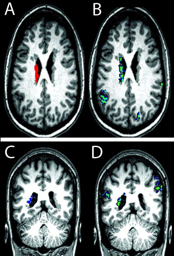
Figure 3. Functional connectivity of periventricular heterotopia to ipsilateral and contralateral cortical regions.
With an extensive left-sided region of heterotopia identified in one subject (top) as a seed region for resting-state functional connectivity analysis (red in A; heterotopia 1–6 in Supporting Table), a highly functionally correlated region of overlying cortex was seen within the left supramarginal gyrus (blue/green in B; peak correlation coefficient 0.61). In another subject (bottom), a left posterior region of heterotopia was identified as a seed region for connectivity analysis (blue in C; heterotopia 8-3 in Supporting Table); highly functionally correlated regions were seen in the left superior temporal gyrus and right supramarginal gyrus (blue-green areas in D; peak correlation coefficients 0.53 and 0.56, respectively).

