Abstract
A key survival mechanism of Clostridium botulinum, the notorious neurotoxic food pathogen, is the ability to form heat-resistant spores. While the genetic mechanisms of sporulation are well understood in the model organism Bacillus subtilis, nothing is known about these mechanisms in C. botulinum. Using the ClosTron gene-knockout tool, sigK, encoding late-stage (stage IV) sporulation sigma factor K in B. subtilis, was disrupted in C. botulinum ATCC 3502 to produce two different mutants with distinct insertion sites and orientations. Both mutants were unable to form spores, and their elongated cell morphology suggested that the sporulation pathway was blocked at an early stage. In contrast, sigK-complemented mutants sporulated successfully. Quantitative real-time PCR analysis of sigK in the parent strain revealed expression at the late log growth phase in the parent strain. Analysis of spo0A, encoding the sporulation master switch, in the sigK mutant and the parent showed significantly reduced relative levels of spo0A expression in the sigK mutant compared to the parent strain. Similarly, sigF showed significantly lower relative transcription levels in the sigK mutant than the parent strain, suggesting that the sporulation pathway was blocked in the sigK mutant at an early stage. We conclude that σK is essential for early-stage sporulation in C. botulinum ATCC 3502, rather than being involved in late-stage sporulation, as reported for the sporulation model organism B. subtilis. Understanding the sporulation mechanism of C. botulinum provides keys to control the public health risks that the spores of this dangerous pathogen cause through foods.
INTRODUCTION
Sporulation plays a central role in the survival of bacteria. Spores tolerate extreme environments and are not readily killed by food pasteurization. A great concern in modern food processing is caused by Clostridium botulinum, the spores of which may survive heating and germinate into a neurotoxic culture even at refrigeration, posing a serious public health risk through ready-to-eat foods (17). Another type of botulism risk occurs when spores germinate and grow into toxigenic cultures in vivo, such as in deep wounds or in the gastrointestinal tract of people lacking normal gut microbial populations (18, 20). Although the role of C. botulinum spores in both food contamination and infection is notorious, little is known about the molecular mechanisms behind sporulation of C. botulinum.
Many sporulation factors characterized in Bacillus subtilis, the model organism for spore-forming bacteria, are encoded by clostridial genomes (23). In both genera, spo0A encodes the master switch of sporulation. In Bacillus, Spo0A is converted to an active form (phosphorylated Spo0A [Spo0A-P]) via a phosphorelay system (26). This system is not found in clostridia; however, orphan kinases in C. botulinum (36) and in C. acetobutylicum (32) were proposed to phosphorylate Spo0A (2) and thereby activate the sporulation cascade downstream of spo0A.
In the B. subtilis model, stage 0 of the sporulation pathway is characterized visually by the elongation of the cell. Chromatin filament formation occurs at stage I, although it is often not mentioned as it resembles stage 0 (35). Spo0A-P positively regulates sigF, the first one of four downstream sigma (σ) factors at stage 0 (13). The spoIIAA operon, containing sigF, and sigE are expressed before stage II of the sporulation pathway begins. At stage II, the cell divides asymmetrically into the mother cell and prespore. After division, σF is activated in the prespore, while in the mother cell, pro-σE is cleaved, resulting in active σE. At stage III of sporulation, the mother cell engulfs the prespore. This activates σG, which further regulates sporulation- and germination-related genes, including the one involved in processing of pro-σK. Stage IV of the pathway involves cortex formation around the engulfed prespore. Active σK, found in the mother cell, transcribes spore coat and germination genes, notably, gerE (5). The corresponding proteins are added to the spore cortex during the final stage of the sporulation pathway (13) and are necessary to create viable spores that are able to germinate in response to specific signals. Genes for all four σ factors (σF, σE, σG, and σK) are present in the C. botulinum ATCC 3502 genome, but their role in sporulation has not been characterized.
The σK regulator appears in the sporulation regulatory pathway across different species. In B. subtilis, the mother cell-specific σK regulates spore coat formation during late-stage sporulation (15, 21). A 42-kb intervening element, called skin, separates two halves of sigK in some B. subtilis strains (16). Mutation in either half of sigK in B. subtilis results in unfinished spore formation at a late stage (25). The skin element undergoes site-specific recombination within the mother cell only, allowing sigK to form and be transcribed. The removal of the skin element does not affect growth or sporulation, and the element was shown to not contain any genes essential for survival (16). It has been suggested that it is an evolutionary rudiment left over from its ancestors (34). With the exception of some C. difficile strains, where a skin element is essential for sporulation (8), most clostridial genomes contain an uninterrupted sigK. In C. perfringens, sporulation was halted at an earlier stage in a sigK mutant than in a sigE mutant (9). The sporulation cycle in this C. perfringens sigK mutant had clearly stopped in early-stage sporulation, in contrast to B. subtilis sigK mutants, in which the cycle stops at late-stage sporulation. This would suggest that σK in clostridia may play a role in the early stages of sporulation, as opposed to the late-stage role in B. subtilis.
Here we show that σK is essential in the sporulation of C. botulinum ATCC 3502 and, unlike in B. subtilis, that it seems to act at an early stage of sporulation. Neither of the two sigK single insertional mutant strains constructed using the ClosTron system (10) was able to sporulate, while the parent strain and complemented mutants sporulated and germinated successfully. The sigK mutants showed elongated cell morphology, suggesting that sporulation was prevented prior to asymmetric cell division. A quantitative real-time PCR (qRT-PCR) analysis revealed that sigK was expressed in the late log phase and early stationary phases, corresponding to the early stages of the sporulation pathway. The relative levels of spo0A and sigF expression were significantly lower in a sigK mutant than in the parent strain at late exponential and early stationary growth, suggesting that the sporulation pathway was being blocked at stage 0 of sporulation with the disruption of spo0A transcription. Levels of the stage II gene, sigF, suggest that it was not being significantly transcribed and that the sporulation pathway had been stopped prior to stage II. These findings may suggest a regulatory role for σK in initiation of sporulation.
MATERIALS AND METHODS
Strains, plasmids, and culture.
The bacterial strains and plasmids used are listed in Table 1. The C. botulinum ATCC 3502 parent and the sigK mutant strains were stored as a frozen stock at −70°C in tryptone-peptone-glucose-yeast extract (TPGY) broth (5% tryptone, 0.5% peptone, 2% yeast extract [Difco, BD Diagnostic Systems, Sparks, MD], 0.4% glucose [VWR International, Leuven, Belgium], 0.1% sodium thioglycolate [Merck, Darmstadt, Germany]) supplemented with 20% glycerol. C. botulinum was grown in anaerobic TPGY broth or on anaerobic TPGY plates (1.5% agar) supplemented with thiamphenicol at 7.5 μg/ml (broth) or 15 μg/ml (plates), cycloserine at 250 μg/ml, and erythromycin at 2.5 μg/ml, when appropriate. Cultures were incubated at 37°C in an anaerobic cabinet (MG1000 anaerobic workstation; Don Whitley Scientific Ltd., Shipley, United Kingdom) with an atmosphere of 85% N2, 10% CO2, and 5% H2. Escherichia coli was grown aerobically at 37°C in Luria-Bertani (LB) broth (Difco) or on 2× YT (1% yeast extract, 1.6% tryptone, 0.5% NaCl [J. T. Baker, Deventer, Netherlands]) agar plates supplemented with 25 μg/ml chloramphenicol and 30 μg/ml kanamycin, when appropriate (Sigma-Aldrich).
Table 1.
Strains and plasmids
| Strain or plasmid | Description | Source or reference |
|---|---|---|
| Strains | ||
| C. botulinum ATCC 3502 | Parent strain | ATCC, Manassas, VA |
| C. botulinum ATCC 3502 sigK-427s | Mutant strain with insertion inactivation of sigK gene in sense orientation | This study |
| C. botulinum ATCC 3502 sigK-296a | Mutant strain with insertion inactivation of sigK gene in antisense orientation | This study |
| C. botulinum ATCC 3502/pMTL82151 | Parent strain with empty plasmid vector | This study |
| C. botulinum ATCC 3502 sigK-427s/pMTL82151 | sigK-427s with empty plasmid vector | This study |
| C. botulinum ATCC 3502 sigK-427s/pMTL82151::sigK | sigK-427s with complementation plasmid vector | This study |
| C. botulinum ATCC 3502 sigK-296a/pMTL82151 | sigK-296a with empty plasmid vector | This study |
| C. botulinum ATCC 3502 sigK-296a/pMTL82151::sigK | sigK-296a with complementation plasmid vector | This study |
| E. coli TOP10 | Electrocompetent cloning strain | Invitrogen, Carlsbad, CA |
| E. coli CA434 | Conjugation donor strain containing fertility plasmid | Purdy et al. (27) |
| Plasmids | ||
| pMTL007 | Clostridial mutagenesis vector | Heap et al. (10) |
| pMTL007::Cbo-sigK-427s | pMTL007 targeting sigK (sense) | This study |
| pMTL007C-E2 | Clostridial mutagenesis vector | Heap et al. (12) |
| pMTL007C-E2::Cbo-sigK-296a | pMTL007C-E2 targeting sigK (antisense) | This study; DNA 2.0 |
| pMTL82151 | Modular plasmid vector | Heap et al. (12) |
| pMTL82151::sigK | pMTL82151 containing sigK gene for complementation | This study |
All experiments were performed with triplicate cultures of the parent and mutant strains. Overnight starting cultures were grown in TPGY broth at 37°C, spread on TPGY plates, and restreaked for single colonies on fresh TPGY plates. Three colonies were then individually inoculated in 10 ml of TPGY broth and cultured overnight at 37°C before use in the experiments described below.
Construction of insertional knockout mutants with ClosTron.
The ClosTron system has proven effective for genetic studies in C. botulinum (1, 4, 30, 31). The sigK locus (CBO2541) was disrupted by inserting a mobile group II intron from Lactococcus lactis (Ll.ltrB) in sigK using the ClosTron system developed by Heap et al. (10). The target sites were designed to make two mutants with mutations of the same gene: one with the insertion in the sense orientation and another one with the insertion in the antisense orientation. In both instances, the target sites were determined by using the online tool provided by the University of Nottingham, United Kingdom (12), based on the algorithm of Perutka et al. (24). For the sense mutant, sigK-427s, the insertion was constructed using splicing by overlap extension PCR (all primers are listed in Table S1 in the supplemental material and were provided by Oligomer Oy, Helsinki, Finland). The HindIII/BsrGI-digested product was inserted into pMTL007, creating pMTL007::Cbo-sigK-427s. This was electrotransformed (capacitance, 25 μF; resistance, 200 Ω: voltage, 2,500 V; Gene Pulser II; Bio-Rad, Brussels, Belgium) into electrocompetent E. coli TOP10, and the plasmids were purified by a GeneJET plasmid miniprep kit (Fermentas, St. Leon-Rot, Germany). The design of the antisense mutant, sigK-296a, was the same as that described above. The plasmid, however, was constructed in the updated pMTL007C-E2 backbone (12) by the company DNA 2.0 (Menlo Park, CA). A key difference between the pMTL007 and pMTL007C-E2 plasmids is that pMTL007 (used for the sigK-427s mutant) requires expression induction for the intron with 1 mM isopropyl-β-d-thiogalactopyranoside (IPTG), whereas pMTL007C-E2 (used for the sigK-296a mutant) does not require this step, as the C. sporogenes fdx promoter drives intron expression (12). After confirmation of the correct sequences for both plasmids, they were chemically transformed into E. coli CA434 (27). The transformed E. coli isolates were grown overnight in LB broth supplemented with kanamycin and chloramphenicol to select for their own fertility plasmid and the ClosTron plasmid, respectively. Each plasmid was introduced into the C. botulinum ATCC 3502 parent strain by conjugation. Aliquots of 100 μl of phosphate-buffered saline (PBS)-suspended mixed cultures of E. coli CA434 and C. botulinum ATCC 3504 were plated onto TPGY plates supplemented with thiamphenicol and cycloserine. The plates were incubated overnight, and colonies were restreaked onto TPGY plates supplemented with fresh antibiotics. Cells from sigK-296a plates and IPTG-induced cells of mutant sigK-427s were plated onto TPGY plates supplemented with erythromycin to select for successful integration of the intron. PCR confirmation of the correct insertion was performed using primers flanking the insertion site and EBS universal primers (see Table S1 in the supplemental material). Additionally, a Southern blot analysis, as performed by Palonen et al. (22), was performed using probes targeted to the group II intron to confirm that only one copy had inserted into the genome.
Construction of complementation plasmids.
The pMTL82151 plasmid designed by Heap et al. (11) was used to complement the sigK mutations. A section of the genome was amplified with the primers sK1268F and sK1268R, giving a fragment of 1,268 bp which contained the coding sequence of sigK, 425 bp upstream of it, considered to include the natural promoter for sigK, and 138 bp downstream of the coding sequence. This was purified from the agarose gel using a GeneJET gel extraction kit and a GeneJET PCR purification kit (Fermentas). The fragment was restriction digested with NdeI/NotI and ligated into pMTL82151. As with the ClosTron plasmid, it was purified from E. coli TOP10, sequenced, and conjugated into the two mutants. Thiamphenicol was used to select for successful transconjugants.
Sporulation assay.
The C. botulinum ATCC 3502 parent strain, the sigK mutants, and the complemented strains were incubated at 37°C under anaerobic conditions for 7 days, considered sufficient for efficient sporulation (3). Two 1-ml aliquots of each culture were taken, and one aliquot was heated at 80°C for 20 min to kill any vegetative cells. The other aliquot was left unheated. The heated and nonheated aliquots were each serially diluted (10−1 to 10−5) in 9 ml of fresh TPGY broth supplemented with thiamphenicol, where appropriate.
Spore staining.
To visualize the spores, colonies of the parent and sigK mutant strains were grown on TPGY plates for 7 days and stained on a glass slide with malachite green and safranin (28).
RNA isolation.
The parent strain and sigK-427s mutant were each inoculated from diluted overnight cultures (optical density at 600 nm [OD600], approximately 1.0) into fresh TPGY broth at a 1:100 dilution. Samples of 1 ml were collected at 8, 10, 12, and 14 h after inoculation. A 200-μl stop solution containing phenol (95%) and ethanol (99.5%) at a ratio of 1:9 was added to each sample, and the samples were stored on ice for 30 min. The samples were then spun down and the supernatant was discarded. The pellets were stored at −70°C until RNA isolation. The frozen cell pellets were resuspended in a lysis buffer of 250 μl containing lysozyme (50 mg/ml; Sigma-Aldrich), mutanolysin (1,000 U/ml; Sigma-Aldrich), and Tris-EDTA (10 mM Tris-HCl, 1 mM EDTA, pH 8.0) and incubated at 37°C for 30 min. RNA isolation and purification were performed using an RNeasy minikit (Qiagen, Hilden, Germany) with a DNase treatment (Qiagen). RNA was eluted twice with RNase-free water, and a second DNase treatment was performed according to the instructions in the Ambion DNA-free kit (Applied Biosystems, Life Technologies Corporation, Carlsbad, CA). The RNA concentration was measured at 260 nm with a NanoDrop 1000 spectrophotometer (Thermo Fisher Scientific, Waltham, MA).
Reverse transcription.
A total of 800 ng of RNA was used for each reverse transcription reaction. A DyNAmo cDNA synthesis kit (Finnzymes, Espoo, Finland) was used as instructed. Reverse transcription reactions were performed in triplicate for each RNA sample. Controls of RNA with no reverse transcriptase were included to control DNA contamination. The cDNA synthesized was stored at −20°C before use in qRT-PCR.
qRT-PCR.
A DyNAmo Flash SYBR green qRT-PCR kit (Finnzymes) was used for qRT-PCR. Primers were designed for the target genes, sigK, spo0A, and sigF, and for the reference gene, 16S rrn (see Table S1 in the supplemental material) using Primer3 (http://frodo.wi.mit.edu/primer3/). Each reaction mixture contained 10 μl of master mix (Finnzymes), 5 μl of ultrapure water, 0.5 μM each primer, and 4 μl of cDNA template. The thermal run (Rotor Gene 3000 real-time thermal cycler; Qiagen) consisted of an enzyme activation step for 7 min at 95°C and 45 cycles of denaturation at 95°C for 10 s and annealing and extension at 60°C for 20 s, followed by a final extension at 60°C for 1 min and melt curve analysis from 60°C to 98°C in 0.5°C steps for 10 s. Standard curves for testing primer efficiency were made using dilution series of pooled cDNA as a template. Efficiencies for the primer pairs for 16S rrn, sigK, spo0A, and sigF were 0.97, 0.94, 0.95, and 1.03, respectively. Relative gene expression values were calculated according to Livak and Schmittgen (19).
RESULTS
Construction of insertional sigK-knockout mutants with ClosTron.
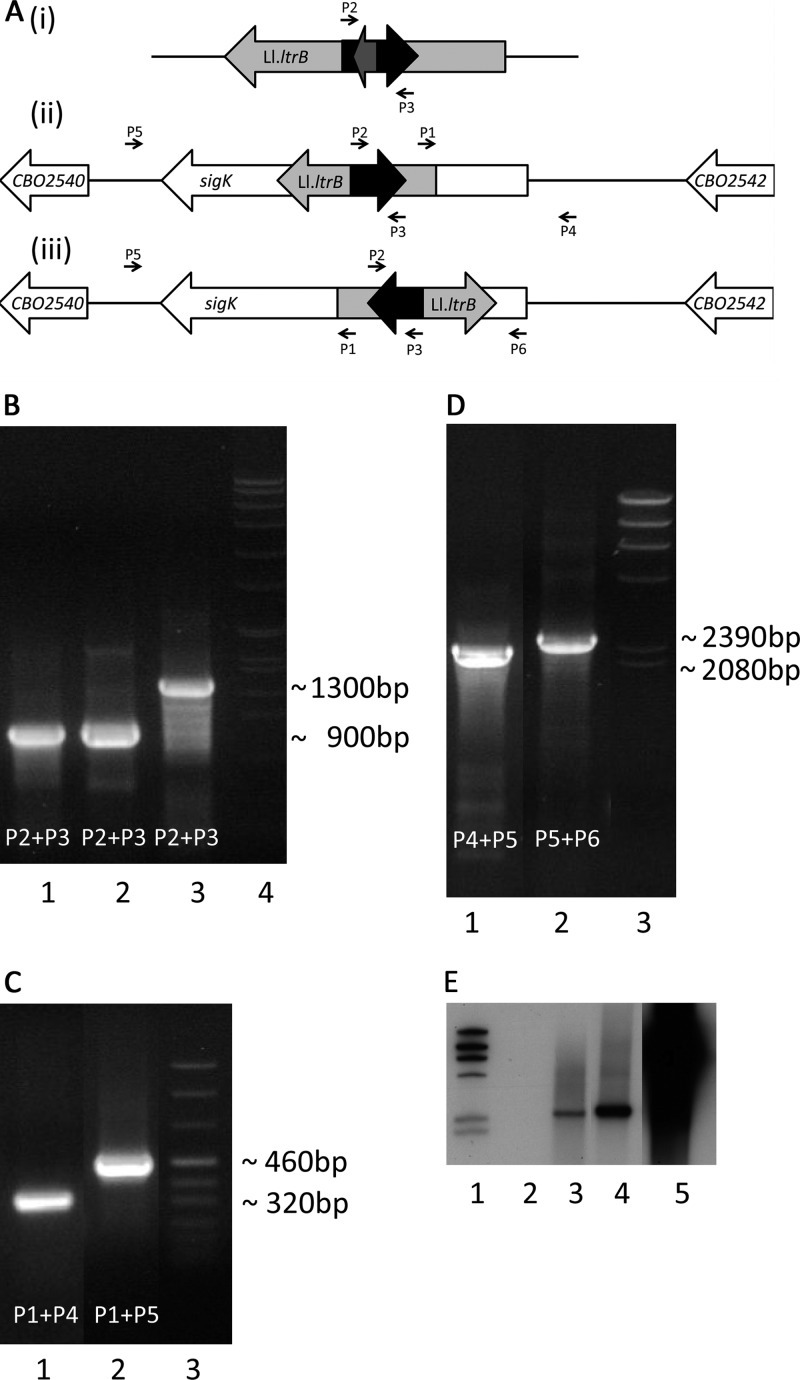
An overview of the sigK region and ClosTron insertion with primer target regions is represented in Fig. 1A. Retrotransposon-activated marker (RAM) insertion into sigK was confirmed with PCR. The RAM was amplified (Fig. 1B) to show that the group I intron had spliced itself out from the erythromycin resistance gene in the integrants by comparison of the PCR fragments with the RAM containing the intron. To confirm that our insertions were at the correct sites, we performed a PCR with primers placed within the insertion and within the gene. The insertion was successful for bands of the correct sizes of 320 bp and 460 bp, respectively, for sigK-427s and sigK-296a (Fig. 1C). We also amplified the site with primers flanking the insertion, giving fragments of 2,080 bp and 2,390 bp, respectively, for sigK-427s and sigK-296a for the correct insertions (Fig. 1D). To show that the insertion occurred only once in the genome, a Southern blot with an intron-specific probe performed on digested DNA produced only one hybridization signal (Fig. 1E).
Fig 1.
Confirmation of successful mutation of sigK. (A) Schematic diagram identifying the locations of primers (P1 to P6; see Table S1 in the supplemental material) used for mutant confirmation. The Ll.ltrb group II intron (i) contains the erythromycin resistance marker (black) and a group I intron (gray); insertional mutations for sigK-427s (ii) and sigK-296a (iii) are shown in the sigK region (white) containing the group II intron and erythromycin resistance marker; the group I intron spliced out upon successful insertion. (B) PCR amplification of RAM showing that the group I intron has been spliced out in integrants. Lane 1, sigK-427s; lane 2, sigK-296a; lane 3, pMTL007C-E2; lane 4, molecular mass marker. (C) PCR with primers outside and inside the integration region confirming correct intron integration. Lane 1, sigK-427s; lane 2, sigK-296a; lane 3, molecular mass marker. (D) PCR with intron-flanking primers to show intron integration within the gene. Lane 1, sigK-427s; lane 2, sigK-296a; lane 3, molecular mass marker. (E) Southern blot with intron probe to show single integration of the intron. Lane 1, molecular mass marker; lane 2, parent strain; lane 3, sigK-427s; lane 4, sigK-296a; lane 5, pMTL007C-E2.
Sporulation assay.
After a 7-day incubation at 37°C and heating at 80°C for 20 min, the parent strain showed growth in fresh medium, demonstrating the presence of heat-stable spores (Table 2). Both of the sigK mutants failed to show any growth after the heat treatment, indicating a lack of sporulation, germination, or both. The complemented sigK-427s containing pMTL82151::Cbo-sigK and the parent control containing pMTL82151 alone showed growth after heating, demonstrating successful sporulation and germination. The complemented sigK-296a also demonstrated growth after heating, although the number of viable spores was 2 log units smaller than that for the parent strain. As expected, the mutant strains containing pMTL82151 alone showed no growth after heating. Unheated controls of each strain demonstrated growth at all dilutions, showing that they were all capable of similar vegetative growth. To confirm that the complemented mutants still contained the inserted RAM, a sample from each heat-treated culture of the complemented strains was spread on TPGY agar plates supplemented with erythromycin. The mutant strains grew in the presence of erythromycin, confirming that the mutations were stable, while the parent strain, as expected, did not grow.
Table 2.
Results of sporulation assay
| Strain tested | Growth observed postheating at a dilution of: |
|
|---|---|---|
| 10−1 | 10−5 | |
| C. botulinum ATCC 3502 | Yes | Yes |
| C. botulinum ATCC 3502 sigK-427s | No | No |
| C. botulinum ATCC 3502 sigK-296a | No | No |
| C. botulinum ATCC 3502/pMTL82151 | Yes | Yes |
| C. botulinum ATCC 3502 sigK-427s/pMTL82151 | No | No |
| C. botulinum ATCC 3502 sigK-427s/pMTL82151::sigK | Yes | Yes |
| C. botulinum ATCC 3502 sigK-296a/pMTL82151 | No | No |
| C. botulinum ATCC 3502 sigK-296a/pMTL82151::sigK | Yes | Noa |
Growth was observed at the 10−3 dilution but not at the 10−4 dilution.
Spore staining.
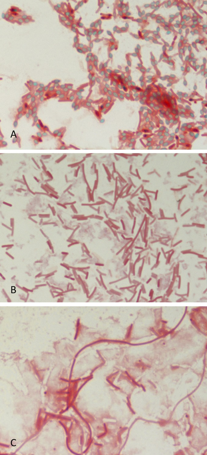
Along with rod-shaped cell bodies counterstained with safranin, spores were detected for the parent strain (Fig. 2A). The mutant strains, sigK-427s (Fig. 2B) and sigK-296a (Fig. 2C), showed no visible spores. Additionally, some of the cells, those of the sigK-296a mutant in particular, appeared to be more elongated than those of the parent strain.
Fig 2.
Spore detection by malachite green and safranin staining after a 7-day incubation on TPGY plates. The parent strain (A) sporulated normally (spores are in green), while the mutants sigK-427s (B) and sigK-296a (C) did not form spores at all.
Growth, sampling, and RNA isolation.
The OD600 was measured for a growth curve and showed similar growth for both the parent and sigK-427s strains (data not shown). The most-probable-number cell counts of both strains showed similar growth at the final time point of 14 h: 1.6 × 10⁹ ± 7 × 10⁸ cells per ml for the parent and 7.6 × 10⁸ ± 3 × 10⁸ cells per ml for the mutant sigK-427s.
qRT-PCR.
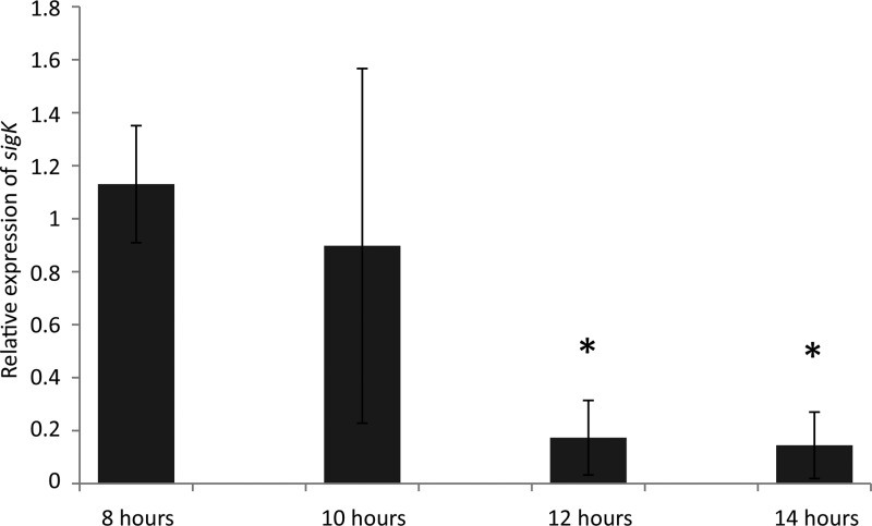
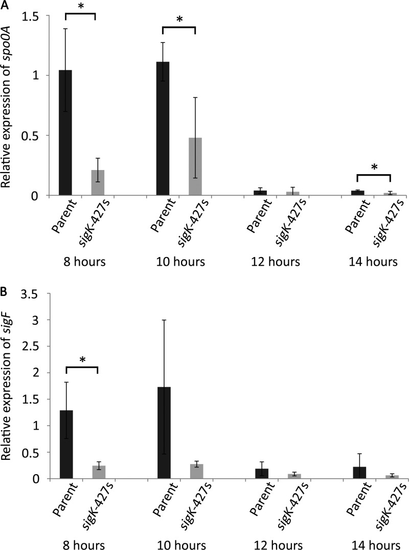
The relative sigK transcription levels for C. botulinum ATCC 3502 cultures were tested at the late exponential phase (8 h of growth) to the stationary growth phases (10 to 14 h) (Fig. 3). The relative sigK transcription levels at the late exponential phase were 6.5- and 7.8-fold higher (P < 0.05) than those at the stationary growth phases at 12 and 14 h, respectively. At 10 h, corresponding to the entry into stationary phase, large variation was noticed among the three biological replicates. The relative spo0A transcription levels were tested for the parent and sigK-427s mutant strains at late exponential phase (8 h of growth) to stationary growth phases (10 to 14 h of growth) (Fig. 4A). Significantly (P < 0.05) higher levels of spo0A transcription were observed in the parent strain than in the sigK-427s mutant at 8 h (4.9-fold) and 10 h (2.3-fold), but they were also observed at 14 h (1.9-fold) after inoculation into fresh medium. At between 10 and 12 h, a notable drop in transcription levels was observed in both the parent (28-fold) and sigK-427s (16.2-fold) strains. No difference between the parent and mutant spo0A transcript levels was observed at 12 h. The relative sigF transcription levels (Fig. 4B) were significantly (P < 0.05) higher in the parent than in the sigK-427s mutant strain at 8 h (5.3-fold) after inoculation into fresh medium. A similar result was observed at 10 h, although this was not statistically significant. At between 10 and 12 h, another notable drop in relative transcription rates in both the parent strain (9.2-fold) and the mutant sigK-427s (3.1-fold) was observed. No major differences between the parent and mutant sigF transcript levels were observed at 12 and 14 h.
Fig 3.
qRT-PCR analysis of sigK transcription levels at 8, 10, 12, and 14 h of growth calibrated to the transcription level at 8 h. The 16S rrn gene was used as a normalization reference. Error bars indicate the standard deviation of three replicate cultures. *, statistically significant differences (P < 0.05, two-tailed Student's t test) between time points relative to 8 h.
Fig 4.
qRT-PCR analysis of the relative spo0A (A) and sigF (B) transcription levels at 8, 10, 12, and 14 h of growth calibrated to the transcription level at 8 h. The 16S rrn gene was used as a normalization reference. The error bars indicate the standard deviation of three replicate cultures. *, statistically significant differences (P < 0.05, two-tailed Student's t test) between mutant and parent strains.
DISCUSSION
Characterization of the molecular mechanisms of C. botulinum is at its infancy. The recent availability of genetic engineering tools (10, 12) finally enables study of gene function in C. botulinum, and these tools are likely to reveal functional differences between Bacillus spp. and clostridia, two differently adapted spore-forming bacteria (33). The sporulation pathway of C. botulinum ATCC 3502 differs largely from that of many B. subtilis strains, in that it lacks a phosphorelay system for Spo0A activation and possesses sigK at a single locus rather than in two parts separated by a skin element. Disruption of sigK in C. botulinum ATCC 3502, as this study shows, results in the halt of sporulation at an early stage. In contrast, sporulation in B. subtilis sigK mutants is blocked at a late stage.
After a sporulation time of 7 days, which has been demonstrated to be adequate for efficient sporulation of clostridia (3), our sigK mutants failed to produce viable, germinating spores. In stage 0 of sporulation, the cell elongates prior to asymmetric cell division typical for stage II. Our sigK mutants exhibited elongated cells and no asymmetric cell division, characteristic of sporulation being disrupted at stage 0. Heap et al. (10) demonstrated elongated cell phenotypes of spo0A mutants in C. difficile, C. acetobutylicum, and C. sporogenes. Early-stage disruption of sporulation in a sigK mutant has been noted before in C. perfringens (9). The investigators proposed that sigK is essential in the logarithmic and early stationary growth phases for successful sporulation (9). They suggested that sigK transcription is self-driven in the early phase, while σE regulates sigK in the later stages. This results in biphasic expression of sigK during early sporulation and again at the later stage typical for B. subtilis (6). Although the sporulation sigma factors of C. acetobutylicum were shown to follow the expression pattern seen in B. subtilis (29), a study by Jones et al. (14) showed with qRT-PCR that sigK was expressed in C. acetobutylicum in the early stages of sporulation, before collapsing and peaking again in the later stages of sporulation, similar to the biphasic expression seen by Harry et al. (9). However, the authors questioned whether sigK was functional in C. acetobutylicum due to the small amounts of sigK transcript detected. Our qRT-PCR results also showed low levels of sigK transcripts comparable to the levels of transcripts of other sigma factors tested from similar cDNA samples; however, sigK was clearly present at significantly larger quantities in the late log phase than the early stationary growth phases. This strongly suggests that sigK is functional and plays a role in early sporulation in C. botulinum. Given the previous observations of sigK transcription in other clostridia, it is tempting to speculate that the roles of other sigma factors in the sporulation pathway may also differ between the two spore-forming genera.
The sporulation cascade in Bacillus spp. and clostridia commonly begins with the activation of Spo0A. Relative levels of spo0A and sigF transcription in C. botulinum ATCC 3502 and in the sigK-427s mutant were examined by qRT-PCR over the course of transition into stationary phase. Considerably lower spo0A and sigF expression levels were observed in the sigK-427s mutant than in the parent strain at late exponential and early stationary growth phases. The results provide further evidence that σK plays a crucial role in the early stages of sporulation in C. botulinum. The results suggest that σK may directly or indirectly positively regulate spo0A, which is supported by the finding that sigK is also transcribed at the late log growth phase. A failure to activate spo0A explains the lack of sigF induction, normally transcribed by Spo0A-P, in the sigK-427s mutant. The lack of sigF transcription indicates that the pathway has been blocked prior to this point, in agreement with our visual observation of the cells being blocked before stage II asymmetric cell division.
A sharp drop in spo0A and sigF expression levels at between 10 and 12 h in the ATCC 3502 parent strain and, for spo0A, in the sigK-427s mutant strain suggests that early sporulation stage 0 is essentially over when the cultures enter the stationary phase. A collapse of spo0A expression was also observed in B. subtilis cultures at the time when Spo0A-P was formed and accumulated to levels high enough to cause a negative-feedback loop (7). The similar, very low level of transcription of spo0A and sigF at 12 and 14 h between the mutant and parent strains may also be explained by temporal regulation of the sporulation pathway by such a negative-feedback loop.
Our experimental analysis showed that mutation of sigK in C. botulinum ATCC 3502 caused a nonsporulating phenotype. Analysis by qRT-PCR showed expression of sigK at the late log and early stationary growth phases. Further analysis of spo0A and sigF expression showed a lower transcription rate for both genes in the sigK mutant than the parent strain. The consistently low levels of sigF transcription suggest that the sporulation pathway had already halted by this point. These qRT-PCR results, in consideration with the observed nonsporulating elongated cell phenotype, show an essential role of σK in the early stages of sporulation in C. botulinum ATCC 3502, in contrast to its late-stage role in B. subtilis.
Supplementary Material
ACKNOWLEDGMENTS
The work was performed in the Finnish Centre of Excellence in Microbial Food Safety Research and supported by the Academy of Finland (grant 141140), the European Community's Seventh Framework Program FP7/2007-2013 (grant 237942), and the Finnish Graduate School on Applied Bioscience.
Esa Penttinen and Hanna Korpunen are thanked for technical assistance.
Footnotes
Published ahead of print 27 April 2012
Supplemental material for this article may be found at http://aem.asm.org/.
REFERENCES
- 1. Bradshaw M, et al. 2010. Construction of a nontoxigenic Clostridium botulinum strain for food challenge studies. Appl. Environ. Microbiol. 76:387–393 [DOI] [PMC free article] [PubMed] [Google Scholar]
- 2. Burbulys D, Trach KA, Hoch JA. 1991. Initiation of sporulation in B. subtilis is controlled by a multicomponent phosphorelay. Cell 64:545–552 [DOI] [PubMed] [Google Scholar]
- 3. Burns DA, Heap JT, Minton NP. 2010. The diverse sporulation characteristics of Clostridium difficile clinical isolates are not associated with type. Anaerobe 16:618–622 [DOI] [PubMed] [Google Scholar]
- 4. Cooksley CM, et al. 2010. Regulation of neurotoxin production and sporulation by a putative agrBD signaling system in proteolytic Clostridium botulinum. Appl. Environ. Microbiol. 76:4448–4460 [DOI] [PMC free article] [PubMed] [Google Scholar]
- 5. Eichenberger P, et al. 2004. The program of gene transcription for a single differentiating cell type during sporulation in Bacillus subtilis. PLoS Biol. 2:e328 doi: 10.1371/journal.pbio.0020328 [DOI] [PMC free article] [PubMed] [Google Scholar]
- 6. Errington J. 1993. Bacillus subtilis sporulation: regulation of gene expression and control of morphogenesis. Microbiol. Rev. 57:1–33 [DOI] [PMC free article] [PubMed] [Google Scholar]
- 7. Fujita M, Sadaie Y. 1998. Feedback loops involving Spo0A and AbrB in in vitro transcription of the genes involved in the initiation of sporulation in Bacillus subtilis. J. Biochem. 124:98–104 [DOI] [PubMed] [Google Scholar]
- 8. Haraldsen JD, Sonenshein AL. 2003. Efficient sporulation in Clostridium difficile requires disruption of the sigmaK gene. Mol. Microbiol. 48:811–821 [DOI] [PubMed] [Google Scholar]
- 9. Harry KH, Zhou R, Kroos L, Melville SB. 2009. Sporulation and enterotoxin (CPE) synthesis are controlled by the sporulation-specific sigma factors SigE and SigK in Clostridium perfringens. J. Bacteriol. 191:2728–2742 [DOI] [PMC free article] [PubMed] [Google Scholar]
- 10. Heap JT, Pennington OJ, Cartman ST, Carter GP, Minton NP. 2007. The ClosTron: a universal gene knock-out system for the genus Clostridium. J. Microbiol. Methods 70:452–464 [DOI] [PubMed] [Google Scholar]
- 11. Heap JT, Pennington OJ, Cartman ST, Minton NP. 2009. A modular system for Clostridium shuttle plasmids. J. Microbiol. Methods 78:79–85 [DOI] [PubMed] [Google Scholar]
- 12. Heap JT, et al. 2010. The ClosTron: mutagenesis in Clostridium refined and streamlined. J. Microbiol. Methods 80:49–55 [DOI] [PubMed] [Google Scholar]
- 13. Hilbert DW, Piggot PJ. 2004. Compartmentalization of gene expression during Bacillus subtilis spore formation. Microbiol. Mol. Biol. Rev. 68:234–262 [DOI] [PMC free article] [PubMed] [Google Scholar]
- 14. Jones SW, et al. 2008. The transcriptional program underlying the physiology of clostridial sporulation. Genome Biol. 9:R114 doi: 10.1186/gb-2008-9-7-r114 [DOI] [PMC free article] [PubMed] [Google Scholar]
- 15. Kodama T, et al. 2011. A novel small protein of Bacillus subtilis involved in spore germination and spore coat assembly. Biosci. Biotechnol. Biochem. 75:1119–1128 [DOI] [PubMed] [Google Scholar]
- 16. Kunkel B, Losick R, Stragier P. 1990. The Bacillus subtilis gene for the development transcription factor sigma K is generated by excision of a dispensable DNA element containing a sporulation recombinase gene. Genes Dev. 4:525–535 [DOI] [PubMed] [Google Scholar]
- 17. Lindström M, Kiviniemi K, Korkeala H. 2006. Hazard and control of group II (non-proteolytic) Clostridium botulinum in modern food processing. Int. J. Food Microbiol. 108:92–104 [DOI] [PubMed] [Google Scholar]
- 18. Lindström M, Korkeala H. 2006. Laboratory diagnostics of botulism. Clin. Microbiol. Rev. 19:298–314 [DOI] [PMC free article] [PubMed] [Google Scholar]
- 19. Livak KJ, Schmittgen TD. 2001. Analysis of relative gene expression data using real-time quantitative PCR and the 2−ΔΔCT method. Methods 25:402–408 [DOI] [PubMed] [Google Scholar]
- 20. McCroskey LM, Hatheway CL. 1988. Laboratory findings in four cases of adult botulism suggest colonization of the intestinal tract. J. Clin. Microbiol. 26:1052–1054 [DOI] [PMC free article] [PubMed] [Google Scholar]
- 21. Oke V, Losick R. 1993. Multilevel regulation of the sporulation transcription factor sigma K in Bacillus subtilis. J. Bacteriol. 175:7341–7347 [DOI] [PMC free article] [PubMed] [Google Scholar]
- 22. Palonen E, Lindström M, Karttunen R, Somervuo P, Korkeala H. 2011. Expression of signal transduction system encoding genes of Yersinia pseudotuberculosis IP32953 at 28°C and 3°C. PLoS One 6:e25063 doi: 10.1371/journal.pone.0025063 [DOI] [PMC free article] [PubMed] [Google Scholar]
- 23. Paredes CJ, Alsaker KV, Papoutsakis ET. 2005. A comparative genomic view of clostridial sporulation and physiology. Nat. Rev. Microbiol. 3:969–978 [DOI] [PubMed] [Google Scholar]
- 24. Perutka J, Wang W, Goerlitz D, Lambowitz AM. 2004. Use of computer-designed group II introns to disrupt Escherichia coli DExH/D-box protein and DNA helicase genes. J. Mol. Biol. 336:421–439 [DOI] [PubMed] [Google Scholar]
- 25. Piggot PJ, Coote JG. 1976. Genetic aspects of bacterial endospore formation. Bacteriol. Rev. 40:908–962 [DOI] [PMC free article] [PubMed] [Google Scholar]
- 26. Piggot PJ, Hilbert DW. 2004. Sporulation of Bacillus subtilis. Curr. Opin. Microbiol. 7:579–586 [DOI] [PubMed] [Google Scholar]
- 27. Purdy D, et al. 2002. Conjugative transfer of clostridial shuttle vectors from Escherichia coli to Clostridium difficile through circumvention of the restriction barrier. Mol. Microbiol. 46:439–452 [DOI] [PubMed] [Google Scholar]
- 28. Reynolds J, Moyes R, Breakwell DP. 2009. Differential staining of bacteria: endospore stain. Curr. Protoc. Microbiol. Appendix 3:3J. [DOI] [PubMed] [Google Scholar]
- 29. Santangelo JD, Kuhn A, Treuner-Lange A, Dürre P. 1998. Sporulation and time course expression of sigma-factor homologous genes in Clostridium acetobutylicum. FEMS Microbiol. Lett. 161:157–164 [DOI] [PubMed] [Google Scholar]
- 30. Selby K, et al. 2011. Important role of class I heat shock genes hrcA and dnaK in the heat shock response and the response to pH and NaCl stress of group I Clostridium botulinum strain ATCC 3502. Appl. Environ. Microbiol. 77:2823–2830 [DOI] [PMC free article] [PubMed] [Google Scholar]
- 31. Söderholm H, et al. 2011. cspB encodes a major cold shock protein in Clostridium botulinum ATCC 3502. Int. J. Food Microbiol. 146:23–30 [DOI] [PubMed] [Google Scholar]
- 32. Steiner E, et al. 2011. Multiple orphan histidine kinases interact directly with Spo0A to control the initiation of endospore formation in Clostridium acetobutylicum. Mol. Microbiol. 80:641–654 [DOI] [PMC free article] [PubMed] [Google Scholar]
- 33. Stephenson K, Lewis RJ. 2005. Molecular insights into the initiation of sporulation in Gram-positive bacteria: new technologies for an old phenomenon. FEMS Microbiol. Rev. 29:281–301 [DOI] [PubMed] [Google Scholar]
- 34. Takemaru K, Mizuno M, Sato T, Takeuchi M, Kobayashi Y. 1995. Complete nucleotide sequence of a skin element excised by DNA rearrangement during sporulation in Bacillus subtilis. Microbiology 141:323–327 [DOI] [PubMed] [Google Scholar]
- 35. Waites, et al. 1970. Sporulation in Bacillus subtilis. Correlation of biochemical events with morphological changes in asporogenous mutants. Biochem. J. 118:667–676 [DOI] [PMC free article] [PubMed] [Google Scholar]
- 36. Wörner K, Szurmant H, Chiang C, Hoch JA. 2006. Phosphorylation and functional analysis of the sporulation initiation factor Spo0A from Clostridium botulinum. Mol. Microbiol. 59:1000–1012 [DOI] [PubMed] [Google Scholar]
Associated Data
This section collects any data citations, data availability statements, or supplementary materials included in this article.


