Abstract
Bacterial communities use “quorum sensing” (QS) to coordinate their population behavior through the action of extracellular signal molecules, such as the N-acyl-l-homoserine lactones (AHLs). The versatile and ubiquitous opportunistic pathogen Pseudomonas aeruginosa is a well-studied model for AHL-mediated QS. This species also produces an intercellular signal distinct from AHLs, 3,4-dihydroxy-2-heptylquinoline (PQS), which belongs to a family of poorly characterized 4-hydroxy-2-alkylquinolines (HAQs) previously identified for their antimicrobial activity. Here we use liquid chromatography (LC)/MS, genetics, and whole-genome expression to investigate the structure, biosynthesis, regulation, and activity of HAQs. We show that the pqsA-E operon encodes enzymes that catalyze the biosynthesis of five distinct classes of HAQs, and establish the sequence of synthesis of these compounds, which include potent cytochrome inhibitors and antibiotics active against human commensal and pathogenic bacteria. We find that anthranilic acid, the product of the PhnAB synthase, is the primary precursor of HAQs and that the HAQ congener 4-hydroxy-2-heptylquinoline (HHQ) is the direct precursor of the PQS signaling molecule. Significantly, whereas phnAB and pqsA-E are positively regulated by the virulence-associated transcription factor MvfR, which is also required for the expression of several QS-regulated genes, the conversion of HHQ to PQS is instead controlled by LasR. Finally, our results reveal that HHQ is itself both released from, and taken up by, bacterial cells where it is converted into PQS, suggesting that it functions as a messenger molecule in a cell-to-cell communication pathway. HAQ signaling represents a potential target for the pharmacological intervention of P. aeruginosa-mediated infections.
In nature, most bacteria live not as individual cells but as pseudomulticellular organisms that coordinate their population behavior by means of small extracellular signal molecules. Under appropriate conditions, these molecules are released into the environment and taken up and responded to by surrounding cells (1–3). “Quorum sensing” (QS), is the archetypal intercellular communication system used by many bacterial species to regulate their gene expression in response to cell density. This regulation allows all of the individual cells to behave coordinately and synergistically as a community, for instance, in growth dynamics and resource utilization (4). A common feature of all QS systems is the transcriptional activation and repression of a large regulon of QS-controlled genes when a minimal threshold concentration of a specific autoinducer is reached.
The well-characterized QS system used by Gram-negative bacteria is mediated by N-acyl-l-homoserine lactones (AHLs) as extracellular signaling molecules (1, 3). The versatile and ubiquitous opportunistic pathogen Pseudomonas aeruginosa is one of the best-studied models of AHL-mediated QS. In this species, two separate autoinducer synthase/transcriptional regulator pairs, LasRI and RhlRI, modulate the expression of several genes, including many virulence factors, in response to increasing concentrations of the specific signaling molecules oxo-C12-HSL and C4-HSL (5, 6).
P. aeruginosa also produces a cell-to-cell signal distinct from AHLs: 3,4-dihydroxy-2-heptylquinoline, called PQS (7). PQS serves as a signaling molecule regulating the expression of a subset of genes belonging to the QS regulon, including the phz and hcn operons (E.D., S. Gopalan, F.L., A. N. Remick, A. P. Tampakaki, M.N.M., and L.G.R., unpublished work). PQS functions in the QS hierarchy by linking a regulatory cascade between the las and the rhl systems (8). That maximal PQS production occurs at the end of the exponential growth phase (9) supports the hypothesis that PQS acts as a secondary regulatory signal for a subset of QS-controlled genes. Although PQS has no antibiotic activity (7), it belongs to a family of poorly characterized antimicrobial P. aeruginosa products, the “pyo” compounds, originally described in 1945, which are derivatives of 4-hydroxy-2-alkylquinolines (HAQs) (10, 11). We have also identified a QS-associated P. aeruginosa transcriptional regulator, MvfR, which is required for the production of several secreted compounds, including virulence factors, and PQS (12, 13). Indeed, MvfR controls the synthesis of anthranilic acid (AA), a PQS precursor (14), by positively regulating the transcription of phnAB, which encodes an anthranilate synthase (12). In addition, mutations in five genes, designated pqsA-E, result in loss of pyocyanin and PQS production (15, 16). These genes likely mediate HAQ synthesis.
Here, we use liquid chromatography (LC)/MS to show that the pqsA-E operon encodes enzymes that direct the biosynthesis of five classes of HAQs, including molecules that function as antibiotics and cytochrome inhibitors and, significantly, as intercellular communication molecules. Furthermore, via genome-wide expression studies using the GeneChip P. aeruginosa oligonucleotide array, we demonstrate that the MvfR transcriptional regulator controls pqsA-E expression. These results reveal the HAQ biosynthesis pathway and furthermore show that one HAQ congener, 4-hydroxy-2-heptylquinoline (HHQ), is the direct precursor of PQS and is itself a message molecule involved in cell-to-cell communication. This pathway represents a candidate target for the pharmacological intervention of P. aeruginosa-mediated infections.
Materials and Methods
Bacterial Strains, Plasmids, and Media. P. aeruginosa strains include wild-type PA14 (17); an mvfR mutant (12); 8C12, a TnphoA-insertion mutant of pqsB (18); and an lasR::Gm mutant, which was generated by allelic exchange using pSB219.9A as described (19). A pqsE deletion mutant was generated via pEX18Ap allelic replacement by using sucrose selection, resulting in a 570-bp nonpolar deletion covering 65% of the sequence (20). The pqsA (U479) TnphoA mutant was obtained from the PA14 Transposon Insertion Mutant Database. For complementation analysis, mvfR was cloned into pDN18 (21). The reporter fusions phzABC-lacZ and hcnA′-lacZ have been described (22, 23). Plasmids were transformed into PA14 by electroporation (24). Specific β-galactosidase activity was determined as reported (25).
Bacteria were grown in LB broth or on 1.5% Bacto-agar (Difco) LB plates. Freshly plated cells served as inoculum. For pyocyanin production, bacteria were grown in King's A broth (26), and the pyocyanin was quantified as OD520 after supernatant extraction (27). Tetracycline (75 mg/liter), carbenicillin (300 mg/liter), kanamycin (200 mg/liter), and gentamicin (100 mg/liter) were included as required.
LC/MS Analysis. Analyses were performed by using a Micromass Quattro II triple quadrupole mass spectrometer (Micromass Canada, Pointe-Claire, Canada) in positive electrospray ionization mode, interfaced to an HP1100 HPLC equipped with a 4.5 × 150-mm reverse-phase C8 column. Culture supernatants were twice extracted with ethyl acetate, the solvent was evaporated, and the residue was dissolved in a water/acetonitrile mixture containing the internal standard. Alternatively, culture samples were directly diluted with a methanolic solution of the internal standard, as reported (9).
Synthesis of Labeled HAQ. 4-Hydroxy-2-heptylquinoline N-oxide (HQNO) was from Sigma. 2,3,4,5-Tetradeuteroanthranilic acid (AA-d4) was from CDN isotopes (Pointe-Claire, Canada). The internal standards, 5,6,7,8-tetradeutero-3,4-dihydroxy-2-heptylquinoline (PQS-d4), and 5,6,7,8-tetradeutero-4-hydroxy-2-heptylquinoline (HHQ-d4) were synthesized as reported (9). 5,6,7,8-Tetradeutero-4-hydroxy-2-heptylquinoline N-oxide (HQNO-d4) was synthesized from HHQ-d4 (28).
RNA Isolation and Transcriptome Analysis. Whole genome expression profiles were produced in duplicate for PA14 and the mvfR mutant. Cultures were grown in 1-liter Erlenmeyer flasks with 100 ml of LB at 37°C and shaking at 200 rpm. Cells were sampled at OD600 = 1.5, 2.5, 3.5, and 4.5, and their RNA was immediately stabilized with RNAprotect Bacteria Reagent (Qiagen, Valencia, CA) and stored at –80°C. Total RNA was isolated with the RNeasy spin column (including an on-column DNase digestion step) according to the manufacturer (Qiagen), treated with RQ1 DNase I (Promega) for 1 h at 37°C, and repurified through an RNeasy column.
Samples were labeled according to the manufacturer (Affymetrix, Santa Clara, CA) and hybridized to the Affymetrix GeneChip P. aeruginosa genome array for 24 h at 50°C by using the GeneChip hybridization oven at 60 rpm. Washing, staining, and scanning were performed according to Affymetrix. The original data files, obtained from the array scans hybridized with the different probes, were converted to cell intensity files (.CEL files) by using microarray suite 5.0. Data analysis/clustering was performed with the dna-chip analyzer (dchip) software (29).
Antimicrobial Activity Assay. HAQ antimicrobial activity was evaluated on well-plates. An overnight culture (30 μl) was plated to produce a bacterial lawn, and 5-mm-diameter holes were punched in the agar and filled with 60 μl of a 25% methanol solution of test extract or pure HAQ. Plates were incubated overnight at 37°C and scored for growth inhibition zones around the test wells.
Cell-to-Cell Communication Assay. To test whether P. aeruginosa cells produce PQS in response to HHQ released by other cells, we compared the concentrations of PQS in cultures of an lasR mutant, grown in 30 ml of LB in 250-ml flasks, with cocultures containing 50% of an lasR mutant and an mvfR mutant. pDN18mvfR was introduced into the lasR mutant to compensate for the lower expression of mvfR in this background. The effect on gene expression of exogenous HHQ was assayed by comparing the β-galactosidase activity of PA14 vs. lasR– cells carrying the phzABC-lacZ or hcnA′-lacZ fusions, grown in the absence or presence of 10 mg/liter of HHQ.
Results
HAQ Identification: mvfR Is Required for the Production of Five Distinct Series of HAQs from the Common Precursor Anthranilic Acid. Calfee et al. (14) recently reported that 14C-labeled AA is incorporated into PQS, but that this PQS represents only 12% of the newly synthesized compounds, indicating that the ethyl acetate extract contains additional AA-derived molecules. Because HHQ biosynthesis proceeds from the coupling of AA and an α-keto fatty acid (30), we hypothesized that these unidentified AA-derived molecules might correspond to HAQs related to PQS and HHQ.
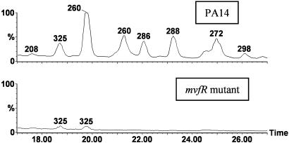
To this end, we fed AA or deuterated AA (AA-d4) to PA14 cultures and analyzed the culture supernatants by using LC/MS (9). The resulting chromatograms exhibit several peaks in the vicinity of PQS, and the mass spectra of these compounds all show the addition of 4 Da in the cultures fed AA-d4, demonstrating that AA is their common precursor (Fig. 8, which is published as supporting information on the PNAS web site). Because we previously reported that PQS production is abrogated in an mvfR mutant (12), we investigated the synthesis of these compounds in this mutant. Fig. 1 shows that all of the deuterium-labeled peaks are absent from the mvfR mutant culture supernatant, with the only residual peaks found at HAQ retention times corresponding to two conformers of the siderophore pyochelin (31), which give M+H ions at m/z 325 and are structurally unrelated to HAQs.
Fig. 1.
LC/MS analysis of PA14 and mvfR mutant culture supernatants. Shown are MS chromatograms of PA14 (Upper) and the mvfR mutant (Lower). The numbers above the peaks represent the m/z values of the most intense ions. Intensities are normalized to the most abundant ion in Upper.
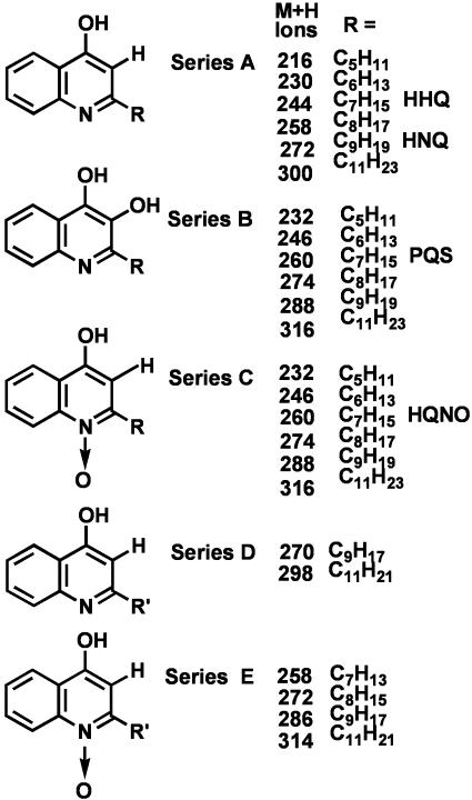
The mass spectra of these labeled peaks show that they correspond to five distinct series of HAQs (Fig. 2). All these congeners share the common basic 4-hydroxyquinoline structure of series A with an additional hydroxyl at the 3-position, as in series B, or with an N-oxide group as in series C and E. Within each series, the 2-position alkyl chain varies in length. Also, the series D and E alkyl side chain is unsaturated. The most abundant congeners contain an odd carbon number alkyl chain, with seven or nine carbons preponderant.
Fig. 2.
Chemical structures of five distinct series of HAQ compounds isolated from the PA14 culture supernatant. Shown is a detailed MS analysis of the peaks in Fig. 1. R, Alkyl side chain length.
The HAQ congeners include both previously identified and previously uncharacterized compounds. The C7 (HHQ) and C9 (HNQ) congeners, shown in Fig. 2, were reported by Wells in 1952 (11) whereas the structures of the other series A congeners were later determined by using GC/MS (32). In contrast, the only reported series B congener is 3,4-dihydroxy-2-heptylquinoline, first isolated in 1959 (33), and later fully characterized and named PQS (7). For series C, the C7, C9, C8, and C11 congeners have been reported (28, 34) and are further discussed below whereas the series E and the series B PQS congeners are identified in this study. The simultaneous mass spectrometric detection of all these HAQ congeners, notably those of series C and E, which have polar N-oxide functions, could not be achieved by GC/MS (32, 35). Positive electrospray ionization MS is better suited than GC/electron impact-MS for the detection of such relatively basic and/or polar compounds.
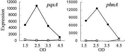
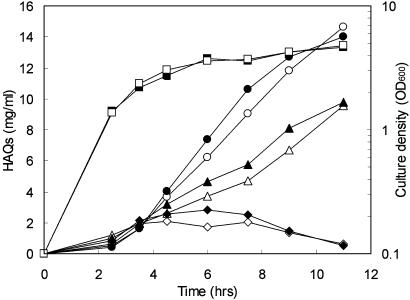
HAQ Regulation: MvfR Controls the Expression of the phnAB and pqsA-E Operons, Which Are Required for HAQ Synthesis. That MvfR regulates phnAB expression (12) suggests that it might also direct HAQ biosynthesis by regulating genes that encode anabolic pathway enzymes. As part of a project to identify MvfR-regulated genes, we carried out a transcriptome comparative analysis between PA14 and its isogenic mvfR mutant at set time points during a growth time course, by using the Affymetrix P. aeruginosa GeneChip oligonucleotide array (E.D., S. Gopalan, F.L., A. N. Remick, A. P. Tampakaki, M.N.M., and L.G.R., unpublished work). The expression profiles of the five genes just upstream from the anthranilate synthase phnAB operon tightly cluster with phnAB expression (Fig. 3 and Fig. 9, which is published as supporting information on the PNAS web site), suggesting they are coregulated. Fig. 4 shows that the expression patterns of these seven genes correlate with the kinetic rates of HAQ production, which are maximal at the end of exponential/early stationary phase (i.e., OD600 ≈ 2.5) (9). Because phnAB expression is under the control of MvfR (12), it is not surprising that the transcription of these seven genes is almost completely abolished in the mvfR mutant (Figs. 3 and 9).
Fig. 3.
Expression profiles of pqsA and phnA in PA14 vs. the mvfR mutant using the GeneChip P. aeruginosa array. •, PA14; □, the mvfR mutant. Shown are signal intensity values calculated by dchip software. OD, optical density at 600 nm of the cultures when harvested.
Fig. 4.
HAQ production kinetics in PA14 vs. the isogenic pqsE mutant. Bacteria were grown in LB at 37°C, and their extracellular HAQ concentrations were analyzed by MS at regular time intervals. Filled symbols, PA14; open symbols, pqsE mutant. ▪ and □, optical density of the culture (OD600); • and ○, PQS; ▴ and ▵, HQNO; ♦ and ⋄, HHQ.
Table 1 shows that knockout inactivation of pqsA or pqsB results in the complete elimination of HAQ production and the striking accumulation of AA in culture supernatants. AA likely accumulates because these mutants fail to consume AA for HAQ synthesis, further supporting AA as the HAQ precursor. In contrast, a pqsE mutant produces wild-type levels of HAQ and AA (Fig. 4 and data not shown), in agreement with the observation that pqsE inactivation does not affect PQS production (15). Genetic complementation suggests that pqsABCDE is a single operon (15). Our expression profiling data corroborate the LC/MS results and further indicate that MvfR controls the transcription of the coregulated pqsABCDE and phnAB operons.
Table 1. Percent relative concentration of extracellular compounds in culture supernatants of mutant strains vs. wild-type isogenic PA14.
| % of PA14
|
|||||
|---|---|---|---|---|---|
| Compound | mvfR- | pqsA- | pqsB- | mvfR- compl. | lasR- |
| HHQ | 0 | 0 | 0 | 1017 ± 247 | 250 ± 11 |
| HHQ N-oxide | 0 | 0 | 0 | 200 ± 41 | 45 ± 8 |
| PQS | 0 | 0 | 0 | 247 ± 65 | 23 ± 3 |
| HNQ | 0 | 0 | 0 | 502 ± 106 | 551 ± 262 |
| HNQ N-oxide | 0 | 0 | 0 | 286 ± 52 | 41 ± 3 |
| diHNQ | 0 | 0 | 0 | 156 ± 49 | 19 ± 3 |
| AA | 0 | 458 ± 221 | 188 ± 102 | 0 | 6 ± 1 |
| Pyocyanin | <10 | <10 | <10 | 177 ± 26 | NT |
Cells were cultivated in LB medium at 37°C to OD600 = 4.0-4.5. Data are averages of triplicate experiments ± SD. diHNQ, 3,4-Dihydroxy-2-nonylquinoline; NT, not tested. mvfR compl., the mutant strain complemented with pDN18mvfR.
HAQ Activity: MvfR Regulates HQNO Antimicrobial Activity Against Gram-Positive Bacteria. Fluorescent pseudomonads, and perhaps just P. aeruginosa, are the only microorganisms identified to produce HAQs (9, 36). Although their biological functions are unknown, many HAQs were initially isolated as antibiotics (10, 11). We assayed the antimicrobial activity of total organic extracts from PA14 and mvfR, pqsA, and pqsE mutant strains, and purified PQS, HHQ, and HQNO, against the Gram-positive species Staphylococcus aureus and Bacillus subtilis. PA14 and pqsE– extracts clearly inhibit both species whereas the pqsA– and mvfR– extracts have low or no antibacterial activity (Fig. 10, which is published as supporting information on the PNAS web site). Thus, MvfR regulates the production of antibiotics that can function in niche competition against other bacteria. Because the pqsE mutant and PA14 produce roughly the same levels of antimicrobial activity, these antibiotic compounds are probably HAQs, instead of other nonpolar compounds whose synthesis is under PQS control.
Although it has been known for many decades that P. aeruginosa produces low molecular weight antibiotics, later found to be HAQs (10, 37), these compounds have been little characterized. Although the HAQ N-oxides C7, C9, and C11 were originally isolated as streptomycin and dihydrostreptomycin antagonists (28, 38, 39), their mode of action remains unknown. We confirm here the specific antibacterial activity of HQNO (Fig. 10), in agreement with Machan et al. (35). This C7 congener, which is a widely used cytochrome inhibitor (40), and also inhibits Na+-translocating NADH-quinone oxidoreductases (41), might function in nature as a virulence factor.
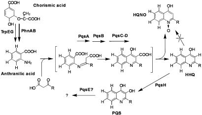
HAQ Biosynthetic Machinery: PQS Is Not a Product of the mvfR Regulated Synthetic Pathway. The similarity of the HAQ structures, along with their colabeling from deuterated AA, suggests they are produced via a common biosynthetic pathway. To this end, we added various known or putative labeled HAQ precursors and derivatives to cultures of PA14 or isogenic pqs and mvfR mutants, to generate the pathway in Fig. 5. Addition of HHQ-d4 to PA14 cultures results in overproduction and labeling of PQS, indicating that HHQ is an intermediate in PQS biosynthesis. Also, because no overproduction or labeling of HQNO is detected, HHQ is not a precursor of HQNO. By analogy, the series A compounds, which include HHQ, are the probable precursors of the corresponding series B compounds, but not the other series. To examine whether PQS is an intermediate in HAQ biosynthesis, PQS-d4 was added to PA14 cultures. Because none of the chromatographic peaks are labeled, PQS must be an end-product in HAQ biosynthesis, or at least is not substantially converted into an extracellular compound. Also, because all HHQ-d4 added to pqs/mvfR mutant cultures is completely converted into PQS, MvfR does not control the final step(s) of PQS synthesis.
Fig. 5.
Proposed HAQ biosynthetic pathway in P. aeruginosa. The sequence of synthesis was determined by supplementing cultures of PA14 and various pqs/mvfR mutants with deuterium-labeled intermediates. Bracketed structures are hypothetical.
We are unable to determine the precise origin(s) of the N-oxides (series C and E). Nevertheless, these HAQs clearly belong to the above pathway because AA-d4 addition results in labeled N-oxides, and they are absent in mutants that fail to synthesize HAQs; however, as proposed in Fig. 5, N-oxides are probably not synthesized downstream of HHQ or PQS because cultures supplemented with these deuterated compounds do not produce the corresponding labeled N-oxides. The N-oxides do not seem to be HAQ precursors, by means of simple reduction of their N-oxide function, because adding HQNO-d4 to PA14 cultures neither results in decreased labeled N-oxide concentrations in the culture medium nor labeling of any HAQ congeners. Because HQNO is a cytochrome inhibitor and has antimicrobial activity, it seems likely that it is actively exported, which would mask its role in HAQ biosynthesis in our assay. Nevertheless, N-oxides are probably the end-products of a branch pathway that is not under mvfR regulation. Indeed, we have shown that series A, B, and D congener concentrations decrease in culture supernatants after peaking whereas the concentrations of the N-oxides (series C and E) remain stable (9).
Overexpression of mvfR results in the excessive accumulation of series A compounds (the series B precursors; HHQ and HNQ in Table 1) in the supernatant but leaves series B, C, and E (PQS congeners and N-oxides) concentrations largely unaffected (Table 1 and data not shown), indicating that the series A (and D) congeners are end-products of the mvfR-regulated synthetic pathway. These data also support the conclusions that MvfR does not directly control PQS production, and that the branch pathways leading to series B, C, and E are saturated when the mvfR-regulated pathway is overactivated. Similarly, that a lasR mutant produces significant amounts of HAQs (Table 1), with the overaccumulation of series A congeners (the PQS analogue precursors) suggests that the QS transcriptional regulator LasR controls the series A to series B conversion. This step is likely mediated by the PqsH-encoded FAD-dependent monoxygenase, a QS-controlled gene that is required for PQS synthesis (15) and is under LasR regulation (42).
Collectively, our results show that the series A compounds are the end-product of the mvfR-controlled biosynthetic pathway and are subsequently converted into the series B PQS analogues via a lasR-dependent pathway, likely via the PqsH enzyme, which is not under MvfR regulation. These results suggest that the final synthesis of the active PQS signal is highly regulated and under additional controls beyond those of the primary HAQ pathway.
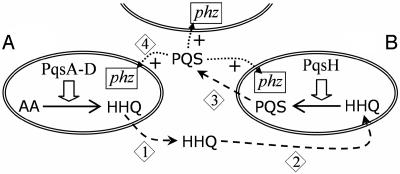
HHQ, the PQS Precursor, Functions in Cell-to-Cell Communication. PQS is an extracellular signal that participates in the QS circuitry. Several observations suggest that HHQ also functions as an intercellular messenger: (i) it is released by bacteria; (ii) its concentration rises during exponential growth phase and then decreases during PQS production (Fig. 4); (iii) it is taken up by bacterial cells, converted into PQS, and then released into the extracellular milieu, as shown in the labeling experiments; and (iv) its synthetic pathway, by means of PsqA-D, is distinct and under different regulation than that of HHQ-to-PQS conversion, by means of PqsH. These results collectively suggest the model depicted in Fig. 6, in which HHQ is released by cells and acts as a messenger that is subsequently converted into the PQS signal by the cells that take it up. To test this hypothesis, we compared PQS production in a lasR mutant culture vs. that of a mixed culture of lasR and mvfR mutant cells. Table 1 shows that the lasR mutant produces low levels of PQS and accumulates high concentrations of HHQ due to its low PqsH activity and the mvfR mutant produces no PQS because it is unable to synthesize the HHQ precursor. Thus, using these two mutants, one able to produce HHQ but unable to process it into PQS and the other unable to produce HHQ but able to convert it into PQS, should allow us to verify whether a P. aeruginosa cell can produce PQS by using HHQ produced by another cell. Indeed, Table 2 shows that, when the two mutants are grown together, PQS concentration is up to five times higher than if the cells fail to exchange the signaling information.
Fig. 6.
HHQ/PQS cell-to-cell communication model. 1, HHQ is synthesized and released by bacterial cells. 2, Extracellular HHQ is taken up by adjacent bacteria and converted into PQS, possibly in the periplasm. 3, PQS is released to act as a signaling molecule for other cells. 4, PQS activates target gene expression, such as the phz1 operon. Note that both HHQ availability and PqsH activity determine the final PQS concentration. In the experimental paradigm used to test the model, cell A and cell B were a lasR mutant and an mvfR mutant, respectively. In the case of a wild-type population, both the A and B cells are producing HHQ and PQS.
Table 2. Concentration of PQS (μg/ml) and phz1 operon expression in an lasR mutant culture and a 1:1 lasR mutant: mvfR mutant culture.
| PQS, μg/ml | β-gal activity, MU* | |
|---|---|---|
| lasR- | 2.1 ± 0.1 | 63.6 ± 2.6 |
| lasR- + mvfR- | 5.2 ± 1.8 | 110.5 ± 4.3 |
| Ratio† | 5.0 ± 1.8 | 3.5 ± 0.2 |
Cultures were assayed at 8-hr sampling time, corresponding to OD600 4.3 to 4.5.
The lasR mutant carries a phzABC-lacZ fusion: Data correspond to averages of triplicates ± SD. MU, Miller units.
Ratios have been corrected by taking into account that the mixed culture contains 50% fewer lasR mutant cells than the lasR culture. mvfR mutant cells do not produce PQS.
phz1 operon expression, which is required for the synthesis of pyocyanin, depends on both PQS signaling and pqsE expression (E.D., S. Gopalan, F.L., A. N. Remick, A. P. Tampakaki, M.N.M., and L.G.R., unpublished work). Therefore, to determine whether the PQS that is produced and released by mvfR– cells, when they are cocultured with the lasR– cells, is biologically active in adjacent cells, we introduced a phzABC-lacZ reporter fusion into the lasR– mutant, where pqsE is expressed, and compared the β-galactosidase activity with and without cocultivation with mvfR mutant cells. Table 2 shows that phz1 operon expression is indeed up-regulated in the presence of mvfR– cells, indicating that the PQS produced by mvfR mutant cells is taken up by the lasR– bacteria where it activates the phz1 operon. Accordingly, the mixed culture, but not the cultures of either mutant alone, also generates pyocyanin (Fig. 7). The lasR mutant is responsible for this pyocyanin production because, whereas the mvfR mutant “sees” PQS, it fails to express pqsE. Indeed, no β-galactosidase induction is obtained in a cocultivation experiment where the phzABC-lacZ reporter is carried by the mvfR– vs. the lasR– cells (data not shown). We note that, although mutants were used for this demonstration, we believe that the “conversational” pathway presented in Fig. 6 occurs in wild-type cell cultures because PA14 cells similarly perform HHQ release, uptake, and PQS conversion.
Fig. 7.
Pyocyanin production in a mixed-mutant culture illustrates the HAQ cell-to-cell communication pathway. Culture suspensions of the mvfR mutant, the lasR mutant, or a 1:1 mixture of both were spotted onto an LB plate and incubated for 18 h at 37°C. Only the mixed culture produces detectable amounts of pyocyanin, presumably from the lasR– cells because mvfR– cells are deficient for phz1 operon expression. This result demonstrates that HHQ, produced by the lasR– cells, is released and taken up by the mvfR– cells and converted into PQS. This PQS signal is then released by the mvfR– cells and taken up by the lasR– cells, where it signals phz1 expression and pyocyanin production.
Does HHQ itself signal gene regulation, independently from PQS activity, or does it function solely as a PQS precursor? To this end, we asked whether exogenous HHQ can stimulate the activity of a phzABC-lacZ reporter carried by wild-type PA14 cells, and by lasR– mutant cells. Although HHQ addition to the culture medium has no effect on this activity in the lasR mutant, it induces significant and consistent levels of β-galactosidase activity in the wild-type strain (Table 3). Similar results are also observed with an hcnA′-lacZ fusion (data not shown). These findings demonstrate that HHQ can induce the expression of genes that are also activated by PQS (15), and this induction seems to require HHQ conversion into PQS because no induction is found in cells that cannot carry out this conversion. Thus, HHQ does not itself function as a signal in our assay, independent of PQS activity, but instead is able to act as a messenger molecule that is converted into PQS by cells other than those that produce it.
Table 3. Effect of HHQ addition on phz1 operon expression in PA14 and lasR– cultures.
| β-gal activity, MU
|
||
|---|---|---|
| PA14 | lasR- | |
| -HHQ | 371 ± 8 | 78.7 ± 1.5 |
| +HHQ | 473 ± 23 | 83.7 ± 0.2 |
Cultures were assayed at 8 hr, corresponding to OD600 4.3-4.5. The bacteria carry a phzABC-lacZ fusion. Data correspond to averages of triplicates ± SD. MU, Miller units.
Discussion
Using LC/MS and DNA microarray analyses, we have determined that the transcriptional regulator MvfR, originally identified via its requirement for full P. aeruginosa broad-host virulence, regulates the expression of pqsABCDE and phnAB, which encode enzymes involved in the synthesis of five distinct families of structurally related HAQ congeners. By adding labeled HAQ precursors to bacterial cultures, we find that AA, mostly the product of the PhnAB synthase, is the precursor of all HAQs, and establish the sequence of their synthesis. Significantly, the last step in PQS synthesis requires an activity whose regulation depends on LasR vs. MvfR. Our results also reveal that one HAQ, HHQ, is the precursor of the PQS-signaling molecule and is itself both released from, and taken up by, bacteria, suggesting that it functions as an intercellular message molecule. These results provide insights into the structure, biosynthesis, regulation, and function of HAQs and have allowed us to uncover a “conversational” cell-to-cell communication pathway used by P. aeruginosa.
At least two branch pathways seem to have evolved from an ancestral synthetic HAQ pathway: one leading to the production of the antibacterial and cytochrome inhibitor N-oxide derivatives and the second leading to PQS signaling congeners. Many HAQs have been previously identified as P. aeruginosa secondary metabolites (36, 43). We confirm that a number of these compounds have Gram-positive antimicrobial activity and attribute some of this activity to HQNO, the most abundant HAQ. Interestingly, this activity has been associated with the clearance of S. aureus lung colonization by P. aeruginosa (35). HHQ is found in cystic fibrosis (CF) lung exudates (35), and PQS occurs in the sputum and bronchoalveolar lavage fluid of CF lungs (44), indicating that HAQs are produced in vivo. Guina et al. (45) have reported that P. aeruginosa isolates from CF patients produce more PQS than isolates from other diseases, suggesting that the PQS pathway could be up-regulated in CF strains. Thus, P. aeruginosa, when colonizing novel infectious niches, might increase HAQ production to inhibit the growth of competing microorganisms.
This study provides insights into PQS signaling, which functions in the expression of QS-regulated genes. Our results demonstrate that this system acts via at least two distinct extracellular molecules: PQS and its precursor, HHQ. As illustrated in Fig. 6, this pathway can be viewed as conversational cell-to-cell communication because an HHQ molecule released by a cell in a population is taken up by another cell and converted into PQS where it is then released into the extracellular milieu to signal cells in the population. Several observations indicate that this signaling is not artifactual and that HHQ and PQS could mean different things to cells in a population, even if their ultimate signaling mechanisms are the same. For instance, their concentrations peak at different growth stages; their production is under different regulation (MvfR for HHQ, LasR for PQS); and they are likely produced in different cellular compartments. Indeed, the psort program (psort.nibb.ac.jp) predicts that the PqsH monoxygenase, which probably mediates the hydroxylation of HHQ into PQS, is localized to the periplasmic space. This result suggests that the HHQ that is converted to PQS typically has an extracellular vs. intracellular origin and engenders the question of whether the intracellular HHQ is destined to serve as the PQS precursor.
Why might P. aeruginosa employ the extra complexity of this PQS and HHQ signaling? Cells in a bacterial community need to tell each other about their different properties, including their density, growth state, and production levels of extracellular compounds, such as antibiotics and virulence factors, to coordinate their activity. Presumably, different signals are required to convey this different information, and P. aeruginosa populations likely employ a panoply of extracellular molecules. For instance, although HHQ is the precursor of PQS, these two molecules could convey different information: HHQ reflects the extracellular levels of HAQ, including antimicrobial and cytochrome inhibitory functions, and indicates the level of MvfR activity whereas PQS reflects LasR activity levels and the status of the AHL-based QS system in population growth regulation. Also, that HAQ and PQS levels peak at different times suggests that PQS-mediated signaling reflects HAQ levels at one growth stage and PQS levels at another.
Finally, we note that HHQ and PQS analogues are largely cell-associated (9), suggesting the possibility that HAQ-based intercellular communication is mediated through cell-to-cell contact. Such contact and intercellular communication may be particularly important in chronic P. aeruginosa infections, including those of the lungs of CF patients because it is beneficial for bacteria to regulate their populations in such niches. Significantly, both HHQ and PQS are produced in the lungs of CF individuals (35, 45), suggesting that these molecules represent a pharmacological target for treating the debilitating P. aeruginosa infections common to CF patients.
Supplementary Material
Acknowledgments
We thank the Massachusetts General Hospital-ParaBioSys/National Heart, Lung, and Blood Institute (MGH–ParaBioSys/NHLBI) Program for Genomic applications, Massachusetts General Hospital and Harvard Medical School, Boston, MA (http://pga.mgh.harvard.edu/cgi-bin/pa14/mutants/retrieve.cgi) for the pqsA mutant; S. Beatson for pSB219.9A; and P. Greenberg and D. Haas for the fusion reporters. We are grateful to Andrea Remick and Guilherme Coelho for technical assistance and to Scott Stachel for comments and editing. We thank Cystic Fibrosis Foundation Therapeutics for subsidizing the P. aeruginosa microarrays. This work was supported by Cystic Fibrosis Foundation Award 02G0 and Shriners Hospitals Award 8800 (to L.G.R.). E.D. is supported by a postdoctoral fellowship from the Canadian Institutes of Health Research.
Abbreviations: QS, quorum sensing; AA, anthranilic acid; HAQ, 4-hydroxy-2-alkylquinolines; PQS, Pseudomonas quinolone signal (3,4-dihydroxy-2-heptylquinoline); HHQ, 4-hydroxy-2-heptylquinoline; HQNO, 4-hydroxy-2-heptylquinoline N-oxide; HNQ, 4-hydroxy-2-nonylquinoline; AHL, N-acyl-l-homoserine lactone; LC/MS, liquid chromatography/MS; CF, cystic fibrosis.
References
- 1.Fuqua, C., Parsek, M. R. & Greenberg, E. P. (2001) Annu. Rev. Genet. 35, 439–468. [DOI] [PubMed] [Google Scholar]
- 2.Miller, M. B. & Bassler, B. L. (2001) Annu. Rev. Microbiol. 55, 165–199. [DOI] [PubMed] [Google Scholar]
- 3.Withers, H., Swift, S. & Williams, P. (2001) Curr. Opin. Microbiol. 4, 186–193. [DOI] [PubMed] [Google Scholar]
- 4.Fuqua, W. C., Winans, S. C. & Greenberg, E. P. (1994) J. Bacteriol. 176, 269–275. [DOI] [PMC free article] [PubMed] [Google Scholar]
- 5.Pesci, E. C. & Iglewski, B. (1999) in Cell-Cell Signaling in Bacteria, eds. Dunny, G. M. & Winans, S. C. (Am. Soc. Microbiol., Washington, DC), pp. 147–155.
- 6.Van Delden, C. & Iglewski, B. H. (1998) Emerg. Infect. Dis. 4, 551–560. [DOI] [PMC free article] [PubMed] [Google Scholar]
- 7.Pesci, E. C., Milbank, J. B. J., Pearson, J. P., McKnight, S., Kende, A. S., Greenberg, E. P. & Iglewski, B. H. (1999) Proc. Natl. Acad. Sci. USA 96, 11229–11234. [DOI] [PMC free article] [PubMed] [Google Scholar]
- 8.McKnight, S. L., Iglewski, B. H. & Pesci, E. C. (2000) J. Bacteriol. 182, 2702–2708. [DOI] [PMC free article] [PubMed] [Google Scholar]
- 9.Lépine, F., Déziel, E., Milot, S. & Rahme, L. G. (2003) Biochim. Biophys. Acta 1622, 36–40. [DOI] [PubMed] [Google Scholar]
- 10.Hays, E. E., Wells, I. C., Katzman, P. A., Cain, C. K., Jacobs, F. A., Thayer, S. A., Doisy, E. A., Gaby, W. L., Roberts, E. C., Muir, R. D., et al. (1945) J. Biol. Chem. 159, 725–750. [Google Scholar]
- 11.Wells, I. C. (1952) J. Biol. Chem. 196, 331–340. [PubMed] [Google Scholar]
- 12.Cao, H., Krishnan, G., Goumnerov, B., Tsongalis, J., Tompkins, R. & Rahme, L. G. (2001) Proc. Natl. Acad. Sci. USA 98, 14613–14618. [DOI] [PMC free article] [PubMed] [Google Scholar]
- 13.Rahme, L. G., Tan, M.-W., Le, L., Wong, S. M., Tompkins, R. G., Calderwood, S. B. & Ausubel, F. M. (1997) Proc. Natl. Acad. Sci. USA 94, 13245–13250. [DOI] [PMC free article] [PubMed] [Google Scholar]
- 14.Calfee, M. W., Coleman, J. P. & Pesci, E. C. (2001) Proc. Natl. Acad. Sci. USA 98, 11633–11637. [DOI] [PMC free article] [PubMed] [Google Scholar]
- 15.Gallagher, L. A., McKnight, S. L., Kuznetsova, M. S., Pesci, E. C. & Manoil, C. (2002) J. Bacteriol. 184, 6472–6480. [DOI] [PMC free article] [PubMed] [Google Scholar]
- 16.D'Argenio, D. A., Calfee, M. W., Rainey, P. B. & Pesci, E. C. (2002) J. Bacteriol. 184, 6481–6489. [DOI] [PMC free article] [PubMed] [Google Scholar]
- 17.Rahme, L. G., Stevens, E. J., Wolfort, S. F., Shao, J., Tompkins, R. G. & Ausubel, F. M. (1995) Science 268, 1899–1902. [DOI] [PubMed] [Google Scholar]
- 18.Mahajan-Miklos, S., Tan, M.-W., Rahme, L. G. & Ausubel, F. M. (1999) Cell 96, 47–56. [DOI] [PubMed] [Google Scholar]
- 19.Beatson, S. A., Whitchurch, C. B., Semmler, A. B. & Mattick, J. S. (2002) J. Bacteriol. 184, 3598–3604. [DOI] [PMC free article] [PubMed] [Google Scholar]
- 20.Hoang, T. T., Karkhoff-Schweizer, R. R., Kutchma, A. J. & Schweizer, H. P. (1998) Gene 212, 77–86. [DOI] [PubMed] [Google Scholar]
- 21.Nunn, D., Bergman, S. & Lory, S. (1990) J. Bacteriol. 172, 2911–2919. [DOI] [PMC free article] [PubMed] [Google Scholar]
- 22.Whiteley, M., Parsek, M. R. & Greenberg, E. P. (2000) J. Bacteriol. 182, 4356–4360. [DOI] [PMC free article] [PubMed] [Google Scholar]
- 23.Pessi, G. & Haas, D. (2000) J. Bacteriol. 182, 6940–6949. [DOI] [PMC free article] [PubMed] [Google Scholar]
- 24.Smith, A. W. & Iglewski, B. H. (1989) Nucleic Acids Res. 17, 10509. [DOI] [PMC free article] [PubMed] [Google Scholar]
- 25.Miller, J. H. (1972) Experiments in Molecular Genetics (Cold Spring Harbor Lab. Press, Plainview, NY), pp. 352–355.
- 26.King, E. O., Ward, M. K. & Raney, D. E. (1954) J. Lab. Clin. Med. 44, 301–307. [PubMed] [Google Scholar]
- 27.Essar, D. W., Eberly, L., Hadero, A. & Crawford, I. P. (1990) J. Bacteriol. 172, 884–900. [DOI] [PMC free article] [PubMed] [Google Scholar]
- 28.Cornforth, J. W. & James, A. T. (1956) Biochem. J. 63, 124–130. [DOI] [PMC free article] [PubMed] [Google Scholar]
- 29.Li, C. & Wong, W. H. (2001) Proc. Natl. Acad. Sci. USA 98, 31–36. [DOI] [PMC free article] [PubMed] [Google Scholar]
- 30.Ritter, C. & Luckner, M. (1971) Eur. J. Biochem. 18, 391–400. [DOI] [PubMed] [Google Scholar]
- 31.Rinehart, K. L., Staley, A. L., Wilson, S. R., Ankenbauer, R. G. & Cox, C. D. (1995) J. Org. Chem. 60, 2786–2791. [Google Scholar]
- 32.Taylor, G. W., Machan, Z. A., Mehmet, S., Cole, P. J. & Wilson, R. (1995) J. Chromatogr. B Biomed. Appl. 664, 458–462. [DOI] [PubMed] [Google Scholar]
- 33.Takeda, R. (1959) Hakko Koyaku Zasshi 37, 59–63. [Google Scholar]
- 34.Luckner, M. & Ritter, C. (1965) Tetrahedron Lett. 12, 741–744. [DOI] [PubMed] [Google Scholar]
- 35.Machan, Z. A., Taylor, G. W., Pitt, T. L., Cole, P. J. & Wilson, R. (1992) J. Antimicrob. Chemother. 30, 615–623. [DOI] [PubMed] [Google Scholar]
- 36.Budzikiewicz, H. (1993) FEMS Microbiol. Rev. 104, 209–228. [DOI] [PubMed] [Google Scholar]
- 37.Bouchard, C. (1889) C. R. Acad. Sci. 108, 713–714. [Google Scholar]
- 38.Sureau, B., Arquie, E., Boyer, F. & Saviard, M. (1948) Ann. Inst. Pasteur Paris 75, 169–171. [PubMed] [Google Scholar]
- 39.Lightbown, J. W. (1954) J. Gen. Microbiol. 11, 477–492. [DOI] [PubMed] [Google Scholar]
- 40.Lightbown, J. W. & Jackson, F. L. (1956) Biochem. J. 63, 130–137. [DOI] [PMC free article] [PubMed] [Google Scholar]
- 41.Häse, C. C., Fedorova, N. D., Galperin, M. Y. & Dibrov, P. A. (2001) Microbiol. Mol. Biol. Rev. 65, 353–370. [DOI] [PMC free article] [PubMed] [Google Scholar]
- 42.Whiteley, M., Lee, K. M. & Greenberg, E. P. (1999) Proc. Natl. Acad. Sci. USA 96, 13904–18909. [DOI] [PMC free article] [PubMed] [Google Scholar]
- 43.Leisinger, T. & Margraff, R. (1979) Microbiol. Rev. 43, 422–442. [DOI] [PMC free article] [PubMed] [Google Scholar]
- 44.Collier, D. N., Anderson, L., McKnight, S. L., Noah, T. L., Knowles, M., Boucher, R., Schwab, U., Gilligan, P. & Pesci, E. C. (2002) FEMS Microbiol. Lett. 215, 41–46. [DOI] [PubMed] [Google Scholar]
- 45.Guina, T., Purvine, S. O., Yi, E. C., Eng, J., Goodlett, D. R., Aebersold, R. & Miller, S. I. (2003) Proc. Natl. Acad. Sci. USA 100, 2771–2776. [DOI] [PMC free article] [PubMed] [Google Scholar]
Associated Data
This section collects any data citations, data availability statements, or supplementary materials included in this article.


