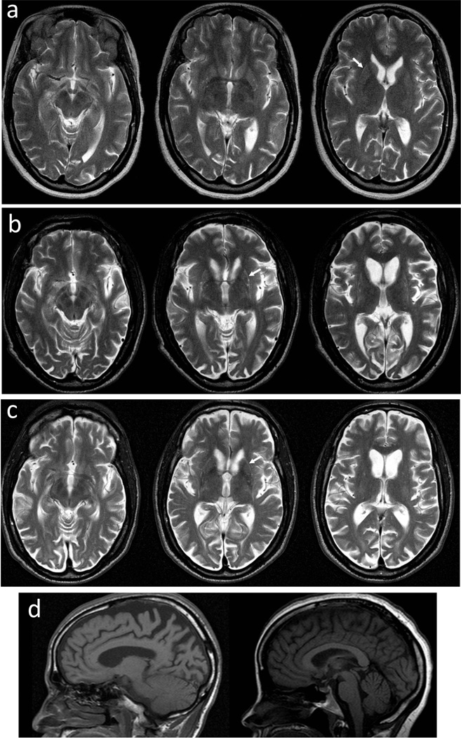
Fig. 2.
Magnetic resonance images (MRIs) of subjects III.1, III.6, and IV.2. a T2-axial images from subject IV.2 two years after disease onset. b T2-axial images from subject III.1 ten years after disease onset. c T2-axial images from subject III.6 two years after disease onset. Putaminal rim hyperintensities are marked with arrows. d Sagittal MRIs show that the brainstems and cerebellae were relatively well preserved in subjects III.6 (left) and IV.2 (right).

