Synaptic modifications in the brain store information. As we experience something new, some synapses get stronger and other synapses become weaker. The memory of this experience is encoded in the pattern of synaptic change distributed among many neurons. A central question in neurobiology concerns the mechanisms that underlie such synaptic modifications. Remarkable progress has been made by using the experimental models of homosynaptic long-term potentiation (LTP) and depression (LTD) in the CA1 region of the hippocampus. Unfortunately, there is far from universal agreement that these models actually reveal the mechanisms of memory. In the case of LTD, contradictory answers have been given to a very basic question, i.e., can LTD be induced in the adult hippocampus under behavioral conditions where learning is possible? Fortunately, a recent study published in the Proceedings (1) seems to clear up much of the confusion. LTD may have finally come of age as a candidate memory mechanism in the adult brain.
To date, the most popular model for the study of activity-dependent synaptic enhancement has been LTP in the hippocampus. Hippocampal LTP originally was induced in anesthetized rabbits with brief bursts of high-frequency synaptic stimulation (2, 3). It became a viable memory mechanism when it was subsequently demonstrated that LTP could also be induced in awake rabbits and that it could be very long lasting (4). Dissection of the molecular basis of LTP became feasible when the phenomenon was described in brain-slice preparations (5). Today, the vast majority of LTP studies are conducted in the CA1 region of hippocampal slices, which, for technical reasons, are usually prepared from young animals.
Homosynaptic LTD in the hippocampus has a different history. This type of synaptic plasticity was established first in brain slice preparations from rats (6). Although progress was rapid in dissecting the mechanisms of LTD in slices prepared from young animals, inconsistent results were soon encountered when the same types of experiments were performed in adult animals, particularly in vivo. Two camps quickly emerged: those who could reliably induce LTD in the adult rat CA1 in vivo (7–9), and those who could not (10–12).
Perhaps differences between labs are related to the behavioral state of the animals when they are prepared for study. The precedent for this idea is that LTP is inhibited in animals that have been stressed (e.g., ref. 13). Indeed, it was soon shown that exposing a rat to a stressful situation renders CA1 synapses suddenly susceptible to LTD (14–16), even when synaptic plasticity is studied ex vivo in slices prepared from the stressed animal. These results imply that one source of the differences between labs is how stressed the animals are at the time of the experiments. However, this explanation alone cannot account for the variability, because well-handled and acclimatized animals show perfectly good LTD in the labs where LTD can be induced routinely (e.g., ref. 9). Thus, although stress can clearly modulate LTD, this cannot be the whole story. This is where the recent work of Manahan-Vaughan and Braunewell (1) sheds new light.
The first important new observation is that the successful induction of LTD depends greatly on the strain of rat. In well handled, acclimatized, awake animals, LTD is produced by the standard induction protocol (1 Hz stimulation for 15 min) in Wistar, but not Hooded Lister, rats. Thus, a likely source of variability between labs is the source of animals. This finding is actually reminiscent of the early days of LTP, when the synaptic plasticity, so readily observed in anesthetized Norwegian rabbits, could not be induced in English rabbits (17).
The strain differences go a long way in explaining differences between labs, but what about the significance of LTD to memory? After all, even LTD-resistant Hooded Lister rats learn. Here’s where the second new finding comes into play. Homosynaptic depression, lasting at least 1 week, can be induced in the Hooded Lister rats when they are exposed to a novel (but nonstressful) environment. Novelty also facilitates LTD in the Wistar rats. However, when the rats are returned to the now-familiar (as assessed by the animals’ behavioral responses) environment 2 weeks later, the facilitation of LTD is lost. Thus, learning to recognize a new environment is correlated with a striking facilitation of the mechanism of homosynaptic LTD in CA1.
The facilitation of synaptic plasticity is so marked that the usual 1-Hz tetanus is no longer required to induce LTD. The low-frequency electrical stimulation normally used to monitor synaptic transmission is enough to significantly depress synaptic transmission for up to 4 hr, depending on the strain, if it is delivered during novelty exposure. Consistent with the findings of an earlier study (18), the effect of baseline electrical stimulation is larger and longer lasting if LTP is induced before the exposure of the animal to the novel environment.
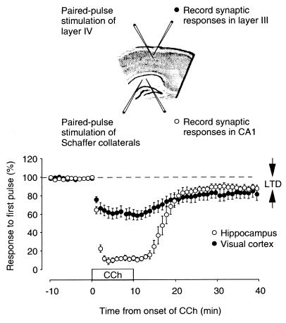
What mechanism could account for this effect of behavioral state on synaptic plasticity? Exposing a rat to a novel environment stimulates (among other things) the release of acetylcholine (ACh) in the hippocampus from fibers originating in the medial septum (19). Recent studies using hippocampal slices have shown that ACh can dramatically facilitate LTD (20) and depotentiation (21) in CA1. Like novelty, ACh can reveal LTD in response to synaptic stimulation that is normally without any lasting effect (Fig. 1). It will be of interest to see whether the effects of novelty on LTD in vivo are sensitive to manipulations of the cholinergic system.
Figure 1.
A potential mechanism for the facilitation of LTD by novelty. Manahan-Vaughan and Braunewell (1) report that exposing a rat to a novel environment produces a striking facilitation of homosynaptic LTD in vivo. Similar to the effect of novelty exposure, transient application of carbachol, an analogue of ACh, enables induction of LTD in visual cortex and hippocampus by synaptic stimulation that is normally without effect. This figure is modified, with permission, from ref. 20.
What does facilitation of LTD during memory acquisition tell us about memory mechanisms? An exciting possibility is that the pattern of electrical stimulation imposed on the brain during the novel experience is incorporated into the memory of that experience and that this memory is stored as LTD of the synapses that were active at that time. Indeed, recordings from neurons in the temporal lobes have consistently revealed that a cellular correlate of recognition memory is a diminished response to the learned stimulus (22). Perhaps this reduced response, and the memory trace, is accounted for by the mechanisms of homosynaptic LTD.
A few loose ends will need to be tied up before this idea can be taken seriously, however. First, although cellular responses to familiar stimuli are depressed in many regions of the temporal lobes, the hippocampus is, unfortunately, not usually regarded as one of them (22). Second, whereas the LTD induced during exposure to the novel environment lasted longer than 1 week, it did not persist for 2 weeks, despite the fact that at 2 weeks the animals demonstrate that they still recognize the environment as familiar. In other words, the memory outlasted the LTD in CA1.
Homosynaptic LTD is not confined to the synapses of the hippocampus; this form of synaptic plasticity is expressed widely in the neocortex (23), including the inferotemporal cortex of humans (24). And, as Fig. 1 shows, the facilitation of LTD by ACh is also widespread. An important question that needs to be addressed in future studies is whether the facilitation of LTD by novelty also occurs in the cortical regions where recognition memories are stored, and, if so, how long the LTD lasts relative to the memory trace.
If LTD is a memory mechanism, could LTP (the reversal of LTD) reflect the process of forgetting? As fun as it is to turn the tables on LTP, the leading synaptic model of memory, this simple logic seems as flawed as the converse reasoning. If memories are encoded as patterns of synaptic change, it seems that the mechanisms of both LTD and LTP have important contributions to make.
Footnotes
The companion to this Commentary begins on page 8739 in issue 15 of volume 96.
References
- 1.Manahan-Vaughan D, Braunewell K-H. Proc Natl Acad Sci USA. 1999;96:8739–8744. doi: 10.1073/pnas.96.15.8739. [DOI] [PMC free article] [PubMed] [Google Scholar]
- 2.Lømo T. Acta Physiol Scand. 1966;68:128. [Google Scholar]
- 3.Bliss T V P, Lømo T. J Physiol (London) 1973;232:331–356. doi: 10.1113/jphysiol.1973.sp010273. [DOI] [PMC free article] [PubMed] [Google Scholar]
- 4.Bliss T V, Gardner-Medwin A R. J Physiol (London) 1971;216:32P–33P. [PubMed] [Google Scholar]
- 5.Schwartzkroin P A, Wester K. Brain Res. 1975;89:107–119. doi: 10.1016/0006-8993(75)90138-9. [DOI] [PubMed] [Google Scholar]
- 6.Dudek S M, Bear M F. Proc Natl Acad Sci USA. 1992;89:4363–4367. doi: 10.1073/pnas.89.10.4363. [DOI] [PMC free article] [PubMed] [Google Scholar]
- 7.Thiels E, Barrionuevo G, Berger T W. J Neurophysiol. 1994;71:3009–3016. doi: 10.1152/jn.1994.72.6.3009. [DOI] [PubMed] [Google Scholar]
- 8.Heynen A J, Abraham W C, Bear M F. Nature (London) 1996;381:163–166. doi: 10.1038/381163a0. [DOI] [PubMed] [Google Scholar]
- 9.Manahan-Vaughan D. J Neurosci. 1997;17:3303–3311. doi: 10.1523/JNEUROSCI.17-09-03303.1997. [DOI] [PMC free article] [PubMed] [Google Scholar]
- 10.Errington M L, Bliss T V P, Richter-Levin G, Yenk K, Doyere V, Laroche S. J Neurophysiol. 1995;74:1793–1799. doi: 10.1152/jn.1995.74.4.1793. [DOI] [PubMed] [Google Scholar]
- 11.Staubli U, Scafidi J. J Neurosci. 1997;17:4820–4828. doi: 10.1523/JNEUROSCI.17-12-04820.1997. [DOI] [PMC free article] [PubMed] [Google Scholar]
- 12.Doyle C A, Cullen W K, Rowan M J, Anwyl R. Neuroscience. 1997;77:75–85. doi: 10.1016/s0306-4522(96)00427-7. [DOI] [PubMed] [Google Scholar]
- 13.Diamond D M, Rose G M. Ann NY Acad Sci. 1994;746:411–414. doi: 10.1111/j.1749-6632.1994.tb39271.x. [DOI] [PubMed] [Google Scholar]
- 14.Kim J J, Foy M R, Thompson R F. Proc Natl Acad Sci USA. 1996;93:4750–4753. doi: 10.1073/pnas.93.10.4750. [DOI] [PMC free article] [PubMed] [Google Scholar]
- 15.Xu L, Anwyl R, Rowan M J. Nature (London) 1997;387:497–500. doi: 10.1038/387497a0. [DOI] [PubMed] [Google Scholar]
- 16.Rowan M J, Anwyl R, Xu L. Mol Psychiatry. 1998;3:472–474. doi: 10.1038/sj.mp.4000429. [DOI] [PubMed] [Google Scholar]
- 17.Bliss T, Lømo T. In: Neuroscience: Exploring the Brain. Bear M F, Paradiso M A, Connors B W, editors. Baltimore: Williams & Wilkins; 1996. pp. 563–564. [Google Scholar]
- 18.Xu L, Anwyl R, Rowan M J. Nature (London) 1998;394:891–894. doi: 10.1038/29783. [DOI] [PubMed] [Google Scholar]
- 19.Aloisi A M, Casamenti F, Scali C, Pepeu G, Carli G. Brain Res. 1997;748:219–226. doi: 10.1016/s0006-8993(96)01304-2. [DOI] [PubMed] [Google Scholar]
- 20.Kirkwood A, Rozas C, Kirkwood J, Perez F, Bear M F. J Neurosci. 1999;19:1599–1609. doi: 10.1523/JNEUROSCI.19-05-01599.1999. [DOI] [PMC free article] [PubMed] [Google Scholar]
- 21.Huerta P T, Lisman J E. J Neurophysiol. 1996;75:877–884. doi: 10.1152/jn.1996.75.2.877. [DOI] [PubMed] [Google Scholar]
- 22.Xiang J Z, Brown M W. Neuropharmacology. 1998;37:657–676. doi: 10.1016/s0028-3908(98)00030-6. [DOI] [PubMed] [Google Scholar]
- 23.Bear M F. Proc Natl Acad Sci USA. 1996;93:13453–13459. doi: 10.1073/pnas.93.24.13453. [DOI] [PMC free article] [PubMed] [Google Scholar]
- 24.Chen W R, Lee S, Kato K, Spencer D D, Shepherd G M, Williamson A. Proc Natl Acad Sci USA. 1996;93:8011–8015. doi: 10.1073/pnas.93.15.8011. [DOI] [PMC free article] [PubMed] [Google Scholar]


