
Figure 4.
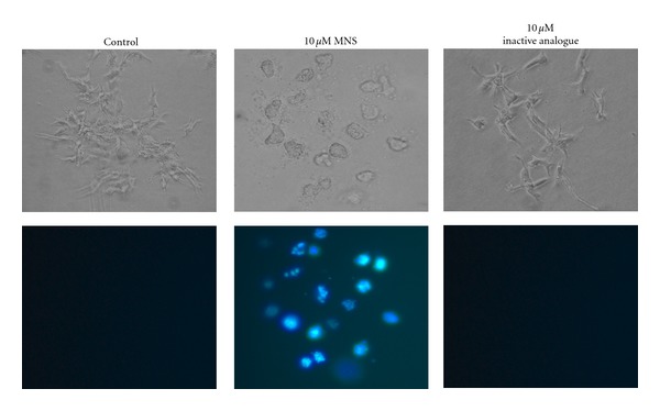
MNS disrupts preformed 143B colonies. Images of the metastatic 143B cell colonies incubated for 24 hours in presence of 10 μM MNS, 10 μM of the inactive analogue, or 1% DMSO as a vehicle control. The membrane-impermanent fluorescent stain 4′, 6-diamidino-2-phenylindole (DAPI, 0.1 μg/mL) was added 8 hours after incubation began. Fluorescent microscopy was used to obtain photomicrographs (bottom panel). This figure shows representative images from three independent experiments, each with two to three wells per group.