

Abstract
The arenavirus envelope glycoprotein (GPC) retains a stable signal peptide (SSP) as an essential subunit in the mature complex. The 58-amino-acid residue SSP comprises two membrane-spanning hydrophobic regions separated by a short ectodomain loop that interacts with the G2 fusion subunit to promote pH-dependent membrane fusion. Small-molecule compounds that target this unique SSP-G2 interaction prevent arenavirus entry and infection. The interaction between SSP and G2 is sensitive to the phylogenetic distance between New World (Junín) and Old World (Lassa) arenaviruses. For example, heterotypic GPC complexes are unable to support virion entry. In this report, we demonstrate that the hybrid GPC complexes are properly assembled, proteolytically cleaved, and transported to the cell surface but are specifically defective in their membrane fusion activity. Chimeric SSP constructs reveal that this incompatibility is localized to the first transmembrane segment of SSP (TM1). Genetic changes in TM1 also affect sensitivity to small-molecule fusion inhibitors, generating resistance in some cases and inhibitor dependence in others. Our studies suggest that interactions of SSP TM1 with the transmembrane domain of G2 may be important for GPC-mediated membrane fusion and its inhibition.
INTRODUCTION
Arenaviruses comprise a diverse family of enveloped negative-strand RNA viruses that are endemic to rodent populations worldwide. Infection can be transmitted to humans to cause severe acute hemorrhagic fevers with high morbidity and mortality. Lassa fever virus (LASV) is prevalent in western Africa, infecting a half-million persons annually (26). Five species of New World (NW) hemorrhagic fever viruses are distributed throughout South America, including the Junín virus (JUNV) in Argentina. New arenavirus species frequently emerge from rodent reservoirs (5, 9, 11). In the absence of effective vaccines or therapies, the hemorrhagic fever arenaviruses are recognized to pose significant threats to public health and biodefense. Accordingly, these viruses are classified as Category A priority pathogens, and JUNV has additionally been determined by the Department of Homeland Security to pose a Material Threat to the U.S. population.
Arenavirus entry into the host cell is mediated by the virus envelope glycoprotein (GPC) (Fig. 1). Upon binding to a cell surface receptor (reviewed in references 10 and 29), the virion is endocytosed, and GPC-mediated fusion of the viral and endosomal membranes is activated upon acidification in the maturing endosome. GPC is synthesized as a precursor glycoprotein and cleaved by the cellular SKI-1/S1P protease in the Golgi (22, 25) to generate the receptor-binding (G1) and transmembrane fusion (G2) subunits. The mature GPC complex is metastable and thus primed to mediate membrane fusion in response to acidic pH. Upon activation, GPC undergoes a series of conformational changes leading to formation of a trimer-of-hairpins structure (14, 20, 41) and fusion of the viral and cellular membranes (reviewed in references 19 and 39). The arenavirus GPC is unique among class I envelope glycoproteins in that it retains its cleaved signal peptide as a third subunit (13, 15, 47).
Fig 1.
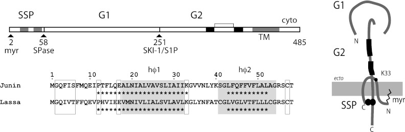
GPC open reading frame and subunit organization, and comparison of JUNV and LASV SSP amino acid sequences. (Top left) Amino acid residues are numbered according to the sequences of JUNV GPC. The mature subunits (SSP, G1, and G2) are labeled, as are the signal peptidase (SPase) and SKI-1/S1P cleavage sites. The myristoylation site at glycine-2 is labeled, and the transmembrane regions in SSP and G2 are shaded. The two heptad-repeat sequences in G2, and the hinge-region disulfide-bonded loop, are drawn in black. (Bottom left) The amino acid sequences of JUNV and LASV SSP are compared. The two hydrophobic membrane-spanning regions (hϕ1 and hϕ2) are shaded, and conserved residues referred to in the text are boxed (myristoylation motif, P12, E17, K33, and C57). Asterisks below the sequences indicate helical folds predicted by the PROFsec algorithm as implemented by PredictProtein (reliability score, ≥7) (31). (Right) Schematic model for the subunit organization of the tripartite GPC complex. SSP is shown spanning the membrane twice (2); the myristoylated N terminus is presumed to be associated with the cytoplasmic face of the membrane, and the penultimate C-terminal C57 residue participates in a binuclear zinc-finger motif in G2 (6, 43). The K33 position in the ectodomain of SSP modulates pH-dependent membrane fusion and sensitivity to small-molecule fusion inhibitors (42). The two heptad-repeat regions in the G2 ectodomain are shown in black. The relative placement of the three transmembrane regions is arbitrary and the drawing is not to scale.
The 58-amino-acid stable signal peptide (SSP) of GPC contains two hydrophobic segments that span the membrane and are joined by a short ectodomain loop (Fig. 1) (2). The cytoplasmic N terminus of SSP is myristoylated, while the penultimate C-terminal cysteine (C57) coordinates with a zinc-binding domain in the cytoplasmic tail of G2 to form an intersubunit structure that anchors SSP in the GPC complex (6, 43). SSP association masks endogenous endoplasmic reticulum (ER) retention/retrieval signals in the cytoplasmic domain of G2 to facilitate GPC transport through the Golgi (1), whereupon the precursor is proteolytically cleaved and transported to the cell surface for virion assembly.
Our studies suggest that pH-induced activation of the mature GPC complex is controlled by a unique interaction between the short ectodomain loop of SSP and the G2 fusion subunit. Side chain substitutions that reduce positive polarity at SSP K33 depress the pH required to trigger membrane fusion (46), and this phenotype can be rescued by secondary mutations in G2 (45). Importantly, this SSP-G2 interaction provides a molecular target for small-molecule compounds that stabilize the prefusion GPC complex, thereby preventing pH-induced activation in the endosome (4, 23, 24, 42). The different classes of fusion inhibitors demonstrate distinct patterns of specificity against New World (NW) and Old World (OW) arenaviruses yet share a binding site on GPC (4, 23, 37, 42). Sequence variation at the nominal SSP-G2 interface likely accounts for the differences in species specificity (37, 42). Several of these fusion inhibitors have recently been shown to protect against lethal arenavirus disease in animal models (4, 8).
Sequence variation between OW and NW arenavirus species may also affect the ability of one SSP to function in the context of a heterotypic GPC complex. For instance, recombinant JUNV virions in which SSP and the G1G2 precursor are heterotypic are not viable (3). We have exploited this interspecies incompatibility between LASV and JUNV GPCs to identify determinants in SSP required for membrane fusion activity. We found that SSP association, proteolytic maturation, and transport to the cell surface are promiscuous in interspecific hybrid GPCs and that heterotypic SSPs support these functions in the context of either JUNV or LASV G1G2 precursors. Preservation of pH-dependent membrane fusion, however, requires a specific homotypic match in the first transmembrane domain (TM1) of SSP. We propose that this amphipathic helical region of SSP interacts with the transmembrane domain of G2 and thus contributes to the pH-dependent membrane fusion activity of arenavirus GPC.
MATERIALS AND METHODS
Plasmids.
GPC from the pathogenic MC2 isolate of JUNV (17) and from the Josiah isolate of LASV (23) was expressed using the minimal T7 promoter sequence in pcDNA 3.1-based vectors (Life Technologies). In order to obviate concerns regarding signal peptidase cleavage of SSP in the chimeric GPC constructs, SSP and G1G2 open reading frames were expressed from separate plasmids, taking advantage of the ability of the two polypeptides to associate in trans to reconstitute the functional GPC complex (12, 44). The G1G2 precursor was directed to the membrane using the conventional signal peptide of human CD4, as previously described (44). An innocuous FLAG affinity tag was appended to the C terminus of LASV G1G2 to facilitate detection (47). SSP chimeras were constructed using conventional PCR procedures, and mutations were introduced using QuikChange methodology (Stratagene).
Antibodies and small-molecule entry inhibitors.
JUNV G1-specific monoclonal antibodies (MAbs) BF11 and BE08 (33) were obtained through the NIAID Biodefense and Emerging Infections Research Resources Program (BEIResources) and the FLAG peptide-specific MAb (M2) was purchased from Sigma. The LASV G1-specific MAb L52 134-23 (32) was kindly provided by Connie Schmaljohn (USAMRIID). The small-molecule fusion inhibitors ST-294, ST-193, ST-161, and ST-761 have been previously described (4, 23, 37, 42) and were kindly provided by SIGA Technologies (Corvallis, OR).
Analysis of GPC expression.
GPC was expressed by transient transfection in Vero cells infected with the vTF-7 vaccinia virus expressing T7 polymerase (16) or in engineered BHK-21 cells expressing T7 polymerase (BSR T7/5) (7), kindly provided by Klaus Conzelmann (Ludwig-Maximilians-University Munich). Proteins were metabolically labeled using 35S-labeled amino acids (Perkin Elmer) and immunoprecipitated with the appropriate MAb as previously described (44). Flow cytometric detection of cell surface GPC was hindered by low levels of expression using our pcDNA-based vectors in BSR T7/5 cells, especially for LASV GPC (see Results and Discussion). Therefore, for these studies GPC was expressed in Vero cells using a pCAGGS-MCS vector (27) provided by Juan Carlos de la Torre (Scripps Research Institute) (38). Cell surface expression was determined using the JUNV G1-specific MAb BE08 (33) or the LASV G1-specific MAb L52 134-23 (32), and a secondary fluorescein isothiocyanate (FITC)-labeled anti-mouse immunoglobulin antibody. The cell populations were stained with propidium iodine to exclude dead cells, fixed with 2% formaldehyde, and analyzed using a FACSCalibur flow cytometer (BD Biosciences).
Analysis of GPC-dependent membrane fusion.
The recombinant vaccinia virus-based assay for GPC-mediated cell-cell fusion was performed as previously described (46). Briefly, Vero cells infected by vTF-7 and expressing GPC are cocultured with cells infected with a recombinant vaccinia virus vCB21R-lacZ bearing the β-galactosidase gene under the control of the T7 promoter (28). Cell-cell fusion is triggered by exposure to medium adjusted to pH 5.0 and detected through β-galactosidase expression in the newly formed syncytia. Fusion is quantitated by chemiluminescence using the GalactoLite Plus substrate (Life Technologies). Fusion inhibition by small-molecule SIGA compounds was determined as previously described (42) and GraphPad Prism software was used for nonlinear regression calculations using a single-slope dose-response model constrained to 100% fusion in the absence of inhibitor.
In order to circumvent biosafety concerns associated with the use of vaccinia viruses, we developed an alternative fusion reporter assay based on expression of T7 polymerase in BSR T7/5 cells. In this format, GPC-expressing BSR T7/5 cells were cocultured with human 293T cells transfected with the internal ribosome entry site (IRES)-containing pT7EMC-luc reporter plasmid expressing luciferase under the control of the T7 promoter (34), which was kindly provided by Yoshiharu Matsuura (Osaka University). Following a 5-min exposure to medium adjusted to pH 5.0, the coculture was continued at neutral pH for 12 h to allow for luciferase expression. Cell-cell fusion was detected using the luciferase assay kit substrate (Promega). Consistency of GPC expression was monitored by immunohistochemical staining. Results from this novel fusion reporter assay were validated in parallel experiments using the well-established vaccinia virus-based assay.
RESULTS AND DISCUSSION
Divergence between JUNV and LASV SSPs.
A comparison of the amino acid sequences of JUNV and LASV SSPs reveals a high degree of sequence divergence, as well as an overall conservation of sequence motifs (Fig. 1). The two hydrophobic domains (hϕ1 and hϕ2) in each are separated by a short region containing the conserved K33 residue (46). A myristoylation motif and zinc-coordinating cysteine (C57) are present in both N- and C-terminal cytoplasmic domains, respectively (2). Both SSPs are also predicted to possess similar secondary structure features, with two helical regions interspersed by an unstructured ectodomain loop. Despite these similarities, fewer than 40% of the amino acids are identically conserved.
Hybrid GPC containing heterologous SSP and G1G2.
Reverse-genetics studies have shown that hybrid GPC complexes are functional for arenavirus infection if and only if SSP is homotypic with the TM and C-terminal cytoplasmic domains of G2 (3). To investigate the molecular basis for this finding, we characterized the assembly, transport, and function of hybrid GPCs. Taking advantage of the observation that SSP can associate in trans with the G1G2 precursor to reconstitute the functional GPC complex (12, 47), we coexpressed JUNV or LASV SSP with the reciprocal G1G2 precursors, which contained the conventional signal peptide of human CD4 (44, 47).
Membrane fusion activity of the homologous and heterologous hybrid GPCs was determined using a biosafe modification of the well-characterized vaccinia virus-based cell-cell fusion assay (47). BSR T7/5 cells expressing the bacteriophage T7 polymerase (7) were cotransfected with pcDNA3.1-based plasmids expressing SSP and the G1G2 precursor under the control of the T7 promoter. These cells were then cocultured with 293T fusion reporter cells expressing luciferase in a T7 polymerase-dependent manner. Cell-cell fusion was initiated by exposing the coculture to medium adjusted to pH 5.0, and luciferase expression in the newly formed syncytia was determined following continued incubation at neutral pH. Using this assay, we verified that neither of the two heterotypic GPC hybrids (JUNV SSP with LASV G1G2 or LASV SSP with JUNV G1G2) was able to mediate membrane fusion (Fig. 2A). Concordant cell-cell fusion results were obtained using the vaccinia virus-based fusion reporter assay (reference 47 and data not shown).
Fig 2.

Membrane fusion activity of JUNV and LASV hybrids and TM1 mutants. Schematic drawings of chimeric SSP constructs are shown to the right of A and B. Regions 1, 2, and 3 (R1, R2, and R3, respectively) are boxed, and JUNV or LASV sequences are indicated by black or white fills, respectively. The regions are drawn to scale, and junction amino acid residues are labeled. Cell-cell fusion by hybrid GPCs containing JUNV or LASV G1G2 and the SSP chimeras is indicated by paired black and white bars, respectively, to the right. Fusion activity was determined using BSR T7/5 cells (7) as described in Materials and Methods and normalized as a percentage of the respective wild-type JUNV or LASV GPC. The symbol ϕ is used to indicate the absence of significant fusion. Error bars represent the standard error of the mean (SEM) of 2 to 6 independent assays. (C) Mutated JUNV SSPs were expressed with JUNV G1G2 and cell-cell fusion was normalized to the wild type. One complete assay representative of at least four experiments with complete or partial collections of the mutants is depicted. Each point is an average of 8 duplicate wells, and error bars (SEM) are shown. The bar denoting S27A cell-cell fusion (284% ± 14%) is truncated at 200% of wild type.
Hybrid GPC assembles and is transported to the cell surface.
To determine the molecular basis of heterotypic incompatibility, we first investigated the ability of SSP to associate with the G1G2 precursor. It is possible that the absence of membrane fusion activity reflects an inability of SSP to bind the heterologous G1G2 precursor, thereby preventing GPC transport through the Golgi, proteolytic maturation, and cell surface expression. To assess GPC biogenesis, SSP and the G1G2 precursor were expressed in trans, and metabolically labeled GPC was immunoprecipitated (44) using MAbs directed to either JUNV G1 (33) or the C-terminal FLAG tag on LASV G2. We found that GPC protein synthesis was markedly reduced in BSR T7/5 cells relative to that typically seen in Vero cells infected with recombinant vaccinia viruses expressing T7 polymerase (16, 47), presumably reflecting the absence of mRNA capping in the cytosol of transfected BSR T7/5 cells. Nevertheless, SDS-PAGE analysis showed that SSP association in the heterotypic GPCs was similar to that in the homotypic complex (Fig. 3A). Furthermore, this association was sufficient to promote a significant degree of proteolytic maturation of the heterologous G1G2 precursor (Fig. 3A).
Fig 3.

JUNV and LASV hybrid GPCs are properly assembled, proteolytically matured, and transported to the cell surface. Combinations of JUNV and LASV SSPs and G1G2 precursor proteins were coexpressed in BSR T7/5 cells, and the metabolically labeled GPCs were immunoprecipitated for SDS-PAGE analysis (46). GPCs containing JUNV G1G2 were precipitated using the G1-directed MAb BF11 (33), and those containing LASV were collected using a FLAG tag appended to the C terminus of G1G2. (A) The immunoprecipitated proteins were resolved using a 4–12% NuPAGE Bis-Tris gel (Invitrogen). The JUNV and LASV G1G2 precursors and coprecipitated SSPs are indicated. The mature G1 and G2 glycoproteins are heterodisperse and are better resolved as polypeptides following deglycosylation using PNGaseF (New England BioLabs) (bottom). The unequal recovery of G1 and G2 subunits seen in some experiments using G1-directed (JUNV) or G2-directed FLAG (LASV) antibodies likely reflects partial dissociation of the complex during immunoprecipitation. While GPC expression in BSR T7/5 cells is adequate for membrane fusion activity, protein levels are low, and carets are used to distinguish background bands manifest in mock-transfected cells. (B) Cell surface accumulation of JUNV and LASV GPC hybrids expressed in Vero cells using a pCAGGS plasmid (27, 38) was determined by flow cytometry. GPC was detected using JUNV or LASV G1-specific MAbs (BE08 [33] or L52 134-23 [32], respectively) and a FITC-conjugated secondary antibody. Cells expressing JUNV SSP (thin line) or LASV SSP (thick line) are compared with those in which the cell surface transport of the respective G1G2 precursor is prevented by the absence of SSP (shaded histogram) (1).
Flow cytometry was used to confirm trafficking of the heterotypic GPC hybrids to the cell surface. Due to the low level of expression of LASV GPC in BSR T7/5 cells (Fig. 3A), we used a well-characterized pCAGGS plasmid vector (27) for these studies of GPC transport (38). Cell surface accumulation of heterologous GPC hybrid was found to be similar to that of the homotypic protein in both cases (Fig. 3B). As GPC transport does not require proteolytic cleavage (1, 36), these findings assess an independent function of SSP association. Conversely, transit of GPC through the Golgi, as evidenced by proteolytic cleavage, is predictive of cell surface expression. Despite substantial sequence divergence, both heterologous SSPs were capable of promoting the assembly and maturation of the hybrid GPC complex, as well as its transport to the plasma membrane. This result agrees with previous studies using a recombinant LASV GPC encoding JUNV SSP (3). Because very low levels of mature GPC are sufficient to support membrane fusion activity (2, 40), we surmise that the partial reductions in SSP association and proteolytic cleavage in the heterotypic GPC hybrids are by themselves insufficient to explain the complete loss of fusogenicity. This conclusion is reinforced by the overall lack of correlation between variations in proteolytic cleavage and membrane fusion activity in studies using chimeric SSP molecules (see below).
Interchange of the ectodomain loop in SSP.
We have previously shown that the short ectodomain loop of SSP is critical for pH-dependent membrane fusion and its inhibition by small-molecule inhibitors (42, 46). This region of JUNV SSP is defined by charged residues at the ectodomain termini of TM1 and TM2 (K33 and K40, respectively) (Fig. 1). We therefore subdivided SSP into three regions for purposes of constructing chimeras: region 1 included the myristoylated N terminus of SSP and TM1 (residues M1 to K33), region 2 comprised the ectodomain loop (K33 to K40 in JUNV, T40 in LASV), and region 3 contained TM2 and the short cytoplasmic domain bearing C57 (to the C-terminal T58). All combinations of the three JUNV and LASV subdomains were constructed (Fig. 2A), and the chimeric SSPs were named according to the three regions. For instance, JJJ represents the wild-type JUNV SSP and JLJ signifies a chimera in which the ectodomain from LASV was fused to regions 1 and 3 of JUNV SSP. For clarity, we will refer to recombinant SSPs as chimeras and reserve the term hybrid for the reconstituted GPC complex.
As anticipated from the fully heterotypic GPC hybrids (Fig. 3A), all of the chimeric SSPs associated with the JUNV G1G2 precursor and supported proteolytic cleavage in BSR T7/5 cells (Fig. 4). Parallel metabolic labeling studies using LASV GPC again showed poor expression but nonetheless allowed similar conclusions (not shown). These findings were further validated using recombinant vaccinia virus to drive GPC expression (reference 47 and data not shown). We conclude that the sequence requirements for SSP association and proteolytic maturation are relatively relaxed in interspecific GPC chimeras.
Fig 4.

JUNV GPCs containing chimeric SSPs are properly assembled and proteolytically matured. JUNV G1G2 was coexpressed with the indicated chimeric SSPs in BSR T7/5 cells, and metabolically labeled GPC was immunoprecipitated for SDS-PAGE analysis as described in Fig. 3. The G1G2 precursors and coprecipitated SSPs are indicated (top). The mature G1 and G2 glycoproteins are heterodisperse and are better resolved as polypeptides following deglycosylation using PNGaseF (New England BioLabs) (bottom).
Despite its critical role in membrane fusion, interspecific exchange of the ectodomain loop had little effect on the fusion activity of hybrid GPCs (Fig. 2A). LJL did not restore fusion activity to the JUNV G1G2 hybrid (∼3% of wild-type JUNV GPC) and likewise the reciprocal JLJ SSP in the LASV G1G2 hybrid (<1% of wild-type LASV GPC). Conversely, replacement of the ectodomain in JUNV SSP with that of LASV (JLJ) had a relatively small effect on the fusion activity of the hybrid JUNV GPC (∼40% of JUNV GPC). The reciprocal hybrid, LASV G1G2 bearing LJL SSP, retained a lower albeit significant level of activity (∼10% of LASV GPC). We conclude that a homotypic ectodomain loop in SSP is neither sufficient nor absolutely necessary to support membrane fusion by the hybrid GPC complex.
Region 1 of SSP is essential for membrane fusion activity.
Analysis of the remaining SSP chimeras did reveal an important role for the N-terminal region 1 in SSP function. Only LASV hybrids containing the homologous region 1 (LJL, LLJ, and LJJ) showed significant membrane fusion activity (Fig. 2A). Whereas the LJL hybrid supported ∼10% of wild-type activity (above), the latter two SSP chimeras promoted cell-cell fusion at levels comparable to those of native LASV GPC. The reciprocal pattern was seen with the JUNV G1G2 precursor and the JLJ, JJL, and JLL chimeras. In JUNV GPC, SSP bearing a mismatch in regions 1 and 2 (LLJ) unexpectedly also exhibited fusion activity (∼20% of the wild type). By comparison, all hybrids displayed similar patterns of GPC expression (Fig. 4). Taken together, these results indicate that homotypic pairing in SSP region 1 is paramount for membrane fusion activity. Region 2 appears to contribute somewhat to activity when the homologous region 1 is present (JJL and LLJ), whereas homology in region 3 is relatively unimportant. The reciprocal relationship between JUNV and LASV hybrids validates the importance of region 1 as a determinant of GPC-mediated membrane fusion.
The apparent indifference to sequence variation in region 3 is consistent with previous results from mutational studies (2). Triplet alanine replacements in TM2 of JUNV SSP (44FQF46 and 47FVF49 mutants) have no affect on fusogenicity. Similarly in the short C-terminal cytoplasmic tail of SSP, only the conserved C57 side chain is essential for membrane fusion activity (44). Collectively, these observations suggest a lack of sequence specificity in the function of region 3. However, the presumed helical nature of TM2 appears to be important, as SSP association is completely abrogated by single amino acid deletions that are expected to alter the register of the helix (2).
TM1 forms an extended helical domain.
To further dissect the role of region 1 in fusogenicity, we bisected the N-terminal cytoplasmic and TM regions using the conserved E17 as the nominal cytosolic junction of TM1. Thus, the (J1L2) exchange in region 1 comprised JUNV residues M1 to E17 and LASV residues E17 to K33 (Fig. 2B). SSPs including the reciprocally exchanged sequences (J1L2 and L1J2) associated with and supported proteolytic maturation of both JUNV (Fig. 4) and LASV precursors (not shown) but were entirely defective in promoting membrane fusion (Fig. 2B). In contrast, the parental SSP chimeras containing the intact region 1 (JJL and LJL) produced functional hybrids with their respective G1G2 precursors. The symmetric loss of fusogenicity at this junction is likely to reflect an internal sequence incompatibility within the SSP chimeras rather than between SSP and G2.
Secondary structure predictions suggested a possible explanation for this intramolecular incompatibility (Fig. 1). For both JUNV and LASV SSP, prediction algorithms (30) suggest that the helical structure of TM1 extends N terminally to the conserved proline at position 12. To test this notion, we generated additional region 1 exchanges in which TM1 was extended N terminally to P12. We found that an SSP chimera including the extreme N-terminal residues of LASV (residues M1 to P12) and the extended helical region of JUNV (residues P12 to K33), referred to as (LexJ)JL, supported cell-cell fusion comparably to the parental JJL chimera in the hybrid JUNV GPC (Fig. 2B). Likewise, SSPs containing the (JexL)LJ and (JexL)JL exchanges promoted detectable fusion activity in LASV G1G2 hybrids. In keeping with our previous finding that replacing E17 with alanine did not disrupt membrane fusion activity (46), we propose that TM1 spans P12 to K33 to form a functional transmembrane subdomain in SSP.
The above results also suggest that the extreme N terminus of SSP, comprising a myristoylation motif (GxxxS/T) and residues through I11, is interchangeable between JUNV and LASV. Indeed, alanine-scanning mutagenesis in this region of LCMV SSP showed only minimal effects on fusogenicity (35). The amino acid sequence of the cytoplasmic portion of region 1 appears relatively unimportant for membrane fusion activity. By contrast, the lack of myristoylation in SSP reduces fusogenicity by ∼80% through an unknown mechanism (35, 47).
Genetic analysis of the extended TM1.
Site-directed mutagenesis was used to probe the extended TM1 helix in JUNV SSP. We reasoned that deletion of a single amino acid residue would alter the register of the helix without disrupting the overall helical fold (2). A JUNV GPC mutant bearing a deletion at TM1 residue A25 was found to be devoid of membrane fusion activity (Fig. 2C), consistent with a requirement for specific interhelical interactions within the GPC complex. Replacing hydrophobic amino acids with arginines in a membrane-spanning region is expected to be highly destabilizing, and the F14R and A25R mutants were indeed defective in membrane fusion (Fig. 2C). The deficiency in F14R supports our previous suggestion that SSP enters the membrane at P12. Neither the deletion nor arginine replacement mutants associated with the G1G2 precursor (not shown). Taken together, these results point to the critical importance of the extended TM1 helix in GPC biology.
Owing to the helix-breaking property of proline, TM1 is unlikely to include residues N terminal to the conserved P12. To determine whether P12 itself is essential for GPC assembly and fusogenicity, we mutated this position in JUNV SSP to alanine. The P12A mutant was found to possess wild-type fusion activity (Fig. 2C). The identical mutant in LCMV SSP also showed significant fusion activity (∼30% of the wild type) but did not promote entry by recombinant virus-like particles (35). Thus, a proline-dependent articulation between the cytosolic N terminus and TM1 is not required for fusogenicity per se.
TM1 forms an amphipathic helix.
A helical-wheel projection diagram reveals distinct hydrophobic and hydrophilic faces to the extended TM1 helices in JUNV and LASV SSPs (Fig. 5). By contrast, TM2 of SSP (K40/T40 to G54) is uniformly hydrophobic. As TM2 was found to be highly tolerant of triplet-alanine substitutions (2), we employed a similar strategy to examine the requirement for side chain-specific interactions in TM1. We replaced three hydrophobic sequences in TM1 of JUNV SSP (13TFL15, 22ALV24, and 29IAI31) with triplet alanines, a small residue with high helical propensity. Alanine substitutions at 13TFL15 and 22ALV24 were found to disrupt membrane fusion activity (Fig. 2C). Fusogenicity was unaffected by alanine replacements at 29IAI31, probably due to its position in the helix and the conservative nature of the change. The defect in the 13TFL15 and 22ALV24 mutants strongly suggests that TM1 participates in side chain-specific interactions in the GPC complex, likely with the transmembrane helical domain of G2.
Fig 5.

Helical-wheel projections of the amphipathic TM1 of JUNV and LASV SSP. The drawing was prepared using DrawCoil 1.0 (18). This view is from the N terminus of TM1 (P12) and extends through K33 in the SSP ectodomain. Polar residues are highlighted in gray.
To probe the contributions of specific side chains in TM1 to these interactions, we individually replaced each polar residue on the hydrophilic face of TM1 with alanine (Fig. 2C). T13A, Q16A, and S27A GPCs were essentially wild type in cell-cell fusion activity, as was E17A (2). The N20A mutation, however, decreased membrane fusion activity to ∼30% of the wild type (Fig. 2C), as did the identical mutant in LCMV GPC (35). Interestingly, structural predictions by the Robetta server (http://robetta.bakerlab.org) (21) consistently position N20 at a kink in the TM1 helix (not shown).
SSP chimeras differ in sensitivity to small-molecule fusion inhibitors.
We have previously shown that both pH-dependent activation and its inhibition by small-molecule fusion inhibitors are mediated through interactions between SSP and the G2 fusion subunit (42, 45, 46). We therefore examined the sensitivity of hybrid GPCs to inhibition by the four chemically distinct fusion inhibitors discovered by SIGA Technologies (Corvallis, OR) (4, 23, 37, 42). These compounds share a binding site (37) but differ in their specificity toward NW and OW arenaviruses: ST-294 and ST-761 are active only against NW viruses, ST-161 is specific for LASV, and ST-193 is broadly inhibitory against both OW and NW arenaviruses (4, 23, 37, 42).
As hybrid GPCs heterotypic in SSP region 1 are nonfunctional, we focused our attention on the ectodomain loop, a region previously shown to affect inhibition (42). Substitution of the JUNV ectodomain loop in LASV GPCs [LJJ and (JexL)JL] abrogated inhibition by the LASV-active compounds ST-161 and ST-193 (not shown). In contrast, replacement of the ectodomain loop in JUNV GPCs (JLL, JLJ) showed no significant effects on sensitivity; all were inhibited by ST-294, ST-761, and ST-193 and resistant to ST-161 (not shown). Structural differences at the hybrid inhibitor-binding site likely contribute to the differing contributions of the heterotypic SSP ectodomain in LASV and JUNV GPC. None of the hybrids displayed de novo sensitivity to inhibition.
We utilized the panel of alanine mutations in JUNV TM1 to further identify specific side chains that may influence sensitivity to inhibition. The triplet-alanine mutant (the 29IAI31 mutant), with substitutions adjacent to the SSP ectodomain and the critical K33 residue, was found to be unchanged in its sensitivity to ST-294 and ST-193 and resistance to ST-161 (Fig. 6 and Table 1). Similarly, individual alanine mutations at Q16, E17, and S27 on the hydrophilic face of TM1 did not significantly alter the pattern of inhibition. By contrast, alanine substitutions toward the cytosolic terminus of TM1 (P12A and T13A) engendered resistance to ST-193 without qualitative or quantitative changes in the effects of ST-294, ST-761, or ST-161 (Fig. 6 and Table 1). Furthermore, the N20A mutant was now strikingly dependent on ST-193 for wild-type fusion activity. Fusion was enhanced by the addition of ST-193 in a dose-dependent manner. Maximal activity approaches that of the wild-type JUNV GPC at ∼10 μM ST-193, at which point inhibitory and/or cytotoxic effects may intervene. Sensitivities to ST-294 and ST-161 remain unaffected. The diversity in the effects of these different amino acid substitutions highlights the multiplicity of determinants for fusion inhibition within the GPC complex (42, 45).
Fig 6.

Mutations in SSP TM1 affect sensitivity to small-molecule fusion inhibitors. The highly sensitive recombinant vaccinia virus-based cell-cell fusion assay was used determine fusion inhibition by the chemically distinct small-molecule entry inhibitors ST-294 (blue), ST-193 (green), ST-161 (red), and ST-761 (cyan). The labels of mutants with altered sensitivity to ST-193 are in bold. Colored data points represent averages of triplicate 96-well microcultures using seven serial dilutions of inhibitor, and data are normalized as a percentage of the untreated control cultures. Lines are best-fit nonlinear regression curves for a dose-response model of inhibition constrained between 0 and 100% of the untreated control and using a single slope (−1) (GraphPad Prism). The line for ST-193 enhancement of fusion by N20A GPC is hand-drawn. nd, inhibition by ST-761 was not tested; wt, wild type. All conclusions reported were confirmed in at least two independent experiments. Inhibitor concentrations required to reduce membrane fusion by 50% (IC50s) are listed in Table 1.
Table 1.
JUNV GPC-mediated fusion IC50sa
| SSP | IC50 (μM) (95% CI) |
|||
|---|---|---|---|---|
| ST-294 | ST-761 | ST-193 | ST-161b | |
| wt | 1.0 (0.8–1.2) | 0.7 (0.6–1.0) | 2.9 (2.1–4.0) | 96 (57–160) |
| IAI | 0.4 (0.4–0.5) | nd | 1.5 (1.1–2.2) | 450 (67–3100) |
| P12A | 1.9 (1.6–2.3) | 1.4 (1.1–1.7) | 50 (9.5–250) | 33 (9.4–120) |
| T13A | 3.2 (2.4–4.3) | 1.4 (1.1–1.9) | 19 (12–33) | 24 (11–51) |
| Q16A | 1.7 (1.5–1.9) | 0.9 (0.8–1.2) | 4.1 (2.4–7.0) | 15 (4.4–54) |
| E17A | 1.4 (1.1–1.8) | 0.9 (0.7–1.2) | 1.0 (0.6–1.6) | 8.4 (4.1–17) |
| N20A | 3.1 (2.0–4.7) | nd | Enhanced | 140 (77–250) |
| S27A | 2.8 (1.7–4.4) | 1.3 (0.9–3.0) | 3.0 (2.3–3.9) | 71 (45–110) |
Best-fit nonlinear regression using dose-response model and data shown in Fig. 6. Significant changes in inhibitor concentrations required to reduce JUNV GPC-mediated fusion by 50% (IC50s) are indicated in bold. Enhanced, dose-dependent enhancement of fusion activity. nd, inhibition by ST-761 was not tested; wt, wild type; CI, confidence interval.
JUNV is nominally resistant (IC50 ≥ 10 μM) to ST-161.
We suggest that ST-193 binding to N20A compensates for structural changes induced by the mutation, thereby facilitating on-path conformational changes during pH-induced activation of membrane fusion. Mutations in G2 previously reported to increase the pH of activation in the K33Q mutant to wild type without themselves affecting pH sensitivity may act similarly (45). Among these compensatory G2 mutations, two (D400A and F427A) also engender resistance to ST-193 and ST-294 (reference 42 and J.Y. and J.H.N., unpublished data). Based on the dependence of the N20A mutant on the presence of inhibitor, we infer that resistance at the two G2 positions, as well as that in P12A and T13A SSP mutants, may reflect a balance of inhibitory and compensatory consequences of ST-193 binding rather than a simple loss in binding affinity.
Conclusions.
Our previous studies have shown that the short ectodomain loop of SSP and its interactions with G2 are important determinants for both pH-dependent membrane fusion and its inhibition by small-molecule compounds (42, 46). The present study identifies the critical role of the first membrane-spanning domain of SSP in these events. By characterizing a series of SSP chimeras containing JUNV and LASV sequences, we demonstrate that a homotypic pairing between TM1 and G2 is required for GPC-mediated membrane fusion. We propose that multiple intersubunit contacts between these transmembrane helices serve to position the critical K33 side chain in the SSP ectodomain for pH-sensitive interactions with the G2 ectodomain. Small-molecule compounds that stabilize these interactions in the prefusion GPC complex have been shown to prevent pH-induced fusion activation in the endosome and thereby inhibit arenavirus entry. Detailed knowledge of the atomic interactions between SSP and G2 in the membrane-anchored GPC trimer will be important for understanding the mechanism of pH-dependent membrane fusion and guiding the design of potent and broadly active small-molecule fusion inhibitors.
ACKNOWLEDGMENTS
We are grateful to Min Lu (Public Health Research Institute, University of Medicine and Dentistry, New Jersey) and Meg Trahey (The University of Montana) for their editorial comments and assistance. We are also grateful to Sean Amberg and Dongcheng Dai (SIGA Technologies, Inc., Corvallis, OR) for providing small-molecule fusion inhibitors and to Celestine Thomas (The University of Montana) for assistance in 3D modeling of SSP. We thank Connie Schmaljohn (USAMRIID) for providing the LASV G1-specific hybridoma L52 134-23, Klaus Conzelmann (Ludwig-Maximilians-University, Munich, Germany) for the BSR T7/5 cell line, and Yoshiharu Matsuura (Osaka University, Japan) and Juan Carlos de la Torre (The Scripps Research Institute, La Jolla, CA), respectively, for the pT7EMC-luc and pCAGGS-MCS expression plasmids. JUNV G1-specific MAbs were provided by the CDC through the NIH Biodefense and Emerging Infections Research Resources Program.
This work was supported by U.S. Public Health Service grants R01 AI074818 and U54 AI065357 (Rocky Mountain Regional Center of Excellence for Biodefense and Emerging Infectious Diseases at Colorado State University) from the National Institutes of Health.
Footnotes
Published ahead of print 21 March 2012
REFERENCES
- 1. Agnihothram SS, York J, Nunberg JH. 2006. Role of the stable signal peptide and cytoplasmic domain of G2 in regulating intracellular transport of the Junin virus envelope glycoprotein complex. J. Virol. 80:5189–5198 [DOI] [PMC free article] [PubMed] [Google Scholar]
- 2. Agnihothram SS, York J, Trahey M, Nunberg JH. 2007. Bitopic membrane topology of the stable signal peptide in the tripartite Junín virus GP-C envelope glycoprotein complex. J. Virol. 81:4331–4337 [DOI] [PMC free article] [PubMed] [Google Scholar]
- 3. Albariño CG, et al. 2011. Reverse genetics generation of chimeric infectious Junin/Lassa virus is dependent on interaction of homologous glycoprotein stable signal peptide and G2 cytoplasmic domains. J. Virol. 85:112–122 [DOI] [PMC free article] [PubMed] [Google Scholar]
- 4. Bolken TC, et al. 2006. Identification and characterization of potent small molecule inhibitor of hemorrhagic fever New World arenaviruses. Antiviral Res. 69:86–89 [DOI] [PMC free article] [PubMed] [Google Scholar]
- 5. Briese T, et al. 2009. Genetic detection and characterization of Lujo virus, a new hemorrhagic fever-associated arenavirus from southern Africa. PLoS Pathog. 5:e1000455. [DOI] [PMC free article] [PubMed] [Google Scholar]
- 6. Briknarova K, Thomas CJ, York J, Nunberg JH. 2011. Structure of a zinc-binding domain in the Junin virus envelope glycoprotein. J. Biol. Chem. 286:1528–1536 [DOI] [PMC free article] [PubMed] [Google Scholar]
- 7. Buchholz UJ, Finke S, Conzelmann KK. 1999. Generation of bovine respiratory syncytial virus (BRSV) from cDNA: BRSV NS2 is not essential for virus replication in tissue culture, and the human RSV leader region acts as a functional BRSV genome promoter. J. Virol. 73:251–259 [DOI] [PMC free article] [PubMed] [Google Scholar]
- 8. Cashman KA, et al. 2011. Evaluation of Lassa antiviral compound ST-193 in a guinea pig model. Antiviral Res. 90:70–79 [DOI] [PMC free article] [PubMed] [Google Scholar]
- 9. Centers for Disease Control and Prevention 2000. Fatal illnesses associated with a new world arenavirus–California, 1999–2000. MMWR Morb. Mortal. Wkly. Rep. 49:709–711 [PubMed] [Google Scholar]
- 10. Choe H, Jemielity S, Abraham J, Radoshitzky SR, Farzan M. 2011. Transferrin receptor 1 in the zoonosis and pathogenesis of New World hemorrhagic fever arenaviruses. Curr. Opin. Microbiol. 14:476–482 [DOI] [PMC free article] [PubMed] [Google Scholar]
- 11. Delgado S, et al. 2008. Chapare virus, a newly discovered arenavirus isolated from a fatal hemorrhagic fever case in Bolivia. PLoS Pathog. 4:e1000047. [DOI] [PMC free article] [PubMed] [Google Scholar]
- 12. Eichler R, et al. 2003. Identification of Lassa virus glycoprotein signal peptide as a trans-acting maturation factor. EMBO Rep. 4:1084–1088 [DOI] [PMC free article] [PubMed] [Google Scholar]
- 13. Eichler R, Lenz O, Strecker T, Garten W. 2003. Signal peptide of Lassa virus glycoprotein GP-C exhibits an unusual length. FEBS Lett. 538:203–206 [DOI] [PubMed] [Google Scholar]
- 14. Eschli B, et al. 2006. Identification of an N-terminal trimeric coiled-coil core within arenavirus glycoprotein 2 permits assignment to class I viral fusion proteins. J. Virol. 80:5897–5907 [DOI] [PMC free article] [PubMed] [Google Scholar]
- 15. Froeschke M, Basler M, Groettrup M, Dobberstein B. 2003. Long-lived signal peptide of lymphocytic choriomeningitis virus glycoprotein pGP-C. J. Biol. Chem. 278:41914–41920 [DOI] [PubMed] [Google Scholar]
- 16. Fuerst TR, Niles EG, Studier FW, Moss B. 1986. Eukaryotic transient-expression system based on recombinant vaccinia virus that synthesizes bacteriophage T7 RNA polymerase. Proc. Natl. Acad. Sci. U. S. A. 83:8122–8126 [DOI] [PMC free article] [PubMed] [Google Scholar]
- 17. Ghiringhelli PD, Rivera-Pomar RV, Lozano ME, Grau O, Romanowski V. 1991. Molecular organization of Junin virus S RNA: complete nucleotide sequence, relationship with other members of the Arenaviridae and unusual secondary structures. J. Gen. Virol. 72:2129–2141 [DOI] [PubMed] [Google Scholar]
- 18. Grigoryan G. 2011. DrawCoil 1.0. arteni.cs.dartmouth.edu/drawcoil/
- 19. Harrison SC. 2008. Viral membrane fusion. Nat. Struct. Mol. Biol. 15:690–698 [DOI] [PMC free article] [PubMed] [Google Scholar]
- 20. Igonet S, et al. 2011. X-ray structure of the arenavirus glycoprotein GP2 in its postfusion hairpin conformation. Proc. Natl. Acad. Sci. U. S. A. 108. [DOI] [PMC free article] [PubMed] [Google Scholar]
- 21. Kim DE, Chivian D, Baker D. 2004. Protein structure prediction and analysis using the Robetta server. Nucleic Acids Res. 32:W526–W531 [DOI] [PMC free article] [PubMed] [Google Scholar]
- 22. Kunz S, Edelmann KH, de la Torre J-C, Gorney R, Oldstone MBA. 2003. Mechanisms for lymphocytic choriomeningitis virus glycoprotein cleavage, transport, and incorporation into virions. Virology 314:168–178 [DOI] [PubMed] [Google Scholar]
- 23. Larson RA, et al. 2008. Identification of a broad-spectrum arenavirus entry inhibitor. J. Virol. 82:10768–10775 [DOI] [PMC free article] [PubMed] [Google Scholar]
- 24. Lee AM, et al. 2008. Unique small molecule entry inhibitors of hemorrhagic fever arenaviruses. J. Biol. Chem. 283:18734–18742 [DOI] [PMC free article] [PubMed] [Google Scholar]
- 25. Lenz O, ter Meulen J, Klenk H-D, Seidah NG, Garten W. 2001. The Lassa virus glycoprotein precursor GP-C is proteolytically processed by subtilase SKI-1/S1P. Proc. Natl. Acad. Sci. U. S. A. 98:12701–12705 [DOI] [PMC free article] [PubMed] [Google Scholar]
- 26. McCormick JB, Webb PA, Krebs JW, Johnson KM, Smith ES. 1987. A prospective study of the epidemiology and ecology of Lassa fever. J. Infect. Dis. 155:437–444 [DOI] [PubMed] [Google Scholar]
- 27. Niwa H, Yamamura K, Miyazaki J. 1991. Efficient selection for high-expression transfectants with a novel eukaryotic vector. Gene 108:193–199 [DOI] [PubMed] [Google Scholar]
- 28. Nussbaum O, Broder CC, Berger EA. 1994. Fusogenic mechanisms of enveloped-virus glycoproteins analyzed by a novel recombinant vaccinia virus-based assay quantitating cell fusion-dependent reporter gene activation. J. Virol. 68:5411–5422 [DOI] [PMC free article] [PubMed] [Google Scholar]
- 29. Rojek JM, Kunz S. 2008. Cell entry by human pathogenic arenaviruses. Cell. Microbiol. 10:828–835 [DOI] [PubMed] [Google Scholar]
- 30. Rost B, Sander C. 1993. Prediction of protein secondary structure at better than 70% accuracy. J. Mol. Biol. 232:584–599 [DOI] [PubMed] [Google Scholar]
- 31. Rost B, Yachdav G, Liu J. 2004. The PredictProtein server. Nucleic Acids Res. 32:W321–W326 [DOI] [PMC free article] [PubMed] [Google Scholar]
- 32. Ruo SL, et al. 1991. Antigenic relatedness between arenaviruses defined at the epitope level by monoclonal antibodies. J. Gen. Virol. 72:549–555 [DOI] [PubMed] [Google Scholar]
- 33. Sanchez A, Pifat DY, Kenyon RHPCJ, McCormick JB, Kiley MP. 1989. Junin virus monoclonal antibodies: characterization and cross-reactivity with other arenaviruses. J. Gen. Virol. 70:1125–1132 [DOI] [PubMed] [Google Scholar]
- 34. Satoh T, et al. 2008. PILRalpha is a herpes simplex virus-1 entry coreceptor that associates with glycoprotein B. Cell 132:935–944 [DOI] [PMC free article] [PubMed] [Google Scholar]
- 35. Saunders AA, et al. 2007. Mapping the landscape of the lymphocytic choriomeningitis virus stable signal peptide reveals novel functional domains. J. Virol. 81:5649–5657 [DOI] [PMC free article] [PubMed] [Google Scholar]
- 36. Schlie K, Strecker T, Garten W. 2010. Maturation cleavage within the ectodomain of Lassa virus glycoprotein relies on stabilization by the cytoplasmic tail. FEBS Lett. 584:4379–4382 [DOI] [PubMed] [Google Scholar]
- 37. Thomas CJ, et al. 2011. A specific interaction of small-molecule entry inhibitors with the envelope glycoprotein complex of the Junín hemorrhagic fever arenavirus. J. Biol. Chem. 286:6192–6200 [DOI] [PMC free article] [PubMed] [Google Scholar]
- 38. Urata S, et al. 2011. Antiviral activity of a small-molecule inhibitor of arenavirus glycoprotein processing by the cellular site 1 protease. J. Virol. 85:795–803 [DOI] [PMC free article] [PubMed] [Google Scholar]
- 39. White JM, Delos SE, Brecher M, Schornberg K. 2008. Structures and mechanisms of viral membrane fusion proteins: multiple variations on a common theme. Crit. Rev. Biochem. Mol. Biol. 43:189–219 [DOI] [PMC free article] [PubMed] [Google Scholar]
- 40. York J, Agnihothram SS, Romanowski V, Nunberg JH. 2005. Genetic analysis of heptad-repeat regions in the G2 fusion subunit of the Junin arenavirus envelope glycoprotein. Virology 343:267–279 [DOI] [PMC free article] [PubMed] [Google Scholar]
- 41. York J, et al. 2010. An antibody directed against the fusion peptide of Junin virus envelope glycoprotein GPC inhibits pH-induced membrane fusion. J. Virol. 84:6119–6129 [DOI] [PMC free article] [PubMed] [Google Scholar]
- 42. York J, Dai D, Amberg SA, Nunberg JH. 2008. pH-induced activation of arenavirus membrane fusion is antagonized by small-molecule inhibitors. J. Virol. 82:10932–10939 [DOI] [PMC free article] [PubMed] [Google Scholar]
- 43. York J, Nunberg JH. 2007. A novel zinc-binding domain is essential for formation of the functional Junín virus envelope glycoprotein complex. J. Virol. 81:13385–13391 [DOI] [PMC free article] [PubMed] [Google Scholar]
- 44. York J, Nunberg JH. 2007. Distinct requirements for signal peptidase processing and function of the stable signal peptide (SSP) subunit in the Junin virus envelope glycoprotein. Virology 359:72–81 [DOI] [PubMed] [Google Scholar]
- 45. York J, Nunberg JH. 2009. Intersubunit interactions modulate pH-induced activation of membrane fusion by the Junin virus envelope glycoprotein GPC. J. Virol. 83:4121–4126 [DOI] [PMC free article] [PubMed] [Google Scholar]
- 46. York J, Nunberg JH. 2006. Role of the stable signal peptide of the Junín arenavirus envelope glycoprotein in pH-dependent membrane fusion. J. Virol. 80:7775–7780 [DOI] [PMC free article] [PubMed] [Google Scholar]
- 47. York J, Romanowski V, Lu M, Nunberg JH. 2004. The signal peptide of the Junín arenavirus envelope glycoprotein is myristoylated and forms an essential subunit of the mature G1–G2 complex. J. Virol. 78:10783–10792 [DOI] [PMC free article] [PubMed] [Google Scholar]