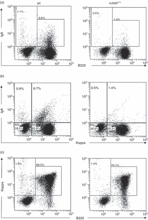
Figure 5.

Expression of IgA in primary cells from Peyer's patches. Peyer's patches from αΔtail+/+ and wild-type (wt) mice were collected and cell suspensions were treated in saponin for intracellular staining with indicated surfaces markers and analysed by flow cytometry. Dot plot showed expression of mIgA+ B220+ B cells (a), κ light chain+ mIgA+ cells (b) and κ+ B220+ B cells (c) in Peyer's patches from αΔtail+/+ mice by comparison with wt.
