ABSTRACT
Spirochaetes is one of a few bacterial phyla that are characterized by a unifying diagnostic feature, namely, the helical morphology and motility conferred by axial periplasmic flagella. Their unique morphology and mode of propulsion also represent major pathogenicity factors of clinical spirochetes. Here we describe the genome sequences of two coccoid isolates of the recently described genus Sphaerochaeta which are members of the phylum Spirochaetes based on 16S rRNA gene and whole-genome phylogenies. Interestingly, the Sphaerochaeta genomes completely lack the motility and associated signal transduction genes present in all sequenced spirochete genomes. Additional analyses revealed that the lack of flagella is associated with a unique, nonrigid cell wall structure hallmarked by a lack of transpeptidase and transglycosylase genes, which is also unprecedented in spirochetes. The Sphaerochaeta genomes are highly enriched in fermentation and carbohydrate metabolism genes relative to other spirochetes, indicating a fermentative lifestyle. Remarkably, most of the enriched genes appear to have been acquired from nonspirochetes, particularly clostridia, in several massive horizontal gene transfer events (>40% of the total number of genes in each genome). Such a high level of direct interphylum genetic exchange is extremely rare among mesophilic organisms and has important implications for the assembly of the prokaryotic tree of life.
IMPORTANCE
Spiral shape and motility historically have been the unifying hallmarks of the phylum Spirochaetes. These features also represent important virulence factors of highly invasive pathogenic spirochetes such as the causative agents of syphilis and Lyme disease. Through the integration of genome sequencing, microscopy, and physiological studies, we conclusively show that the spiral morphology and motility of spirochetes are not universal morphological properties. In particular, we found that the genomes of the members of the recently described genus Sphaerochaeta lack the genes encoding the characteristic flagellar apparatus and, in contrast to most other spirochetes, have acquired many metabolic and fermentation genes from clostridia. These findings have major implications for the isolation and study of spirochetes, the diagnosis of spirochete-caused diseases, and the reconstruction of the evolutionary history of this important bacterial phylum. The Sphaerochaeta sp. genomes offer new avenues to link ecophysiology with the functionality and evolution of the spirochete flagellar apparatus.
Introduction
Spirochaetes is a diverse, deeply branching phylum of Gram-negative bacteria. Members of this phylum share distinctive morphological features, i.e., a spiral shape and axial, periplasmic flagella (1, 2). These traits enable propulsion through highly viscous media and thus are directly associated with the ecological niches spirochetes occupy. For instance, motility mediated by axial flagella represents a major pathogenicity factor that allows strains of the Treponema, Borrelia, and Leptospira genera to invade and colonize host tissues, resulting in important diseases such as Lyme disease and syphilis. Several studies have shown that disruption of the flagellar genes or the chemotaxis genes that control the periplasmic flagella attenuates the pathogenic potential of spirochetes (3–5).
The focus on clinical isolates has biased our understanding of the ecology, physiology, and diversity of the phylum Spirochaetes. Indeed, free-living, nonpathogenic spirochetes are greatly underrepresented in culture collections, while culture-independent studies have revealed that spirochetes are ubiquitous in anoxic environments, implying that they are key players in anaerobic food webs (6–9). Consistent with the latter findings, studies of members of the genus Spirochaeta have demonstrated that environmental isolates possess physiological properties distinct from those of their pathogenic relatives; e.g., they encode a diverse set of saccharolytic enzymes (7), while other members of the genus are alkaliphiles (10) and thermophiles (11). More recently, screening of environmental samples revealed a novel genus of free-living spirochetes, Sphaerochaeta (12). Phylogenetic analysis of 16S rRNA genes identified this group as a member of the phylum Spirochaetes, most closely related to the genus Spirochaeta. Interestingly, Sphaerochaeta pleomorpha strain Grapes and Sphaerochaeta globosa strain Buddy are nonmotile and show the same spherical morphology during laboratory cultivation (12). However, it remains unclear whether this unusual morphology and the lack of motility represent a distinct stage of the cell cycle and/or responses to culture conditions or if these distinguishing features have a genetic basis.
To elucidate the metabolic properties and evolutionary history of environmental, nonpathogenic spirochetes and to provide insights into the unusual morphological features of Sphaerochaeta, we sequenced the genomes of strain Grapes and strain Buddy, which represent the type strains of S. pleomorpha and S. globosa, respectively. Our analyses of the two complete genome sequences suggest that the members of the genus Sphaerochaeta are unique spirochetes that completely lack the genes for the motility apparatus and have acquired nearly half of their genomes from Gram-positive bacteria, an extremely rare event among mesophilic organisms.
RESULTS
Phylogenetic affiliation.
The S. pleomorpha strain Grapes and S. globosa strain Buddy genomes contain about 3,200 and 3,000 putative protein coding sequences and have average G+C contents of 46 and 49% and sizes of 3.5 and 3.2 Mbp, respectively (see Table S1 in the supplemental material). The two genomes share about 1,850 orthologous genes (i.e., 57 to 61% of the total number of genes in the genome, depending on the reference genome), and these genes show, on average, 65% amino acid identity. Therefore, the two genomes represent two divergent species of the genus Sphaerochaeta according to current taxonomic standards (13).
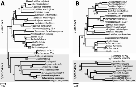
Phylogenetic analysis of the concatenated alignment of 43 highly conserved, single-copy informational genes (see Table S2 in the supplemental material), which showed no obvious horizontal gene transfer (HGT) signal when their individual trees were assessed against the 16S rRNA gene tree, corroborated previous 16S rRNA gene-based findings (12). The genus Sphaerochaeta represents a distinct lineage of the phylum Spirochaetes most closely related to members of the genus Spirochaeta, e.g., Spirochaeta coccoides and Spirochaeta smaragdinae (Fig. 1). The average amino acid identity between S. smaragdinae and S. pleomorpha or S. globosa was 46% (based on 900 shared orthologous genes). This level of genomic relatedness is typically observed between organisms of different families, if not orders (14); hence, Sphaerochaeta and Spirochaeta represent distantly related genera of the phylum Spirochaetes. Other spirochetal genomes had fewer orthologous genes in common with Sphaerochaeta (i.e., 300 to 500), and these genes showed lower levels of amino acid identity than those of S. smaragdinae (e.g., 30 to 45%). No obvious inter- or intraphylum HGT of any of the 43 informational genes was observed when the phylogenetic analysis was expanded to include selected genomes of Proteobacteria and Gram-positive bacteria (see below).
FIG 1 .
Phylogenetic affiliation of S. globosa and S. pleomorpha. Neighbor-joining phylogenetic trees of Sphaerochaeta and selected bacterial species based on 16S rRNA gene sequences (A) and the concatenated alignment of 43 single-copy informational gene sequences (B) are shown. Values at the nodes represent bootstrap support from 1,000 replicates. The scale bar represents the number of nucleotide (A) or amino acid (B) substitutions per site.
Motility and chemotaxis.
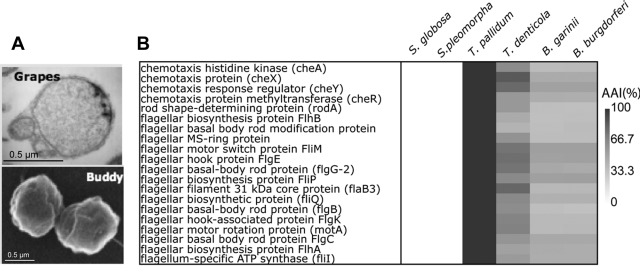
Typical spirochetal flagella are composed of about 30 different proteins (15), and about a dozen additional regulatory and sensory proteins have been demonstrated to interact directly with flagellar proteins, such as the methyl-accepting chemotaxis proteins encoded by the che operon (1). To determine whether or not the Sphaerochaeta genomes possess motility genes, we queried the protein sequences of the Treponema pallidum flagellar and chemotaxis genes against the S. pleomorpha and S. globosa genome sequences (tBLASTn). Although the T. pallidum sequences had clear orthologs in all available spirochetal genomes, none of the motility or chemotaxis genes were present in the S. pleomorpha or S. globosa genome (Fig. 2B). Incomplete sequencing, assembly errors, or low sequence similarity did not present a plausible explanation for these results since the flagellar genes are typically located in three distinct, large gene clusters, each 20 to 30 kbp long, and it is not likely that such clusters were missed in genome sequencing and annotation. Consistent with these interpretations, all of the informational genes encoding ribosomal proteins and RNA and DNA polymerases were recovered in the assembled genome sequences. These results were consistent with previous microscopic observations and corroborated the finding that the spherical morphology characteristic of Sphaerochaeta is related to the absence of axial flagella (12).
FIG 2 .
Absence of flagellar and chemotaxis genes from Sphaerochaeta genomes. (A) Transmission electron micrograph showing the nonspiral shape of S. globosa strain Buddy and S. pleomorpha strain Grapes cells. (B) Heat map showing the presence or absence and the level of amino acid identity (see scale) of T. pallidum chemotaxis, flagellar assembly, and locomotion gene homologs in selected spirochetal genomes.
A unique cell wall structure.
Our analyses revealed additional features of Sphaerochaeta that are unusual among spirochetes and Gram-negative bacteria in general and are probably linked to the lack of axial flagella. Both Sphaerochaeta genomes contain all of the genes required for peptidoglycan biosynthesis, and electron microscopy verified the presence of a cell wall in growing cells (12); however, the genomes lack genes for penicillin-binding proteins (PBP). PBP catalyze the formation of linear glycan chains (transglycosylation) during cell elongation and the transpeptidation of murein glycan chains (see Table S3 in the supplemental material), which confers rigidity on the cell wall (16, 17). Consequently, Sphaerochaeta spp. are resistant to β-lactam antibiotics (ampicillin at up to 250 µg/ml, which was the highest concentration tested). In Gram-negative bacteria without antibiotic resistance mechanisms, including clinical spirochetes, β-lactam antibiotics block PBP functionality, resulting in cell lysis. Often, β-lactam-treated, cell wall-deficient cells can be maintained in isotonic growth media as so-called L forms with characteristic spherical morphologies (18–20). While Sphaerochaeta sp. cells occur in spherical morphologies (Fig. 2A), they possess a cell wall, grow in defined hypertonic and hypotonic media without the addition of osmotic stabilizers (12), and are not L forms. It is conceivable that a rigid cell wall is required for anchoring of the axial flagella. Thus, the absence of both axial flagella and PBP genes presumably explains the atypical spirochete morphology of the members of the genus Sphaerochaeta. The loss of the flagella and PBP genes likely occurred in the ancestor of Sphaerochaeta, since both members of the genus lack these genes.
Extensive gene acquisition from Gram-positive bacteria.
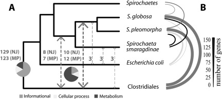
Searching of all Sphaerochaeta protein sequences against the nonredundant (NR) protein database of GenBank revealed that ~700 of the protein-encoding genes had best matches to genes of members of the order Clostridiales (phylum Firmicutes), ~700 had best matches to genes of members of the phylum Spirochaetes, and ~100 had best matches to genes of members of the class Bacilli (see Fig. S1 in the supplemental material). Consistent with the best-match results, S. pleomorpha and S. globosa exclusively shared more unique genes with clostridia than with other members of the phylum Spirochaetes (~110 versus ~70 genes, respectively). Both species exclusively had a substantial number of unique genes in common with Bacilli (phylum Firmicutes, 25 genes) and Escherichia (Gammaproteobacteria, 60 and 10 genes for S. pleomorpha and S. globosa, respectively) (Fig. 3B). Functional analysis based on the COG database showed that the spirochete-like genes of Sphaerochaeta were associated mostly with informational categories, e.g., transcription and translation, whereas the clostridium-like genes were highly enriched in metabolic functions, e.g., carbohydrate and amino acid metabolism and transport (see Fig. S1 and S2 in the supplemental material). Several of the carbohydrate and amino acid metabolism genes, such as the multidomain glutamate synthase (SpiBuddy_0108-0113) and genes related to polysaccharide biosynthesis (SpiBuddy_0254-0259), were found in large gene clusters, indicating that their acquisition likely occurred in single HGT events. Interestingly, many of the clostridium-like genes had high sequence identity to their clostridial homologs (>70% amino acid identity), even though these genes did not encode informational proteins (e.g., ribosomal proteins and RNA/DNA polymerases). While informational genes tend to show high levels of sequence conservation, much lower sequence conservation was expected for (vertically inherited) metabolic genes shared across phyla, revealing that some of the genetic exchange events between Sphaerochaeta and Clostridiales occurred recently relative to the divergence of the Spirochaetes and Firmicutes phyla.
FIG 3 .
HGT between Sphaerochaeta spp. and Clostridiales. The cladogram depicts the 16S rRNA gene phylogeny. Arrows connecting branches represent cases of HGT (A); the values next to the arrows indicate the numbers of genes exchanged (out of a total of 178 genes examined). Pie charts show the distribution of the genes in major COG functional categories (the key at the bottom shows the category designations by color). Orthologous genes shared exclusively by Sphaerochaeta and other taxa are graphically represented by arcs in panel B. The thickness of each arc is proportional to the number of genes shared (see scale bar).
Homology-based (best-hit) bioinformatic analyses are inherently prone to artifacts, including uneven numbers of representative genomes in the database, disparate G+C contents, different rates of evolution, multidomain proteins, and gene loss (21, 22). To provide further insights into the genome fluidity of Sphaerochaeta and the interphylum HGT events, we performed a detailed phylogenetic analysis of 223 orthologous proteins that had at least one homologous sequence in each of the taxa evaluated (i.e., Sphaerochaeta spp., S. smaragdinae, other members of the phylum Spirochaetes, Escherichia coli, and Clostridiales). We evaluated genetic exchange events based on embedded quartet decomposition analysis (23) by using both the maximum-parsimony (MP) and neighbor-joining (NJ) methods and 178 trees with at least 50% bootstrap support in all branches. The gene set contributing to the trees was biased toward informational functions; hence, it was not surprising that the most frequent topology obtained (123 trees [MP] and 129 trees [NJ]) was congruent with the 16S rRNA gene-based topology, denoting no interphylum genetic exchange. Nonetheless, the analysis also provided trees with topologies consistent with genetic exchange between Clostridiales and Sphaerochaeta and identified 19 (MP) and 18 (NJ) genes (i.e., ~10% of the total number of trees evaluated) that were most likely subject to interphylum HGT. This gene set was enriched in genes encoding metabolic functions, e.g., carbohydrate metabolism (Fig. 3A). About half of the 19 trees identified by MP analysis were consistent with genetic exchange between Clostridiales and the ancestor of both S. smaragdinae and Sphaerochaeta, while the other trees were consistent with exchange between the ancestor of Clostridiales and Sphaerochaeta (more recent events; Fig. 3). The phylogenetic distribution of the genes exchanged between Clostridiales and Sphaerochaeta in other spirochetes and Gram-positive bacteria (e.g., see Fig. S3 in the supplemental material) suggested that members of the order Clostridiales were predominantly the donors (>95% of the genes examined) in these genetic exchange events (unidirectional HGT). These findings corroborated those of the best-match analysis and collectively revealed that, with the exception of informational genes, interphylum HGT and gene loss (e.g., flagellar genes) have shaped more than half of the Sphaerochaeta genomes through evolutionary time.
How unique is the case of Sphaerochaeta-Clostridiales gene transfer?
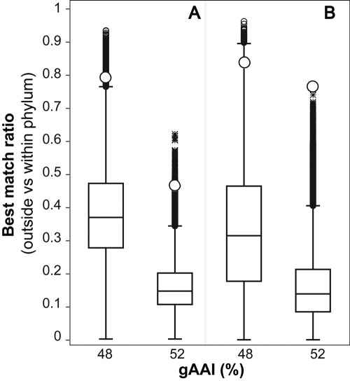
We evaluated how frequently a high level of interphylum gene transfer such as that observed between Clostridiales and Sphaerochaeta genomes occurs within the prokaryotic domain. To this end, the ratio of the number of genes of a reference genome with best matches in a genome of a different phylum versus the number of genes of the reference genome with best matches to a genome of a member of the same phylum was determined. To account for differences in the coverage of phyla with sequenced representatives, the analysis was performed using three genomes at a time (two of the same phylum and one of a different phylum). Further, only genomes of the same phylum that showed genetic relatedness among them, measured by the genome-aggregate average amino acid identity, or gAAI (14), similar to that between Sphaerochaeta and selected Spirochaetes genomes, i.e., Leptospira (48% gAAI) and Treponema (52% gAAI) genomes, were compared. This strategy sidesteps the limitation that the number of genes common to any two genomes depends on the genetic relatedness between the genomes (see Fig. S4 in the supplemental material) (24) and thus can affect estimates of the number of best-match genes and HGT. The sets compared represented 12 different bacterial and 3 archaeal phyla and 308 and 249 different genomes (150,022 and 86,516 unique 3-genome sets) for the 48% and 52% gAAI set comparisons, respectively. The analysis revealed that the extent of genetic exchange between Sphaerochaeta and Clostridiales is highly uncommon relative to that which occurs among other genomes, i.e., the upper 99.74th and 99.99th percentiles for the 48% and 52% gAAI sets, respectively. Similar results were obtained when all of the genes in the genome or only the genes common to the three genomes, which were enriched in conserved housekeeping functions, were evaluated (Fig. 4). Most of the clostridium-like genes in Sphaerochaeta genomes had best matches within a phylogenetically narrow group of clostridia that included fermenters such as Clostridium saccharolyticum and Clostridium phytofermentans, which are associated with anaerobic organic matter decomposition (25), and species such as Eubacterium rectale (26) and Butyrivibrio proteoclasticus (27), which are associated with the animal gut.
FIG 4 .
Comparisons of the extents of interphylum HGT. The ratio of the number of genes of a reference genome with best BLASTP matches in a genome of a different phylum relative to a genome of the same phylum as the reference genome was determined in three-genome comparisons (sets) as described in the text. The graph shows the distribution of the ratios for 150,022 and 86,516 comparisons that included genomes of the same phylum showing ~48% and ~52% gAAI, respectively; the distributions were based on all of the genes common to the three genomes in a comparison (A) and all of the genes in the reference genome (B). Horizontal bars represent the median, the upper and lower box boundaries represent the upper and lower quartiles, and the upper and lower whiskers represent the 99th percentile. Open circles represent the values for the Sphaerochaeta-Clostridiales case.
Metabolic properties of Sphaerochaeta.
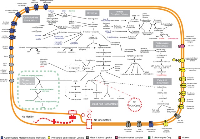
Metabolic genome reconstruction revealed that most of the central metabolic pathways were common to S. pleomorpha and S. globosa (Fig. 5). The complete glycolytic and pentose phosphate pathways were present in both genomes. Only a few genes for the tricarboxylic acid (TCA) cycle, such as those for citrate lyase, 2-oxoglutarate oxidoreductase, and succinate dehydrogenase, were found, suggesting an incomplete TCA cycle. A recent study of Synechococcus sp. strain PCC 7002, a photosynthetic cyanobacterium, identified missing cyanobacterial TCA cycle functions among the uncharacterized genes of this genome. Two proteins, encoded by the SynPCC7002_A2770 and SynPCC7002_A2771 genes, were reported to carry out the (previously) missing functions of 2-oxoglutarate decarboxylase and succinic semialdehyde dehydrogenase, respectively (28). Searches for homology between these two genes and the Sphaerochaeta genomes detected only one homolog, that of SynPCC7002_A2770, with 56% amino acid identity. These results indicate that the missing functions of the TCA cycle in Sphaerochaeta might be found among the uncharacterized genes of the genome.
FIG 5 .
Overview of the metabolic pathways encoded by the S. pleomorpha and S. globosa genomes. Shown are the primary energy generation pathways, diversity of carbohydrate metabolism pathways, biosynthesis genes for amino acids and fatty acids, and cell wall features encoded by both genomes. Pathways not found in the genomes, such as those encoding flagellar and two-component signal transduction systems related to motility, are in red. The substrates and pathways found exclusively in S. pleomorpha are in green. Transporters related to carbohydrate metabolism (in blue), metal ion transport and metabolism (in gray), and phosphate and nitrogen uptake (in yellow) are also shown.
Another important feature of the two genomes was the absence of key components of respiratory electron transport chains such as c-type cytochromes and the ubiquinol-cytochrome c reductase (cytochrome bc1 complex), corroborating physiological test findings that Sphaerochaeta spp. do not respire. Instead, cellular energy conservation (ATP, reducing power) in Sphaerochaeta relies on fermentation, a feature common to several other spirochetes lacking respiratory functions, including members of the Spirochaeta, Borrelia, and Treponema genera (29). In Sphaerochaeta, homofermentation of lactate and mixed-acid fermentation appear to be the dominant fermentation pathways, producing lactate, acetate, formate, ethanol, H2, and CO2, consistent with physiological observations. A few genes possibly related to alternative means of energy generation were also present in the genomes and included the rnf and nqr redox complexes. The rnf and nqr complexes export protons and/or ions (e.g., Na+) by coupling the flow of electrons from a reduced ferredoxin to NAD+ (30). This transmembrane potential can be used by V-type ATPases (e.g., SpiGrapes_0737-0742) for ATP synthesis or energize ion-dependent transporters for the uptake of sugars or amino acids.
The two Sphaerochaeta genomes also encode an assortment of transport proteins for the uptake and utilization of oligo- and monosaccharides. Genes involved in carbohydrate metabolism and amino acid transport and metabolism are also overrepresented relative to those in other spirochete genomes. In contrast, genes involved in signal transduction, intracellular trafficking, motility, posttranscriptional modification, and cell wall and membrane biogenesis are underrepresented in Sphaerochaeta genomes (see Fig. S2 in the supplemental material). Consistent with an anaerobic lifestyle (6, 9), several genes related to oxidative stress and protection from reactive oxygen species were found in the Sphaerochaeta genomes. Genes encoding alkyl hydroperoxide reductase, superoxide dismutase, manganese superoxide dismutase, glutaredoxin, peroxidase, and catalase indicate that Sphaerochaeta spp. are adapted to environments with oxidative stress fluctuations. The genome analysis provided no evidence for the formation of selenocysteine.
Each Sphaerochaeta genome contains about 850 species-specific genes (~25% of the genome), the majority of which have unknown or poorly characterized functions (see Fig. S5 in the supplemental material). Nevertheless, our analyses identified a few genes or pathways that can functionally differentiate the two Sphaerochaeta species and might have implications for the habitat distribution of each species. For example, S. pleomorpha-specific genes were enriched in sugar metabolism and energy production functions, including genes for trehalose and maltose utilization and the complete (TCA cycle-independent) fermentation pathway for citrate utilization (31) (green genes in Fig. 5). Further, the genome of S. pleomorpha uniquely contains several genes involved in cell wall and capsule formation, such as those for phosphoheptose isomerase (capsular heptose biosynthesis) and anhydro-N-acetylmuramic acid kinase (peptidoglycan recycling) (32). These findings revealed that S. pleomorpha has a potential for capsule formation and can use a wider range of carbohydrates than S. globosa, which are both consistent with previously reported experimental observations (12). Almost all of the S. globosa-specific genes have unknown or poorly characterized functions.
Bioinformatic predictions in deeply branching organisms.
Sphaerochaeta spp. probably represent a new family or even an order within the phylum Spirochaetes based on their divergent genomes and unique morphological and phylogenetic features. Bioinformatic functional predictions, particularly for such deeply branching organisms, are often limited by weak sequence similarity and/or uncertainty about the actual function of homologous genes or pathways. Nonetheless, bioinformatic analysis remains a powerful tool for hypothesis generation, as well as for understanding of the phenotypic differences among organisms. For the genus Sphaerochaeta, experimental evidence confirmed all of our bioinformatic predictions. For instance, we have confirmed experimentally (12) the predictions regarding the resistance of Sphaerochaeta to β-lactam antibiotics (based on the lack of PBP), utilization of various oligo- and monosaccharides, an unusual cell wall structure, absence of motility, and tolerance to oxygen. These results revealed that bioinformatic-analysis-based inferences about the metabolism and physiology of deep-branching organisms such as those in the genus Sphaerochaeta can be robust and reliable.
Sphaerochaeta and reductive dechlorination.
Sphaerochaeta spp. commonly co-occur with obligate organohalide respirers of the genus Dehalococcoides (9, 12). The reasons for this association are unclear, but it may have important practical implications for the bioremediation of chloro-organic pollutants. The Sphaerochaeta genomes have provided some clues and led to new hypotheses with respect to the potential interactions between free-living, nonmotile Sphaerochaeta spp. and Dehalococcoides dechlorinators. For instance, it was previously hypothesized that Sphaerochaeta may provide a corrinoid to dechlorinators, an essential cofactor for reductive dechlorination activity (33). However, genome analyses revealed that Sphaerochaeta genomes encode only the cobalamin salvage pathway, which is not in agreement with the corrinoid hypothesis. Alternative intriguing hypotheses include the possibility that the fermentation carried out by Sphaerochaeta provides essential substrates (e.g., acetate and H2) to Dehalococcoides or that Sphaerochaeta spp. help to protect highly redox-sensitive Dehalococcoides cells from oxidants (i.e., oxygen) (34).
DISCUSSION
Genomic analyses revealed the absence of motility genes, the underrepresentation of sensing/regulatory genes (Fig. 2; see Fig. S2 in the supplemental material), and the unusual lack of transpeptidase and transglycosylase genes involved in cell wall formation and explained the resistance of Sphaerochaeta spp. to β-lactam antibiotics and their unusual cell morphology. These findings demonstrate that a spiral shape and motility are not attributes shared by all of the members of the phylum Spirochaetes, breaking with the prevalent dogma in spirochete biology that “spirochetes are one of the few major bacterial groups whose natural phylogenetic relationships are evident at the level of phenotypic characteristics” (35). The reasons for the loss of motility genes in the members of the genus Sphaerochaeta are not clear, but the lack of transpeptidase activity (i.e., loss of cell wall rigidity) may have been associated with the loss of axial flagella. Cell wall rigidity is presumably necessary for anchoring of the two ends of the axial flagellum; hence, permanent loss of cell wall rigidity is likely detrimental to the proper functioning of an axial flagellum. It is also possible that habitats such as anoxic sediments enriched in organic matter and/or characterized by a constant influx of nutrients do not select for motility (36, 37) and favor the loss of genes encoding the motility apparatus; Sphaerochaeta spp. were obtained from such habitats (12).
Their unusual nonrigid cell wall structure likely imposes additional challenges to the maintenance of cell integrity by Sphaerochaeta organisms. A cellular adaptation to maintain membrane integrity, possibly accounting for the lack of a rigid cell wall, is the tight regulation of intracellular osmotic potential. Several genes encoding the biosynthesis of osmoregulating periplasmic glucans, osmoprotectant ABC transporters, an uptake system for betaine and choline, and potassium homeostasis were found in the genomes of S. globosa and S. pleomorpha, suggesting fine-tuned responses to osmotic stressors. The importance of these findings for explaining Sphaerochaeta sp. survival and ecological success in the environment remains to be experimentally verified.
The loss of motility genes imposes new challenges for the identification of nonmotile spirochetes in environmental or clinical samples. Free-living spirochetes are isolated routinely by selective enrichment for spiral motility, using specialized filters and/or solidified media, and by taking advantage of the unique spiral morphology, mode of propulsion, and natural rifampin resistance of spirochetes (38). Therefore, traditional isolation methods have failed to recognize and have likely underestimated the abundance and distribution of nonmotile spirochetes. New isolation procedures should be adopted to expand our understanding of the ecology and diversity of this clinically and environmentally important bacterial phylum. The genome sequences reported here will greatly assist such efforts; for instance, they have revealed that Sphaerochaeta spp. are naturally resistant to β-lactam antibiotics. The Sphaerochaeta genomes also provide a long-needed negative control (i.e., lack of axial flagella) to launch new investigations into the flagellum-mediated infection process of spirochetes causing life-threatening diseases. Further, the recently determined genome sequence of Spirochaeta coccoides (accession number CP002659) also lacks the flagellum, chemotaxis, and PBP genes and is more closely related to Sphaerochaeta than to other members of the genus Spirochaeta (e.g., S. smaragdinae). These findings indicate that, to date, nonspiral cell morphology is phylogenetically restricted to the closely related genera Spirochaeta and Sphaerochaeta within the phylum Spirochaetes and that S. coccoides may justifiably be considered a member of the genus Sphaerochaeta.
Our analyses revealed that more than 10% of the core genes and presumably more than 50% of the auxiliary and secondary metabolism genes of Sphaerochaeta were acquired from Gram-positive members of the phylum Firmicutes. The extensive unidirectional HGT (i.e., Clostridiales to Sphaerochaeta) implied that the two taxa (or their ancestors) have an ecological niche(s) and/or physiological properties in common. Consistent with these interpretations, ecological overlap between Clostridiales and both host-associated and free-living spirochetes was observed previously. For instance, several genes related to carbohydrate metabolism in Brachyspira hyodysenteriae, an anaerobic, commensal spirochete, appear to have been acquired from co-occurring members of the genera Escherichia and Clostridium in the porcine large intestine (29). Among free-living spirochetes, ecological overlap is likely to occur within anaerobic food webs where spirochetes and clostridia coexist (36, 39). For example, the biomass yields of and rates of cellulose degradation by Clostridium thermocellum increase when it is grown in coculture with Spirochaeta caldaria (40). In agreement with these studies, the genes transferred between Sphaerochaeta and Clostridiales were heavily biased toward carbohydrate uptake and fermentative metabolism functions. A more comprehensive phylogenetic analysis that included 35 spirochetal and clostridial genomes (see Table S1 in the supplemental material) indicated that Sphaerochaeta acquired several, but not all, of its clostridium-like genes from the ancestor of the anaerobic cellulolytic bacterium C. phytofermentans (see Fig. S3 in the supplemental material), which was also consistent with the BLASTP-based results of the three-genome comparisons.
Such a high level of interphylum genetic exchange is extremely rare among mesophilic organisms like Sphaerochaeta (Fig. 4) (41). This level of HGT has been reported previously only for thermophilic Thermotoga spp. (i.e., organisms living under extreme environmental selection pressures) (42). On the other hand, we did not observe HGT that affected informational proteins such as ribosomal proteins and DNA/RNA polymerases, suggesting that the reconstruction of spirochetal phylogenetic relationships, and in general, the construction of the bacterial tree of life, can be attained even in cases of extensive genetic exchange of metabolic genes (for a contrasting opinion, see reference 43). In the case of Sphaerochaeta, the massive HGT was apparently favored by an ecological niche overlap with Clostridiales and/or strong functional interactions within anoxic environments. The altered, nonrigid cell wall structure of Sphaerochaeta might have played a role in the high level of genetic exchange observed, e.g., by facilitating DNA transfer across the cell wall, although experimental evidence for this hypothesis is lacking. These findings highlight the importance of both ecology and environment in determining the rates and magnitudes of HGT. The acquisition of quantitative insights into the role of the environment and shared ecological niches in HGT will lead to a more educated assembly of the prokaryotic tree of life based on measurable and quantifiable properties.
MATERIALS AND METHODS
Organisms used in this study.
The genome sequence of each Sphaerochaeta species used in this study is shown in Table S1 in the supplemental material. The accession numbers of the genomes are CP003155 (S. pleomorpha) and CP002541 (S. globosa). Details regarding the conditions used to isolate type species are available elsewhere (12).
Sequence analysis and metabolic reconstruction.
Orthologous proteins between Sphaerochaeta and selected publicly available genomes were identified by a reciprocal best-match approach and a minimum cutoff for a match of 70% coverage of the query sequence and 30% amino acid identity, as described previously (44). For phylogenetic analysis, sequence alignments were constructed using the ClustalW software (45) and trees were built using the NJ algorithm as implemented in the MEGA4 package (46). Central metabolic pathways were reconstructed using Pathway Tools version 14 (47). The annotation files required as the input to Pathway Tools were prepared from the consensus results of two approaches. First, amino acid sequences of predicted proteins were annotated based on their best BLAST matches against the NR (48), KEGG (49), and COG (50) databases. Second, the whole-genome sequences were submitted to the RAST annotation pipeline (51) to ensure that the previous approach did not miss any important genes and to assign protein sequences to functions and enzymatic reactions (EC numbers). The results of both approaches were used to extract gene names and EC numbers. Disagreements between the two approaches were resolved by manual curation.
HGT analysis.
For best-match analysis, strain Buddy protein sequences were searched for using BLASTP against two databases, (i) all completed prokaryotic genomes available in January 2011 (n = 1,445) and (ii) the NR database (release 178). The best match for each query sequence, with better than 70% coverage of the length of the query protein and 30% amino acid identity, was identified, and the taxonomic affiliation of the genome containing the best match was extracted from the NCBI taxonomy browser. HGT events were identified as follows. Orthologous protein sequences present in at least one representative genome from the five groups used (i.e., Sphaerochaeta, S. smaragdinae, other spirochetes, Clostridiales, and E. coli) were identified and aligned as described above. Phylogenetic trees for each alignment were built in PHYLIP v3.6 (J. Felsenstein, University of Washington, Seattle, WA [http://evolution.genetics.Washington.edu/phylip.html]) by using both the MP and NJ algorithms and bootstrapped 100 times using Seqboot. The topology of the resulting consensus tree was compared to the 16S rRNA gene-based tree topology, and conflicting nodes between the two trees which also had bootstrap support higher than 50 were identified as cases of HGT.
To evaluate how unique the case of interphylum gene transfer between and Sphaerochaeta is, the following approach was used. All of the available completed bacterial and archaeal genomes (as of January 2011, n = 1,445) that showed genetic relatedness among them similar to the relatedness among the Sphaerochaeta genomes (i.e., 65% ± 0.5% gAAI) were assigned to the same group. All protein-coding genes common to the genomes of different groups were subsequently determined by using the BLASTP algorithm as described above. The BLASTP results were analyzed by using sets of three genomes at a time, each genome representing one of three distinct groups, (i) a reference group, (ii) a group from the same phylum as the reference group, or (iii) a group from another phylum. The ratio of the number of genes of the reference genome with best matches in the genome of the different phylum versus the number of genes in the reference genome with best matches to the genome of the same phylum was determined for each set and plotted against the gAAI between the reference genome and the genome of the same phylum (Fig. 4). Groups of genomes with fewer than 40 genes in common were removed from further analysis to reduce noisy results from very distantly related or small genomes.
SUPPLEMENTAL MATERIAL
Distribution of best BLAST matches of S. globosa protein sequences. Best-match analysis against all of the publicly available complete genomes revealed that the S. globosa genome has as many best matches in Clostridiales (clostridium-like genes) as in Spirochaetes (spirochete-like genes) (A). The histograms show that the spirochete-like genes are enriched in informational functions, while the clostridium-like genes are enriched in metabolic functions (based on assignment of genes to the COG database) (B). Arrows in panel B highlight the high levels of identity of several clostridium-like metabolic genes (>70% amino acid identity). Download Figure S1, DOCX file, 0.1 MB.
Functional characterization of selected spirochetal and clostridial genomes based on the COG database. All genes found in the genomes were assigned to the COG database, and the graph shows the relative abundance of COG categories in each genome. Arrows mark the relative enrichment of genes for carbohydrate and amino acid metabolism in the S. smaragdinae, S. globosa, and S. pleomorpha genomes. Download Figure S2, DOCX file, 0.2 MB.
Phylogenetic analysis of genes exchanged between the ancestors of Sphaerochaeta spp. and C. phytofermentans. NJ trees of four horizontally exchanged genes are shown. Values next to the branches denote the bootstrap support from 1,000 replicates. Genes include phosphoribosyl-amino-imidazole-succino-carboxamide synthase (A), amidophosphoribosyl transferase (B), arginine biosynthesis bifunctional protein (C), and N-acetyl-gamma-glutamyl-phosphate reductase (D). The genes in panels A and B are found in an operon and are possibly expressed as a single cistronic mRNA by the S. globosa and S. pleomorpha genomes. Similarly, the genes in panels C and D are part of an operon. Download Figure S3, DOCX file, 0.2 MB.
Correlation between shared genes and genetic relatedness for 1,445 completed bacterial and archaeal genomes. All-versus-all comparisons of the complete genomes available (as of January 2011) were performed, and the graph shows the fraction of the total number of genes common to a pair of genomes (y axis) plotted against the gAAI between the two genomes of the pair (x axis). Note that there is a significant correlation between the two parameters. To avoid the influence of genetic relatedness on the number of genes in common, and thus on the estimations of HGT, we focused on genomes with similar gAAI values. The boxed region represents the 64 to 66% range, which approximated the 65% gAAI value of the Sphaerochaeta genomes and was used to define the intragroup diversity in the three-group comparisons of HGT (Fig. 4). Download Figure S4, DOCX file, 0.1 MB.
Functional comparisons of the S. globosa and S. pleomorpha genomes. Numbers of shared and strain-specific genes in the S. globosa and S. pleomorpha genomes are shown in a Venn diagram (A). Distributions of homologous but nonorthologous (i.e., not reciprocal best matches) (B) and strain-specific (C) genes in COG functional categories are also shown. The distributions were significant for strain-specific genes (P < 0.05, Student one-tailed t test) but not significant for homologous, nonorthologous genes. Download Figure S5, DOCX file, 0.1 MB.
Bacterial genomes used in the analysis of HGT.
List of the 43 informational genes used in the genome phylogeny shown in Fig. 1B.
Sphaerochaeta genomes lack several widely conserved genes encoding PBP.
ACKNOWLEDGMENTS
We thank the DOE Joint Genome Institute, particularly the people associated with the Community Sequencing Program, for sequencing and assembling the Sphaerochaeta genomes.
This work was supported by the National Science Foundation under grant 0919251.
Footnotes
Citation Caro-Quintero A, Ritalahti KM, Cusick KD, Löffler FE, Konstantinidis KT. 2012. The chimeric genome of Sphaerochaeta: nonspiral spirochetes that break with the prevalent dogma in spirochete biology. mBio 3(3):e00025-12. doi:10.1128/mBio.00025-12.
REFERENCES
- 1. Charon NW, Goldstein SF. 2002. Genetics of motility and chemotaxis of a fascinating group of bacteria: the Spirochetes. Annu. Rev. Genet. 36:47–73 [DOI] [PubMed] [Google Scholar]
- 2. Paster BJ, Dewhirst FE. 2000. Phylogenetic foundation of Spirochetes. J. Mol. Microbiol. Biotechnol. 2:341–344 [PubMed] [Google Scholar]
- 3. Lux R, Miller JN, Park NH, Shi W. 2001. Motility and chemotaxis in tissue penetration of oral epithelial cell layers by Treponema denticola. Infect. Immun. 69:6276–6283 [DOI] [PMC free article] [PubMed] [Google Scholar]
- 4. Rosey EL, Kennedy MJ, Yancey RJ., Jr. 1996. Dual flaA1 flaB1 mutant of Serpulina hyodysenteriae expressing periplasmic flagella is severely attenuated in a murine model of swine dysentery. Infect. Immun. 64:4154–4162 [DOI] [PMC free article] [PubMed] [Google Scholar]
- 5. Sadziene A, Thomas DD, Bundoc VG, Holt SC, Barbour AG. 1991. A flagella-less mutant of Borrelia burgdorferi. Structural, molecular, and in vitro functional characterization. J. Clin. Invest. 88:82–92 [DOI] [PMC free article] [PubMed] [Google Scholar]
- 6. Franzmann PD, Dobson SJ. 1992. Cell wall-less, free-living spirochetes in Antarctica. FEMS Microbiol. Lett. 76:289–292 [DOI] [PubMed] [Google Scholar]
- 7. Leschine S, Paster B, Canale-Parola E. 2006. Free-living saccharolytic spirochetes: the genus “Spirochaeta,” p 195–210 In The Prokaryotes. Springer Verlag, New York, NY. [Google Scholar]
- 8. Magot M, et al. 1997. Spirochaeta smaragdinae sp. nov., a new mesophilic strictly anaerobic spirochete from an oil field. FEMS Microbiol. Lett. 155:185–191 [DOI] [PubMed] [Google Scholar]
- 9. Ritalahti KM, Löffler FE. 2004. Populations implicated in anaerobic reductive dechlorination of 1,2-dichloropropane in highly enriched bacterial communities. Appl. Environ. Microbiol. 70:4088–4095 [DOI] [PMC free article] [PubMed] [Google Scholar]
- 10. Zhilina TN, et al. 1996. Spirochaeta alkalica sp. nov., Spirochaeta africana sp. nov., and Spirochaeta asiatica sp. nov., alkaliphilic anaerobes from the continental soda lakes in Central Asia and the East African rift. Int. J. Syst. Bacteriol. 46:305–312 [DOI] [PubMed] [Google Scholar]
- 11. Janssen PH, Morgan HW. 1992. Glucose catabolism by Spirochaeta thermophila RI 19.B1. J. Bacteriol. 174:2449–2453 [DOI] [PMC free article] [PubMed] [Google Scholar]
- 12. Ritalahti KM, et al. 2012. Isolation of Sphaerochaeta (gen. nov.), free-living, spherical spirochetes, and characterization of Sphaerochaeta pleomorpha (sp. nov.) and Sphaerochaeta globosa (sp. nov.). Int. J. Syst. Evol. Microbiol. 62:210–216 [DOI] [PubMed] [Google Scholar]
- 13. Goris J, et al. 2007. DNA-DNA hybridization values and their relationship to whole-genome sequence similarities. Int. J. Syst. Evol. Microbiol. 57:81–91 [DOI] [PubMed] [Google Scholar]
- 14. Konstantinidis KT, Tiedje JM. 2005. Towards a genome-based taxonomy for prokaryotes. J. Bacteriol. 187:6258–6264 [DOI] [PMC free article] [PubMed] [Google Scholar]
- 15. Liu R, Ochman H. 2007. Stepwise formation of the bacterial flagellar system. Proc. Natl. Acad. Sci. U. S. A. 104:7116–7121 [DOI] [PMC free article] [PubMed] [Google Scholar]
- 16. Matteï PJ, Neves D, Dessen A. 2010. Bridging cell wall biosynthesis and bacterial morphogenesis. Curr. Opin. Struct. Biol. 20:749–755 [DOI] [PubMed] [Google Scholar]
- 17. Sauvage E, Kerff F, Terrak M, Ayala JA, Charlier P. 2008. The penicillin-binding proteins: structure and role in peptidoglycan biosynthesis. FEMS Microbiol. Rev. 32:234–258 [DOI] [PubMed] [Google Scholar]
- 18. Allan EJ, Hoischen C, Gumpert J. 2009. Bacterial L-forms. Adv. Appl. Microbiol. 68:1–39 [DOI] [PubMed] [Google Scholar]
- 19. Mursic VP, et al. 1996. Formation and cultivation of Borrelia burgdorferi spheroplast-L-form variants. Infection 24:218–226 [DOI] [PubMed] [Google Scholar]
- 20. Wise EM, Jr, Park JT. 1965. Penicillin: its basic site of action as an inhibitor of a peptide cross-linking reaction in cell wall mucopeptide synthesis. Proc. Natl. Acad. Sci. U. S. A. 54:75–81 [DOI] [PMC free article] [PubMed] [Google Scholar]
- 21. Eisen JA. 2000. Horizontal gene transfer among microbial genomes: new insights from complete genome analysis. Curr. Opin. Genet. Dev. 10:606–611 [DOI] [PubMed] [Google Scholar]
- 22. Koski LB, Golding GB. 2001. The closest BLAST hit is often not the nearest neighbor. J. Mol. Evol. 52:540–542 [DOI] [PubMed] [Google Scholar]
- 23. Zhaxybayeva O, Gogarten JP, Charlebois RL, Doolittle WF, Papke RT. 2006. Phylogenetic analyses of cyanobacterial genomes: quantification of horizontal gene transfer events. Genome Res. 16:1099–1108 [DOI] [PMC free article] [PubMed] [Google Scholar]
- 24. Konstantinidis KT, Tiedje JM. 2005. Genomic insights that advance the species definition for prokaryotes. Proc. Natl. Acad. Sci. U. S. A. 102:2567–2572 [DOI] [PMC free article] [PubMed] [Google Scholar]
- 25. Warnick TA, Methé BA, Leschine SB. 2002. Clostridium phytofermentans sp. nov., a cellulolytic mesophile from forest soil. Int. J. Syst. Evol. Microbiol. 52:1155–1160 [DOI] [PubMed] [Google Scholar]
- 26. Mahowald MA, et al. 2009. Characterizing a model human gut microbiota composed of members of its two dominant bacterial phyla. Proc. Natl. Acad. Sci. U. S. A. 106:5859–5864 [DOI] [PMC free article] [PubMed] [Google Scholar]
- 27. Moon CD, et al. 2008. Reclassification of Clostridium proteoclasticum as Butyrivibrio proteoclasticus comb. nov., a butyrate-producing ruminal bacterium. Int. J. Syst. Evol. Microbiol. 58:2041–2045 [DOI] [PubMed] [Google Scholar]
- 28. Zhang S, Bryant DA. 2011. The tricarboxylic acid cycle in cyanobacteria. Science 334:1551–1553 [DOI] [PubMed] [Google Scholar]
- 29. Bellgard MI, et al. 2009. Genome sequence of the pathogenic intestinal spirochete Brachyspira hyodysenteriae reveals adaptations to its lifestyle in the porcine large intestine. PLoS One 4:e4641 [DOI] [PMC free article] [PubMed] [Google Scholar]
- 30. Biegel E, Schmidt S, González JM, Müller V. 2011. Biochemistry, evolution and physiological function of the Rnf complex, a novel ion-motive electron transport complex in prokaryotes. Cell. Mol. Life Sci. 68:613–634 [DOI] [PMC free article] [PubMed] [Google Scholar]
- 31. Bott M. 1997. Anaerobic citrate metabolism and its regulation in enterobacteria. Arch. Microbiol. 167:78–88 [PubMed] [Google Scholar]
- 32. Uehara T, et al. 2005. Recycling of the anhydro-N-acetylmuramic acid derived from cell wall murein involves a two-step conversion to N-acetylglucosamine-phosphate. J. Bacteriol. 187:3643–3649 [DOI] [PMC free article] [PubMed] [Google Scholar]
- 33. He J, Holmes VF, Lee PK, Alvarez-Cohen L. 2007. Influence of vitamin B12 and cocultures on the growth of Dehalococcoides isolates in defined medium. Appl. Environ. Microbiol. 73:2847–2853 [DOI] [PMC free article] [PubMed] [Google Scholar]
- 34. Morris JJ, Kirkegaard R, Szul MJ, Johnson ZI, Zinser ER. 2008. Facilitation of robust growth of Prochlorococcus colonies and dilute liquid cultures by “helper” heterotrophic bacteria. Appl. Environ. Microbiol. 74:4530–4534 [DOI] [PMC free article] [PubMed] [Google Scholar]
- 35. Paster BJ, et al. 1991. Phylogenetic analysis of the spirochetes. J. Bacteriol. 173:6101–6109 [DOI] [PMC free article] [PubMed] [Google Scholar]
- 36. Canale-Parola E. 1978. Motility and chemotaxis of spirochetes. Annu. Rev. Microbiol. 32:69–99 [DOI] [PubMed] [Google Scholar]
- 37. Kimsey RB, Spielman A. 1990. Motility of lyme disease spirochetes in fluids as viscous as the extracellular matrix. J. Infect. Dis. 162:1205–1208 [DOI] [PubMed] [Google Scholar]
- 38. Breznak JA, Canale-Parola E. 1975. Morphology and physiology of Spirochaeta aurantia strains isolated from aquatic habitats. Arch. Microbiol. 105:1–12 [DOI] [PubMed] [Google Scholar]
- 39. Harwood CS, Canale-Parola E. 1984. Ecology of spirochetes. Annu. Rev. Microbiol. 38:161–192 [DOI] [PubMed] [Google Scholar]
- 40. Leschine SB. 1995. Cellulose degradation in anaerobic environments. Annu. Rev. Microbiol. 49:399–426 [DOI] [PubMed] [Google Scholar]
- 41. Beiko RG, Harlow TJ, Ragan MA. 2005. Highways of gene sharing in prokaryotes. Proc. Natl. Acad. Sci. U. S. A. 102:14332–14337 [DOI] [PMC free article] [PubMed] [Google Scholar]
- 42. Zhaxybayeva O, et al. 2009. On the chimeric nature, thermophilic origin, and phylogenetic placement of the Thermotogales. Proc. Natl. Acad. Sci. U. S. A. 106:5865–5870 [DOI] [PMC free article] [PubMed] [Google Scholar]
- 43. Doolittle WF, Bapteste E. 2007. Pattern pluralism and the tree of life hypothesis. Proc. Natl. Acad. Sci. U. S. A. 104:2043–2049 [DOI] [PMC free article] [PubMed] [Google Scholar]
- 44. Konstantinidis KT, et al. 2009. Comparative systems biology across an evolutionary gradient within the Shewanella genus. Proc. Natl. Acad. Sci. U. S. A. 106:15909–15914 [DOI] [PMC free article] [PubMed] [Google Scholar]
- 45. Thompson JD, Gibson TJ, Higgins DG. 2002. Multiple sequence alignment using ClustalW and ClustalX. Curr. Protoc. Bioinformatics Chapter 2:Unit 2.3 [DOI] [PubMed] [Google Scholar]
- 46. Tamura K, Dudley J, Nei M, Kumar S. 2007. MEGA4: molecular evolutionary genetics analysis (MEGA) software version 4.0. Mol. Biol. Evol. 24:1596–1599 [DOI] [PubMed] [Google Scholar]
- 47. Paley SM, Karp PD. 2006. The pathway tools cellular overview diagram and omics viewer. Nucleic Acids Res. 34:3771–3778 [DOI] [PMC free article] [PubMed] [Google Scholar]
- 48. Benson DA, Karsch-Mizrachi I, Lipman DJ, Ostell J, Wheeler DL. 2007. GenBank. Nucleic Acids Res. 35:D21–D25 [DOI] [PMC free article] [PubMed] [Google Scholar]
- 49. Moriya Y, Itoh M, Okuda S, Yoshizawa AC, Kanehisa M. 2007. KAAS: an automatic genome annotation and pathway reconstruction server. Nucleic Acids Res. 35:W182–W185 [DOI] [PMC free article] [PubMed] [Google Scholar]
- 50. Tatusov R, et al. 2003. The COG database: an updated version includes eukaryotes. BMC Bioinform. 4:41 [DOI] [PMC free article] [PubMed] [Google Scholar]
- 51. Aziz RK, et al. 2008. The RAST server: rapid annotations using subsystems technology. BMC Genomics 9:75 [DOI] [PMC free article] [PubMed] [Google Scholar]
Associated Data
This section collects any data citations, data availability statements, or supplementary materials included in this article.
Supplementary Materials
Distribution of best BLAST matches of S. globosa protein sequences. Best-match analysis against all of the publicly available complete genomes revealed that the S. globosa genome has as many best matches in Clostridiales (clostridium-like genes) as in Spirochaetes (spirochete-like genes) (A). The histograms show that the spirochete-like genes are enriched in informational functions, while the clostridium-like genes are enriched in metabolic functions (based on assignment of genes to the COG database) (B). Arrows in panel B highlight the high levels of identity of several clostridium-like metabolic genes (>70% amino acid identity). Download Figure S1, DOCX file, 0.1 MB.
Functional characterization of selected spirochetal and clostridial genomes based on the COG database. All genes found in the genomes were assigned to the COG database, and the graph shows the relative abundance of COG categories in each genome. Arrows mark the relative enrichment of genes for carbohydrate and amino acid metabolism in the S. smaragdinae, S. globosa, and S. pleomorpha genomes. Download Figure S2, DOCX file, 0.2 MB.
Phylogenetic analysis of genes exchanged between the ancestors of Sphaerochaeta spp. and C. phytofermentans. NJ trees of four horizontally exchanged genes are shown. Values next to the branches denote the bootstrap support from 1,000 replicates. Genes include phosphoribosyl-amino-imidazole-succino-carboxamide synthase (A), amidophosphoribosyl transferase (B), arginine biosynthesis bifunctional protein (C), and N-acetyl-gamma-glutamyl-phosphate reductase (D). The genes in panels A and B are found in an operon and are possibly expressed as a single cistronic mRNA by the S. globosa and S. pleomorpha genomes. Similarly, the genes in panels C and D are part of an operon. Download Figure S3, DOCX file, 0.2 MB.
Correlation between shared genes and genetic relatedness for 1,445 completed bacterial and archaeal genomes. All-versus-all comparisons of the complete genomes available (as of January 2011) were performed, and the graph shows the fraction of the total number of genes common to a pair of genomes (y axis) plotted against the gAAI between the two genomes of the pair (x axis). Note that there is a significant correlation between the two parameters. To avoid the influence of genetic relatedness on the number of genes in common, and thus on the estimations of HGT, we focused on genomes with similar gAAI values. The boxed region represents the 64 to 66% range, which approximated the 65% gAAI value of the Sphaerochaeta genomes and was used to define the intragroup diversity in the three-group comparisons of HGT (Fig. 4). Download Figure S4, DOCX file, 0.1 MB.
Functional comparisons of the S. globosa and S. pleomorpha genomes. Numbers of shared and strain-specific genes in the S. globosa and S. pleomorpha genomes are shown in a Venn diagram (A). Distributions of homologous but nonorthologous (i.e., not reciprocal best matches) (B) and strain-specific (C) genes in COG functional categories are also shown. The distributions were significant for strain-specific genes (P < 0.05, Student one-tailed t test) but not significant for homologous, nonorthologous genes. Download Figure S5, DOCX file, 0.1 MB.
Bacterial genomes used in the analysis of HGT.
List of the 43 informational genes used in the genome phylogeny shown in Fig. 1B.
Sphaerochaeta genomes lack several widely conserved genes encoding PBP.


