Abstract
Background
Reduced muscle strength is a cardinal feature in cachexia. We investigated whether weight loss is associated differently with muscle strength in men and women in a large cohort of hospitalized patients.
Methods
One thousand five hundred hospitalized patients (whereof 718 men, mean age 57.6 ± 16.0 years, mean body mass index (BMI) 24.6 ± 4.8 kg/m²) were included in the study. Non-edematous involuntary weight loss was determined with Subjective Global Assessment; isometric maximal muscle strength was evaluated by hand grip strength. Mid-upper arm circumference and triceps skinfold were used to calculate arm muscle area. Interrelationship between sex and weight loss was evaluated by regression analysis performed with the general linear model (GLM) allowing adjustment for continuous and categorical variables and corrected for age, arm muscle area (AMA), BMI, and diagnosis category (benign/malignant disease) as potentially confounding covariates.
Results
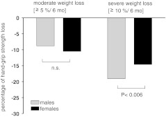
Both men and women exhibited a significant stepwise decrease of hand grip strength with increasing weight loss. Age, sex, moderate and severe weight loss, BMI, and AMA were significant predictors of hand grip strength. The GLM moreover revealed a significant sex × weight loss effect, since grip strength was similarly decreased in moderate weight loss in men and women when compared to control patients without weight loss (8.5% in men and 10.5% in women, not significant (n.s.)), but the further reduction of grip strength in severe weight loss was significantly different between men and women (10.6% vs. 4.1%, P = 0.033).
Conclusions
Our findings indicate sex-specific differences in muscle strength response to weight loss.
Keywords: Weight loss, Muscle strength, Sexual dimorphism
Introduction
Involuntary weight loss or cachexia is frequently observed in chronic disease with a reported prevalence between 5% and 80% depending on clinical population [1, 2]. Loss of muscle mass is a cardinal feature in cachexia [3] which results in measurable impairment of muscle function. When nutritional intake is reduced or requirements are increased, a compensatory loss of whole body protein occurs. It is known that protein is preferably lost from muscle in cachexia as it represents the largest protein reserve [4, 5]. Muscle strength of upper as well as lower extremities is reduced in patients with clinically relevant weight loss [6–8]. In cancer patients, cachexia has even been shown to be an independent predictor of hand grip strength [9]. However, the etiology of muscle dysfunction in weight loss is not yet completely understood.
In disease, several factors may further interact on muscle strength. Bed rest [10, 11], muscle disuse [12], inflammation, infection, endotoxemia, corticosteroids and stress [13, 14], muscle relaxants, hypoxia, as well as oxidative stress all have adverse effects on muscle function [15]. Impaired muscle function has severe consequences affecting functional status, recovery, and outcome. It is therefore not surprising that reduced hand grip strength is an excellent predictor of outcome in the clinical setting. Next to age, sex is one of the major determinants of muscle strength both in healthy and sick individuals [16]. Due to greater muscle mass, men generally exhibit greater grip strength than women [17]. In this large cross-sectional study, we investigated whether involuntary weight loss is differently associated with muscle strength in men and women.
Methods
Patients
One thousand five hundred patients were included in the pooled analysis. Patients were originally consecutively recruited in prospective cross-sectional studies at the Dept. of Gastroenterology, Infectiology and Rheumatology or Dept. of Oncology at the University Hospital Charite [9, 18–20] with the same method protocol. Patients were assessed within 48 h of admission to hospital. Patients under the age of 18 years or with neuromuscular disease, hemiplegia, and osteoarthritis were a priori not considered for inclusion due to bias in hand grip strength measurements.
All patients gave written informed consent and the Ethics Committee of the Charite Universitätsmedizin Berlin approved each study. Demographic characteristics, age and sex, diagnosis, and comorbidities as well as length of hospital stay were recorded.
Anthropometric measurements
Body weight was measured in light clothes with a portable electronic scale (Seca 910, Hamburg, Germany) to the nearest 0.1 kg and height was measured with a portable stadiometer (Seca 220 telescopic measuring rod) to the nearest 0.1 cm. Weight and height were used to calculate body mass index (BMI; weight (kg)/height (m)²).
Mid-upper arm circumference (of the nondominant arm) was measured to the nearest 0.1 cm with a nonelastic tape measure and triceps skinfold was measured to the nearest 0.1 mm with a Holtain caliper (Crymych, UK) on the nondominant relaxed arm midway between the tip of the acromion and the olecranon process. Arm muscle area (AMA) was calculated applying the formula by Gurney [21].
Nutritional status
Involuntary, non-edematous weight loss was determined with the validated Subjective Global Assessment as described by Detsky et al. [22]. In brief, the method relies on the patient’s history regarding weight loss in the last 6 months, nutritional intake, gastrointestinal symptoms, functional capacity, and physical signs of malnutrition (loss of subcutaneous fat or muscle mass, edema, and ascites). Patients were classified as without weight loss (A), with moderate weight loss in case of involuntary weight loss ≥5%/6 months (B) or severe weight loss in case of involuntary weight loss ≥10%/6 months (C).
Maximal isometric skeletal muscle strength
Hand grip strength as indicator of muscle strength of the upper extremities was measured in the nondominant hand with a Jamar dynamometer (Sammons Preston Rolyan, Chicago, USA). The patients performed the test while sitting comfortably with shoulder adducted and neutrally rotated forearm, elbow flexed to 90°, and forearm and wrist in neutral position. The patients were instructed to perform a maximal isometric contraction. The test was repeated within 30 s and the highest value of three tests was used for the analysis.
Inflammation
C-reactive protein as indicator of inflammation was determined by standard laboratory methods.
Statistics
Statistical analysis was carried out using the software package PASW 18, SPSS Inc., Chicago, USA.
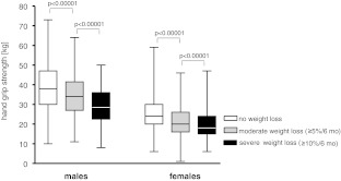
All data are given as mean and standard deviation. Box plots displaying minimum, maximum and 25th, 50th as well as 75th percentiles were used in order to portray hand grip strength. Pearson’s correlation was calculated to assess the relationship between variables. Multiple comparison between the patients with no, moderate and severe weight loss was performed with one-way between-groups analysis of variance.
In order to investigate the interrelationship between sex and disease-related malnutrition, a regression analysis was performed with the general linear model (GLM) allowing adjustment for continuous and categorical variables and corrected for age, AMA, BMI, and diagnosis category (benign/malignant) as covariates. Estimated marginal means, i.e., means adjusted for confounding covariates were calculated for hand grip strength for men and women, respectively. An acceptable level of statistical significance was established a priori at p < 0.05.
Results
One thousand five hundred patients (whereof 718 men, 47.9%) were included. Mean age was 57.6 ± 16.0 years, mean BMI was 24.6 ± 4.8 kg/m². Five hundred ninety-seven patients (39.8%) had malignant disease. Four hundred twenty-one patients (28.1%) exhibited moderate weight loss (mean weight loss, −8.5 ± 4.9%) and 290 patients (19.3%) suffered severe weight loss (mean weight loss, −14.7 ± 6.2%).
Fifty percent of male patients with moderate weight loss had malignant diseases compared to 46.3% in women (n.s.) whereas 47% of male patients with severe weight loss exhibited malignant disease compared to 50% in women (n.s). Diagnoses, demographic, and clinical characteristics stratified according to sex and nutritional status are given in Tables 1 and 2.
Table 1.
Diagnoses in the study population
| Type | Percent | |
|---|---|---|
| Malignant disease (n = 597) | Colorectal cancer | 19.6 |
| Head and neck cancer | 13.2 | |
| Hematologic disease | 11.4 | |
| Urogenital and mamma cancer | 9.6 | |
| Pancreatic cancer | 9.4 | |
| Gastric cancer | 8.0 | |
| Hepatic cancer | 7.7 | |
| Lung | 6.2 | |
| Biliary cancer | 4.5 | |
| other | 10.4 | |
| Benign disease (n = 903) | Inflammatory bowel disease | 33.8 |
| Hepatic disease | 20.3 | |
| Benign colon disease | 10.4 | |
| Heart disease | 8.1 | |
| Gastro-oesophageal disease | 4.6 | |
| Biliary disease | 3.4 | |
| Pancreatic disease | 2.3 | |
| Lung | 1.6 | |
| Diabetes | 1.5 | |
| Other | 14.0 |
Table 2.
Demographic and clinical characteristics of the study population
| All (n = 1,500) | Males (n = 718) | Females (n = 782) | P value | |
|---|---|---|---|---|
| Age (years) | 57.9 ± 16.0 | 58.1 ± 15.3 | 57.6 ± 16.6 | n.s. |
| Malignant disease (%) | 39.8 | 42.2 | 37.6 | n.s. |
| Moderate and severe weight loss: n (%) | 424/290 (28.3/19.3) | 208/158 (29/22) | 216/132 (27/16.9) | 0.016 |
| BMI (kg/m²) | 24.6 ± 4.8 | 24.8 ± 4.5 | 24.4 ± 5.1 | n.s. |
| Arm muscle area (cm²) | 47.3 ± 14.9 | 51.7 ± 14.5 | 43.3 ± 14.2 | <0.0001 |
| Hand grip strength (kg) | 34.6 ± 11.1 | 35.1 ± 11.2 | 23.1 ± 8.2 | <0.0001 |
| CRP (mg/dl) (subcohort: 53%) | 3.0 ± 4.6 | 3.3 ± 4.8 | 2.7 ± 4.4 | n.s. |
Both men and women with moderate and severe weight loss exhibited significant lower values of hand grip strength compared to patients without weight loss (see Fig. 1 and Table 3). There was a significant reduction of hand grip strength with increasing age (males: r = −0.403, p < 0.00001; females: r = −0.363, p < 0.00001). Hand grip strength was moreover correlated with AMA (males: r = 0.365, p < 0.00001; females: r = 0.173, p < 0.00001), but only very weakly with BMI (males: r = −0.085, p = 0.025; females: r = −0.111, p = 0.002). C-reactive protein (CRP) was only available in a subcohort of patients (793 patients), but as expected, hand grip strength was inversely associated with CRP (males: r = −0.2, p < 0.00001; females: r = −0.224, p < 0.00001).
Fig. 1.
Absolute unadjusted hand grip strength values in cachexia, stratified according to sex. Multiple comparisons between the groups was performed with one-waybetween groups analysis of variance
Table 3.
Hand grip strength according to nutritional status: absolute means and means adjusted for confounding factors in males and females
| Nutritional status | Grip strength (kg) | |
|---|---|---|
| Unadjusted means | Means adjusted for age, BMI, arm muscle area and malignant vs. benign disease | |
| Males | ||
| No weight loss | 38.3 ± 11.3 | 36.5 ± 0.5 (35.5–37.4) |
| Moderate weight loss | 34.1 ±10.4 | 33.3 ± 0.6 (32.2–34.6) |
| Severe weight loss | 29.2 ± 9.5 | 29.8 ± .07 (28.5–31.2) |
| Females | ||
| No weight loss | 25.2 ± 7.9 | 25.4 ± 0.4 (24.6–26.2) |
| Moderate weight loss | 21.1 ± 8.0 | 22.7 ± 0.6 (21.5–23.9) |
| Severe weight loss | 19.2 ± 7.3 | 21.7 ± 0.8 (20.4–23.3) |
In order to investigate possible sex-related impact of malnutrition-related muscle weakness, a GLM regression analysis adjusted for confounding variables such as age, sex, and BMI was performed. The GL model revealed that sex had a significant impact on the response of grip strength to weight loss (see Table 4). Grip strength was similarly decreased in moderate weight loss in men and women when compared to patients without weight loss (8.5% in males and 10.5% in females), but the further reduction of grip strength in severe weight loss was significantly different between men and women (10.6% vs. 4.1%, p < 0.001; see Fig. 2). Thus men experienced a much greater reduction of muscle strength (18.2%) in severe weight loss compared to good nutritional status than women (14.2%), corresponding to approximately 3.1 kg greater loss of grip strength in men than women (see Table 4). When the GLM regression analysis was stratified according to younger and higher age (<70 and >70 years), this phenomenon was only seen in younger patients (n = 1,142, 76%) with a significantly greater overall grip strength reduction of 21.7% in severely weight-losing men compared to 15.9% in weight-losing women, p < 0.0001 (data not shown).
Table 4.
Significant interrelationship between sex and cachexia with regard to hand grip strength
| Hand grip strength (kg) | ||
|---|---|---|
| Β coefficient | P value | |
| Age (years) | −0.250 | <0.0001 |
| Male vs. female sex | 7.958 | <0.0001 |
| Moderate weight lossa | −2.795 | <0.001 |
| Severe weight lossa | −3.753 | <0.001 |
| BMI (kg/m²) | −0.228 | <0.0001 |
| AMA (cm²) | 0.20 | <0.0001 |
| Sex-specific impact | ||
| Nutritional status × male sex | ||
| Moderate weight loss vs. no weight loss | −0.415 | n.s. |
| Severe weight loss vs. moderate weight loss | −2.732 | 0.033 |
| Severe weight loss vs. no weight loss | −3.145 | 0.006 |
aVersus good nutritional status
Fig. 2.
Percentage hand grip strength reduction in moderate and severe cachexia, stratified according to sex
Discussion
In this large cross-sectional study, we observed a greater discrepancy in hand grip strength values between well-nourished and weight-losing men than between well-nourished and weight-losing women. In severe weight loss in particular, our male study participants exhibited a larger percentage reduction in hand grip strength values than women did. When analyzing the data stratified according to age, however, this applied only to patients younger than 70 years, whereas there were no differences in grip strength response to weight loss in higher age between men and women.
It has previously been shown that weight loss or underweight has different impact in men and women. Wolf reported sex-related differences regarding the response of the hormone ghrelin and the adipocytokine leptin to weight loss in cancer patients [23].
In cachexia or underweight, testosterone levels are frequently decreased in men [24]. Smith et al. observed low testosterone with higher-than-normal values of luteinizing hormone (LH) in severely malnourished but otherwise healthy men [25]. Similarly, Chlebowski observed lower levels of free and total testosterone in cancer patients with the greatest weight deficit relative to their ideal weight, although LH levels were normal or increased [26]. Lado Abeal et al., moreover, revealed sex differences regarding the impact of disease-related malnutrition on the hypothalamic−pituitarygonadal axis. Again, underweight men (defined by a BMI < 18.5 kg/m²) had low testosterone levels with normal or above normal LH levels, whereas malnourished women had depressed gonadotropin levels [27].
It is therefore tempting to speculate that the greater loss of muscle strength in men which we observe in this study population is due to decreased free testosterone, which correlates with muscle strength in men [28]. We, moreover, did not find these sex-related differences in older patients which might be explained by the already reduced testosterone levels in higher age.
There is much evidence that age-associated loss of muscle strength is different in men and women. Shepard found greater loss of hand grip strength in elderly men than women in a cross-sectional study [29]. This has been attributed to the reduction of sexual hormones. In the elderly, sex hormone status is an important factor for muscle mass in men but not in women [30]. This partly explains why muscle strength is more preserved in women than men in higher age, although estrogen, which also exerts a protective effect on muscle, declines after menopause as well [31]. Kirchengast et al. observed that men appear to be more prone to sarcopenia, loss of muscle mass and strength, in higher age, whereas sarcopenia appears to be more prevalent in women under the age of 70 [32]. Another potential influencing factor which cannot be disregarded is inflammation, as increased levels of C-reactive protein correlated with lower muscle strength values in a subgroup of our study population. The association between CRP and grip strength in elderly in particular has been reported by others [33, 34].
Our findings are invariably limited due to their cross-sectional design and lack of hormone values, but clearly imply that men experience a greater loss of more grip strength in weight loss of more than 10%. Hand grip strength correlates well with functional status and quality of life [9]. Reduced grip strength is a predictor of impaired outcome, such as increased postoperative complications, increased length of hospitalization, higher rehospitalization rate, and decreased physical status [35]. In men in particular, low grip strength in health predicts increased long-term risk of functional limitations and disability as well as all-cause mortality [36, 37]. In contrast to women, high grip strength also appears to be protective against premature mortality in men [17, 38]. These findings suggest that higher strength signifies greater physiologic and functional reserve.
In conclusion, our results show sex-specific differences in muscle strength in severe weight loss. This implies that sex-specific aspects might also be relevant for anticatabolic treatment such as nutritional or physical therapy. Further studies should therefore investigate sex-related response to nutritional repletion or physical exercise in cachectic patients.
Acknowledgments
We thank U. Grittner, PhD., Institute for Biometry and Clinical Epidemiology, Charité-University Medicine for statistical advice. The authors have read and complied with the guidelines of ethical authorship and publishing in the Journal of Cachexia, Sarcopenia and Muscle [39].
Open Access
This article is distributed under the terms of the Creative Commons Attribution Noncommercial License which permits any noncommercial use, distribution, and reproduction in any medium, provided the original author(s) and source are credited.
References
- 1.von Haehling S, Anker SD. Cachexia as a major underestimated and unmet medical need: facts and numbers. J Cachex Sarcopenia Muscle. 2010;1:1–5. doi: 10.1007/s13539-010-0002-6. [DOI] [PMC free article] [PubMed] [Google Scholar]
- 2.Norman K, Pichard C, Lochs H, Pirlich M. Prognostic impact of disease-related malnutrition. Clin Nutr. 2008;27:5–15. doi: 10.1016/j.clnu.2007.10.007. [DOI] [PubMed] [Google Scholar]
- 3.Lenk K, Schuler G, Adams V. Skeletal muscle wasting in cachexia and sarcopenia: molecular pathophysiology and impact of exercise training. J Cachex Sarcopenia Muscle. 2010;1:9–21. doi: 10.1007/s13539-010-0007-1. [DOI] [PMC free article] [PubMed] [Google Scholar]
- 4.Daniel PM. The metabolic homoeostatic role of muscle and its function as a store of protein. Lancet. 1977;2:446–8. doi: 10.1016/S0140-6736(77)90622-5. [DOI] [PubMed] [Google Scholar]
- 5.Heymsfield SB, McManus C, Stevens V, Smith J. Muscle mass: reliable indicator of protein-energy malnutrition severity and outcome. Am J Clin Nutr. 1982;35:1192–9. doi: 10.1093/ajcn/35.5.1192. [DOI] [PubMed] [Google Scholar]
- 6.Norman K, Smoliner C, Valentini L, Lochs H, Pirlich M. Is bioelectrical impedance vector analysis of value in the elderly with malnutrition and impaired functionality? Nutrition. 2007;23:564–9. doi: 10.1016/j.nut.2007.05.007. [DOI] [PubMed] [Google Scholar]
- 7.Vaz M, Thangam S, Prabhu A, Shetty PS. Maximal voluntary contraction as a functional indicator of adult chronic undernutrition. Br J Nutr. 1996;76:9–15. doi: 10.1079/BJN19960005. [DOI] [PubMed] [Google Scholar]
- 8.Norman K, Schutz T, Kemps M, Josef LH, Lochs H, Pirlich M. The Subjective Global Assessment reliably identifies malnutrition-related muscle dysfunction. Clin Nutr. 2005;24:143–50. doi: 10.1016/j.clnu.2004.08.007. [DOI] [PubMed] [Google Scholar]
- 9.Norman K, Stobaus N, Smoliner C, et al. Determinants of hand grip strength, knee extension strength and functional status in cancer patients. Clin Nutr. 2010;29:586–91. doi: 10.1016/j.clnu.2010.02.007. [DOI] [PubMed] [Google Scholar]
- 10.Ferrando AA, Lane HW, Stuart CA, Davis-Street J, Wolfe RR. Prolonged bed rest decreases skeletal muscle and whole body protein synthesis. Am J Physiol. 1996;270:E627–33. doi: 10.1152/ajpendo.1996.270.4.E627. [DOI] [PubMed] [Google Scholar]
- 11.Yasuda N, Glover EI, Phillips SM, Isfort RJ, Tarnopolsky MA. Sex-based differences in skeletal muscle function and morphology with short-term limb immobilization. J Appl Physiol. 2005;99:1085–92. doi: 10.1152/japplphysiol.00247.2005. [DOI] [PubMed] [Google Scholar]
- 12.Lindboe CF, Platou CS. Disuse atrophy of human skeletal muscle. An enzyme histochemical study. Acta Neuropathol (Berl) 1982;56:241–4. doi: 10.1007/BF00691253. [DOI] [PubMed] [Google Scholar]
- 13.Paddon-Jones D, Sheffield-Moore M, Urban RJ, Aarsland A, Wolfe RR, Ferrando AA. The catabolic effects of prolonged inactivity and acute hypercortisolemia are offset by dietary supplementation. J Clin Endocrinol Metab. 2005;90:1453–9. doi: 10.1210/jc.2004-1702. [DOI] [PubMed] [Google Scholar]
- 14.Paddon-Jones D, Sheffield-Moore M, Cree MG, et al. Atrophy and impaired muscle protein synthesis during prolonged inactivity and stress. J Clin Endocrinol Metab. 2006;91:4836–41. doi: 10.1210/jc.2006-0651. [DOI] [PubMed] [Google Scholar]
- 15.Wagenmakers AJ. Muscle function in critically ill patients. Clin Nutr. 2001;20:451–4. doi: 10.1054/clnu.2001.0483. [DOI] [PubMed] [Google Scholar]
- 16.Budziareck MB, Pureza Duarte RR, Barbosa-Silva MC. Reference values and determinants for handgrip strength in healthy subjects. Clin Nutr. 2008;27:357–62. doi: 10.1016/j.clnu.2008.03.008. [DOI] [PubMed] [Google Scholar]
- 17.Lindle RS, Metter EJ, Lynch NA, et al. Age and gender comparisons of muscle strength in 654 women and men aged 20–93 yr. J Appl Physiol. 1997;83:1581–7. doi: 10.1152/jappl.1997.83.5.1581. [DOI] [PubMed] [Google Scholar]
- 18.Norman K, Pirlich M, Sorensen J, et al. Bioimpedance vector analysis as a measure of muscle function. Clin Nutr. 2009;28:78–82. doi: 10.1016/j.clnu.2008.11.001. [DOI] [PubMed] [Google Scholar]
- 19.Norman K, Smoliner C, Kilbert A, Valentini L, Lochs H, Pirlich M. Disease-related malnutrition but not underweight by BMI is reflected by disturbed electric tissue properties in the bioelectrical impedance vector analysis. Br J Nutr. 2008;100:590–5. doi: 10.1017/S0007114508911545. [DOI] [PubMed] [Google Scholar]
- 20.Norman K, Kirchner H, Lochs H, Pirlich M. Malnutrition affects quality of life in gastroenterology patients. World J Gastroenterol. 2006;12:3380–5. doi: 10.3748/wjg.v12.i21.3385. [DOI] [PMC free article] [PubMed] [Google Scholar]
- 21.Gurney JM, Jelliffe DB. Arm anthropometry in nutritional assessment: nomogram for rapid calculation of muscle circumference and cross-sectional muscle and fat areas. Am J Clin Nutr. 1973;26:912–5. doi: 10.1093/ajcn/26.9.912. [DOI] [PubMed] [Google Scholar]
- 22.Detsky AS, McLaughlin JR, Baker JP, et al. What is subjective global assessment of nutritional status? JPEN J Parenter Enteral Nutr. 1987;11:8–13. doi: 10.1177/014860718701100108. [DOI] [PubMed] [Google Scholar]
- 23.Wolf I, Sadetzki S, Kanety H, et al. Adiponectin, ghrelin, and leptin in cancer cachexia in breast and colon cancer patients. Cancer. 2006;106:966–73. doi: 10.1002/cncr.21690. [DOI] [PubMed] [Google Scholar]
- 24.Del FE, Hui D, Dalal S, Dev R, Noorhuddin Z, Bruera E. Clinical outcomes and contributors to weight loss in a cancer cachexia clinic. J Palliat Med 2011;14(9):1004–8 [DOI] [PMC free article] [PubMed]
- 25.Smith SR, Chhetri MK, Johanson J, Radfar N, Migeon CJ. The pituitary–gonadal axis in men with protein–calorie malnutrition. J Clin Endocrinol Metab. 1975;41:60–9. doi: 10.1210/jcem-41-1-60. [DOI] [PubMed] [Google Scholar]
- 26.Chlebowski RT, Heber D. Hypogonadism in male patients with metastatic cancer prior to chemotherapy. Cancer Res. 1982;42:2495–8. [PubMed] [Google Scholar]
- 27.Lado-Abeal J, Prieto D, Lorenzo M, et al. Differences between men and women as regards the effects of protein-energy malnutrition on the hypothalamic–pituitary–gonadal axis. Nutrition. 1999;15:351–8. doi: 10.1016/S0899-9007(99)00051-9. [DOI] [PubMed] [Google Scholar]
- 28.van den Beld AW, de Jong FH, Grobbee DE, Pols HA, Lamberts SW. Measures of bioavailable serum testosterone and estradiol and their relationships with muscle strength, bone density, and body composition in elderly men. J Clin Endocrinol Metab. 2000;85:3276–82. doi: 10.1210/jc.85.9.3276. [DOI] [PubMed] [Google Scholar]
- 29.Shephard RJ, Montelpare W, Plyley M, McCracken D, Goode RC. Handgrip dynamometry, cybex measurements and lean mass as markers of the ageing of muscle function. Br J Sports Med. 1991;25:204–8. doi: 10.1136/bjsm.25.4.204. [DOI] [PMC free article] [PubMed] [Google Scholar]
- 30.Baumgartner RN, Waters DL, Gallagher D, Morley JE, Garry PJ. Predictors of skeletal muscle mass in elderly men and women. Mech Ageing Dev. 1999;107:123–36. doi: 10.1016/S0047-6374(98)00130-4. [DOI] [PubMed] [Google Scholar]
- 31.Enns DL, Tiidus PM. The influence of estrogen on skeletal muscle: sex matters. Sports Med. 2010;40:41–58. doi: 10.2165/11319760-000000000-00000. [DOI] [PubMed] [Google Scholar]
- 32.Kirchengast S, Huber J. Gender and age differences in lean soft tissue mass and sarcopenia among healthy elderly. Anthropol Anz. 2009;67:139–51. doi: 10.1127/0003-5548/2009/0018. [DOI] [PubMed] [Google Scholar]
- 33.Hamer M, Molloy GJ. Association of C-reactive protein and muscle strength in the English Longitudinal Study of Ageing. Age (Dordr) 2009;31:171–7. doi: 10.1007/s11357-009-9097-0. [DOI] [PMC free article] [PubMed] [Google Scholar]
- 34.Tiainen K, Hurme M, Hervonen A, Luukkaala T, Jylha M. Inflammatory markers and physical performance among nonagenarians. J Gerontol A Biol Sci Med Sci. 2010;65:658–63. doi: 10.1093/gerona/glq056. [DOI] [PubMed] [Google Scholar]
- 35.Norman K, Stobaus N, Gonzalez MC, Schulzke JD, Pirlich M. Hand grip strength: outcome predictor and marker of nutritional status. Clin Nutr 2010; 30(2):135–42 [DOI] [PubMed]
- 36.Rantanen T, Guralnik JM, Foley D, et al. Midlife hand grip strength as a predictor of old age disability. JAMA. 1999;281:558–60. doi: 10.1001/jama.281.6.558. [DOI] [PubMed] [Google Scholar]
- 37.Rantanen T, Harris T, Leveille SG, et al. Muscle strength and body mass index as long-term predictors of mortality in initially healthy men. J Gerontol A Biol Sci Med Sci. 2000;55:M168–73. doi: 10.1093/gerona/55.3.M168. [DOI] [PubMed] [Google Scholar]
- 38.Sasaki H, Kasagi F, Yamada M, Fujita S. Grip strength predicts cause-specific mortality in middle-aged and elderly persons. Am J Med. 2007;120:337–42. doi: 10.1016/j.amjmed.2006.04.018. [DOI] [PubMed] [Google Scholar]
- 39.von Haehling S, Morley JE, Coats AJ, Anker SD. Ethical guidelines for authorship and publishing in the Journal of Cachexia, Sarcopenia and Muscle. J Cachex Sarcopenia Muscle. 2010;1:7–8. doi: 10.1007/s13539-010-0003-5. [DOI] [PMC free article] [PubMed] [Google Scholar]


