
Figure 5. Role of BCRP in LN229 cell proliferation.
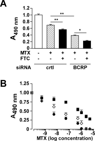
(A) LN229 cells were transfected with control or BCRP siRNA for 24 h followed by the addition of 500 nM MTX in the absence or presence of 5 μM FTC for 48 h. A cell proliferation colorimetric assay based on MTS reduction was then performed. Values are means ± SEM from 3 independent experiments performed in triplicate. Statistical analysis by ANOVA with Dunnett's Multiple comparison test was performed, where *- p < 0.05; **- p <0.01. (B) Control siRNA-transfected LN229 cells were incubated with the indicated concentrations of mitoxantrone (MTX) in the absence (filled square) or the presence of 5μM fumitremorgin C (FTC) (open diamond) for 48 h. Another group consisted of BCRP siRNA-treated LN229 cells incubated with MTX plus FTC (filled circle). Values are means ± SEM from 3 independent experiments performed in triplicate.