Abstract
Bacillus cereus is infrequently associated with invasive central nervous system (CNS) disease. Infection is associated with conditions that lead to reduced host immunity and provide direct access to the CNS, such as spinal anesthesia and ventricular tubes and shunts. A case of ventriculitis secondary to B cereus in a patient receiving intrathecal chemotherapy is reported, along with a review of the current literature. B cereus can colonize medical devices, thus posing a risk for invasive disease. Despite aggressive treatment with broad-spectrum anti-infectives, the mortality of CNS invasive B cereus is high. Clinicians should not dismiss Gram-positive rods resembling Bacillus species from normally sterile sites as contaminants in critically ill patients. Appropriate antibiotic therapy should be promptly initiated to limit morbidity and mortality.
Keywords: Bacillus cereus meningitis
Abstract
Le Bacillus cereus s’associe rarement à une maladie invasive du système nerveux central (SNC). L’infection est liée à des pathologies qui réduisent l’immunité de l’hôte et procurent un accès direct au SNC, telles qu’une rachianesthésie et des tubes et dérivations ventriculaires. Les auteurs rendent compte d’un cas de ventriculite secondaire à un B cereus chez un patient sous chimiothérapie intratéchale et présentent une analyse des publications à jour. Le B cereus peut coloniser les dispositifs médicaux, posant ainsi un risque de maladie invasive. Malgré un traitement énergique au moyen d’anti-infectieux à large spectre, la mortalité attribuable au B cereus invasif du SNC est élevée. Les cliniciens ne devraient pas rejeter la possibilité que des bacilles Gram positif évocateurs d’espèces de Bacillus dans les foyers normalement stériles soient des contaminants chez les patients gravement malades. Il faudrait amorcer rapidement une antibiothérapie pertinente pour limiter la morbidité et la mortalité.
Bacillus cereus is a spore-forming, Gram-positive, or Gram-variable, rod bacterium that is ubiquitous in the environment. It is a well-known cause of gastrointestinal illness, but has been associated with extraintestinal disease, as well (1). Central nervous system (CNS) involvement with B cereus is rare, but has been described in association with both immunosuppression and CNS invasive devices. B cereus is intrinsically resistant to beta-lactam antibiotics (1), and CNS infections with this organism are associated with high mortality. A case involving a 73-year-old woman with chronic myelogenous leukemia and an Ommaya reservoir who developed ventriculitis with B cereus is reported, along with a review of the literature on invasive CNS infections with B cereus.
CASE PRESENTATION
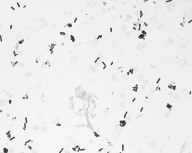
A 73-year-old Afghani woman with a history of chronic myelogenous leukemia, complicated by leukemic meningitis, on therapy with bosutinib (an experimental tyrosine kinase inhibitor) and monthly intrathecal methotrexate and hydrocortisone administered via an Ommaya reservoir, presented to her oncology clinic for a scheduled intrathecal methotrexate/hydrocortisone infusion. On presentation, the patient was clinically well and her cerebrospinal fluid (CSF) revealed no white blood cells. One day later, she developed headache, generalized weakness and confusion and presented to the emergency department. She was tachycardic, febrile (38.7°C), lethargic, had nuchal rigidity and a systolic murmur (grade 3/6) at the left sternal border. She had leukocytosis with a leukocyte count of 19.2×10⁹ cells/L with 96% neutrophils, and CSF analysis revealed 4.02×10⁹/L white blood cells with 99% neutrophils, a protein level of 1.21 g/L, and a glucose level of 4.88 mmol/L. A gram-stain of teh CSF revealed Gram-variable rod bacteria (Figure 1). Blood and CSF cultures were sent for analysis and empirical therapy with vancomycin and cefepime was initiated. By hospital day 2, both the headache and her mental status had improved. Blood and CSF cultures grew B cereus, which was susceptible to vancomycin. The Ommaya reservoir was removed on hospital day 3. Repeat blood cultures were negative and she underwent transesophageal echocardiography that revealed no vegetations. The patient completed five weeks of therapy with intravenous vancomycin with subsequent microbiological cure and clinical recovery.
Figure 1).

Gram stain of cerebrospinal fluid showing Gram-variable rod bacteria
METHODS
A literature review of B cereus meningitis was performed using the MEDLINE/PubMed database. All searches were limited to English-language articles published since 1950. The first search term “Bacillus cereus meningitis” yielded 32 articles. A second search using the search term “Bacillus cereus central nervous system infections” yielded 32 articles. A third search was performed using the terms “Bacillus cereus meningoencephalitis”, which yielded eight articles. The abstracts of the articles retrieved from all three searches were reviewed, and the references from pertinent articles were examined to identify additional reports. A total of 21 relevant publications were identified and details on 32 patients with CNS infection with B cereus were available for review.
DISCUSSION
Members of the Bacillus genus are Gram-positive or Gram-variable, spore-forming rod bacteria, which are ubiquitous in the environment. Clinical infections caused by B cereus fall into six broad groups: local infections of wounds, burns, or the eye; bacteremia; CNS infections; respiratory infections; endocarditis; and food poisoning.
We report a case of B cereus CNS infection associated with the use of an Ommaya reservoir and intrathecal chemotherapy. Since 1950 there have been 22 (including the present case) published case reports of CNS infections with B cereus (Table 1). Reported manifestations of CNS B cereus infection include ventriculitis, meningitis, leptomeningitis, hydrocephalus, intraparenchymal, subarachnoid subdural hemorrhage and brain abscess (2–24).
TABLE 1.
Central nervous system manifestations of Bacillus cereus
| Authors (reference), year | Age*/sex | Clinical picture | Treatment | Outcome | Risk factors |
|---|---|---|---|---|---|
| Present case 2008 | 73/F | Leukemic meningitis | Vancomycin | Recovered | Ommaya reservoir |
| Garcia et al (5), 1984 | 32/M | Leptomeningitis | Penicillin, chloramphenicol | Recovered | Ommaya reservoir |
| Hendrickx et al (6), 1981 | 8d/F | Complete hemorrhagic necrosis of the brain | Ampicillin, gentamicin | Died | Ventricular puncture |
| Barrie et al (4), 1992 | 55/F | Hydrocephalus | Vancomycin | Died | Ventricular drain |
| 41/F | Hematoma | Chloramphenicol | |||
| Berke et al (7), 1981 | 25/F | Bilateral papilledema, bilateral hemianopsia | Chloramphenicol, vancomycin | Recovered | Ventriculostomy |
| Heep et al (8), 2004 | 14d/M | Ventriculitis, hemorrhagic necrotizing lesions | Vancomycin, gentamicin, meropenem | Not reported | Ventriculostomy tube |
| Leffert et al (9), 1970 | 126d/M | Meningitis | Gentamicin, ampicillin | Recovered | Ventriculoarterial shunting |
| Haase et al (10), 2005 | 19/M | Meningoencephalitis, flaccid hemiparesis | Cyclosporine, methotrexate then ceftazidime then teicoplanin, ampicillin, amikacin, ciprofloxacin, clindamycin | Recovered | Broviac catheter |
| Tokieda et al (11), 1999 (2 cases) | 4d/F | Multiple brain parenchyma, subdural, epidural, and subarachnoid hemorrhage; and wide-spread softening and hemorrhagic necrosis of the brain | Ampicillin, gentamicin, cefotaxime | Died | Peripheral venous catheter; nasal feeding tube |
| 5d/F | |||||
| Feder et al (12), 1988 (2 cases) | 51/M | Sequelae included | Chloramphenicol | Recovered | Gun shot wound; contaminated intravenous catheter |
| 47d/F | Brain damage, hydrocephalus, hypotenia and hyper-reflexia | ||||
| Manickam et al (13), 2008 | 8d/M | Hemorrhagic necrosis and liquefaction of brain tissue | Ampicillin, gentamicin | Died | Not identified |
| Akiyama et al (14), 1997 | 64/M | Leptomeningitis, subarachnoid hemorrhage, bacterial infiltration; liver and stomach necrosis | Piperacillin, gentamicin, cefoperazone, cefotaxime, ampicillin | Died | Immunosuppression post chemotherapy |
| Marely et al (15), 1995 | 26/M | Meningoencephalitis, subarachnoid hemorrhage, bacterial infiltration; liver and myocardium necrosis | Ceftazidime | Died | Immunosuppression post chemotherapy |
| Lequin et al (16), 2005 (3 cases) | 5d/F | Meningoencephalitis, ventriculitis | Amoxicillin/clafuran, vancomycin, amikacin, clindamicin | Died | Preterm delivery, central line catheter |
| 5d/F | |||||
| 13d/F | |||||
| Jenson et al (17), 1989 | 3/M | Cerebritis, hemorrhagic necrosis, meningitis | Chloramphenicol, vancomycin, gentamicin, rifampin | Recovered | Immunosuppression postchemotherapy |
| Musa et al (18), 1999 (3 cases) | 30/M | Leptomeningeal and neural necrosis | Ceftazidime, amikacin, vancomycin, gentamicin, ampicillin, tazobactam | Died | Immunosuppression postchemotherapy |
| 43/M | |||||
| 14/M | |||||
| de Almeida et al (19), 2003 | 16/F | Meningitis | Ceftazidime, imipenem | Died | Immunosuppression post marrow transplant |
| Tuladhar et al (20), 2000 | 14d/M | Intraventricular hemorrhage | Vancomycin, gentamicin, imipenem, clindamycin, ciprofloxacillin, immunoglobulin therapy | Died | Not identified |
| Gaur et al (21), 2001 (4 cases) | 20/F | Meningitis, leptomeningitis, brain abscess, meningoencephalitis, hydrocephalus | Vancomycin | Died | Intrathecal chemotherapy |
| 10/F | |||||
| 13/F | |||||
| 15/F | |||||
| Marshman et al (22), 2000 | 41/F | Meningitis | Teicoplanin | Recovered | Cerebrospinal fluid fistula repair |
| Weisse et al (23), 1991 (2 cases) | 5d/M | Meningitis | Vancomycin, gentamicin, chloramphenicol | Recovered | Myelomeningocele; none |
| 21d/M | |||||
| Chu et al (24), 2001 | 28d/M | Meningitis | Vancomycin, amikacin | Died | Bronchopulmonary dysplasia, dexamethasone used |
Age is presented as years, unless otherwise indicated with d, which denotes days. F Female; M Male
The ubiquitous presence of B cereus makes environmental contamination of hospitals and clinics inevitable (2). Van der Zwet et al (3) reported an outbreak of B cereus infections in a neonatal intensive care unit associated with contaminated manual ventilation balloons, and Barrie et al (4) reported contamination of hospital linen by B cereus. Although rare, CNS infections with B cereus occur when host defense mechanisms are compromised, either by invasive CNS/ vascular devices and/or by underlying immunodeficiency. We suspect that, becasue our patient was undergoing intrathecal chemotherapy, the frequent manipulation of the Ommaya reservoir led to bacterial contamination with B cereus and subsequent invasive disease. Previous cases of B cereus CNS disease have reported ventriculostomy tubes, ventricular punctures, central venous catheters, nasal feeding tubes and immunosuppression as risk factors (4,5,6–12,14–19). Only Garcia et al (5) reported the presence of an Ommaya reservoir as a risk factor. B cereus produces several toxins, including hemolysins and phospholipases, both believed to be important virulence determinants (1).
Despite aggressive treatment with broad-spectrum anti-infectives, the crude mortality is high (66%, including the present case, and excluding a case without a documented outcome). The high mortality is likely a reflection of underlying disease severity and comorbid illnesses. B cereus produces beta-lactamases and is resistant to beta-lactam antibiotics, including third-generation cephalosporins. B cereus is typically susceptible to aminoglycosides, clindamycin, vancomycin, chloramphenicol and erythromycin (1). Guidance on management of CNS B cereus infections is from case reports because no prospective trials or treatment guidelines exist.
The present case adds to the body of literature on CNS infections with B cereus, and is the second case to describe an Ommaya reservoir as a potential risk factor. We underscore the potential risk that a foreign body may pose for invasive disease with B cereus. Intraocular foreign bodies have been significantly associated with severe B cereus endophthalmitis (25,26). CNS invasive devices, such as Ommaya reservoirs, may pose a similar risk. These devices should be placed and accessed with strict attention to aseptic technique to minimize bacterial contamination and invasion. Additionally, when Gram stain or culture reveals Gram-positive rods resembling Bacillus species from normally sterile sites, such as blood or CSF, in critically ill immunosuppressed patients with invasive devices, these should not be dismissed as contaminants. Clinicians should be aware of the potential for B cereus CNS infection, and initiate prompt and appropriate antimicrobial therapy in an attempt to limit both morbidity and mortality.
SUMMARY
CNS disease with B cereus is an infrequent occurrence. Reports of invasive CNS disease exist in both pediatric and adult populations. Risk factors for CNS invasive disease include immunosuppression and invasive devices. Because B cereus is a ubiquitous environmental organism, CNS invasive devices, such as Ommaya reservoirs, should be placed and accessed with strict attention to aseptic technique to minimize bacterial contamination and invasion. The crude mortality rate is high, underscoring the clinical significance of CNS invasive disease with B cereus. For critically ill patients with invasive devices and immunosuppression, the presence of Bacillus species from normally sterile sites should not be dismissed as contaminants. To limit morbidity and mortality, clinicians should have a high index of suspicion for B cereus CNS infection, and initiate prompt and appropriate antimicrobial therapy.
REFERENCES
- 1.Drobniewski FA. Bacillus cereus and related species. Clin Microbiol Rev. 1993;6:324–38. doi: 10.1128/cmr.6.4.324. [DOI] [PMC free article] [PubMed] [Google Scholar]
- 2.Berthelot P, Dietemann J, Fascia P, et al. Bacterial contamination of nonsterile disposable gloves before use. Am J Infect Control. 2006;34:128–30. doi: 10.1016/j.ajic.2005.08.017. [DOI] [PubMed] [Google Scholar]
- 3.Van Der Zwet WC, Parlevliet GA, Savelkoul PH, et al. Outbreak of bacillus cereus infections in a neonatal intensive care unit traced to balloons used in manual ventilation. J Clin Microbiol. 2000;38:4131–6. doi: 10.1128/jcm.38.11.4131-4136.2000. [DOI] [PMC free article] [PubMed] [Google Scholar]
- 4.Barrie D, Wilson JA, Hoffman PN, Kramer JM. Bacillus cereus meningitis in two neurosurgical patients: An investigation into the source of the organism. J Infect. 1992;25:291–7. doi: 10.1016/0163-4453(92)91579-z. [DOI] [PubMed] [Google Scholar]
- 5.Garcia I, Fainstein V, McLaughlin P. Bacillus cereus meningitis and bacteremia associated with an ommaya reservoir in a patient with lymphoma. South Med J. 1984;77:928–9. doi: 10.1097/00007611-198407000-00036. [DOI] [PubMed] [Google Scholar]
- 6.Hendrickx B, Azou M, Vandepitte J, Jaeken J, Eggermont E. Bacillus cereus meningo-encephalitis in a pre-term baby. Acta Paediatr Belg. 1981;34:107–12. [PubMed] [Google Scholar]
- 7.Berke E, Collins WF, von Graevenitz A, Bia FJ. Fulminant postsurgical Bacillus cereus meningitis: Case report. J Neurosurg. 1981;55:637–9. doi: 10.3171/jns.1981.55.4.0637. [DOI] [PubMed] [Google Scholar]
- 8.Heep A, Schaller C, Rittmann N, Himbert U, Marklein G, Bartmann P. Multiple brain abscesses in an extremely preterm infant: Treatment surveillance with interleukin-6 in the CSF. Eur J Pediatr. 2004;163:44–5. doi: 10.1007/s00431-003-1333-5. [DOI] [PubMed] [Google Scholar]
- 9.Leffert HL, Baptist JN, Gidez LI. Meningitis and bacteremia after ventriculoatrial shunt-revision: Isolation of a lecithinase-producing Bacillus cereus. J Infect Dis. 1970;122:547–52. doi: 10.1093/infdis/122.6.547. [DOI] [PubMed] [Google Scholar]
- 10.Haase R, Sauer H, Dagwadordsch U, Foell J, Lieser U. Successful treatment of Bacillus cereus meningitis following allogenic stem cell transplantation. PediatrTransplant. 2005;9:338–40. doi: 10.1111/j.1399-3046.2004.00286.x. [DOI] [PubMed] [Google Scholar]
- 11.Tokieda K, Morikawa Y, Maeyama K, Mori K, Ikeda K. Clinical manifestations of Bacillus cereus meningitis in newborn infants. J Paediatr Child Health. 1999;35:582–4. doi: 10.1046/j.1440-1754.1999.00405.x. [DOI] [PubMed] [Google Scholar]
- 12.Feder HM, Garibaldi RA, Nurse BA, Kurker R. Bacillus species isolates from cerebrospinal fluid in patients without shunts. Pediatrics. 1988;82:909–13. [PubMed] [Google Scholar]
- 13.Manickam N, Knorr A, Muldrew K. Neonatal meningoencephalitis caused by Bacillus cereus. Pediatr Infect Dis J. 2008;27:843–6. doi: 10.1097/INF.0b013e31816feec4. [DOI] [PubMed] [Google Scholar]
- 14.Akiyama N, Mitani K, Tanaka Y, et al. Fulminant septicemic syndrome of Bacillus cereus in a leukemic patient. Intern Med. 1997;36:221–6. doi: 10.2169/internalmedicine.36.221. [DOI] [PubMed] [Google Scholar]
- 15.Marley EF, Saini NK, Venkatraman C, Orenstein JM. Fatal Bacillus cereus meningoencephalitis in an adult with acute myelogenous leukemia. South Med J. 1995;88:969–72. doi: 10.1097/00007611-199509000-00017. [DOI] [PubMed] [Google Scholar]
- 16.Lequin M, Vermeulen J, van Elburg R, et al. Bacillus cereus meningoencephalitis in preterm infants: Neuroimaging characteristics. AJNR, Am J Neuroradiol. 2005;26:2137–43. [PMC free article] [PubMed] [Google Scholar]
- 17.Jenson HB, Levy SR, Duncan C, McIntosh S. Treatment of multiple brain abscesses caused by Bacillus cereus. Pediatr Infect Dis J. 1989;8:795–8. doi: 10.1097/00006454-198911000-00013. [DOI] [PubMed] [Google Scholar]
- 18.Musa MO, Al Douri M, Khan S, Shafi T, Al Humaidh A, Al Rasheed AM. Fulminant septicemic syndrome of Bacillus cereus: Three case reports. J Infect. 1999;39:154–6. doi: 10.1016/s0163-4453(99)90009-9. [DOI] [PubMed] [Google Scholar]
- 19.de Almeida S, Teive HAG, Brandi I, et al. Fatal Bacillus cereus meningitis without inflammatory reaction in cerebral spinal fluid after bone marrow transplantation. Transplantation. 2003;76:1533–4. doi: 10.1097/01.TP.0000079251.82361.99. [DOI] [PubMed] [Google Scholar]
- 20.Tuladhar R, Patole SK, Koh TH, Norton R, Whitehall JS. Refractory Bacillus cereus infection in a neonate. Int J Clin Pract. 2000;54:345–7. [PubMed] [Google Scholar]
- 21.Gaur AH, Patrick CC, McCullers JA, et al. Bacillus cereus bacteremia and meningitis in immunocompromised children. Clin Infect Dis. 2001;32:1456–62. doi: 10.1086/320154. [DOI] [PubMed] [Google Scholar]
- 22.Marshman LA, Hardwidge C, Donaldson PM. Bacillus cereus meningitis complicating cerebrospinal fluid fistula repair and spinal drainage. Br J Neurosurg. 2000;14:580–2. doi: 10.1080/02688690050206774. [DOI] [PubMed] [Google Scholar]
- 23.Weisse ME, Bass JW, Jarrett RV, Vincent JM. Non-anthrax bacillus infections of the central nervous system. Pediatr Infect Dis J. 1991;10:243–6. doi: 10.1097/00006454-199103000-00014. [DOI] [PubMed] [Google Scholar]
- 24.Chu WP, Que TL, Lee WK, Wong SN. Meningoencephalitis caused by Bacillus cereus in a neonate. Hong Kong Med J. 2001;7:89–92. [PubMed] [Google Scholar]
- 25.Yang C, Lu CK, Lee FL, et al. Treatment and outcome of traumatic endophthalmitis in open globe injury with retained intraocular foreign body. Ophthalmologica. 2010;224:79–85. doi: 10.1159/000235725. [DOI] [PubMed] [Google Scholar]
- 26.Miller JJ, Scott IU, Flynn, et al. Endophthalmitis caused by Bacillus species. Am J Ophthalmol. 2007;145:883–8. doi: 10.1016/j.ajo.2007.12.026. [DOI] [PubMed] [Google Scholar]


