Abstract
The light chain of botulinum neurotoxin A (BoNT/A-LC) is a Zn-dependent protease that specifically cleaves SNAP25 of the SNARE complex, thereby impairing vesicle fusion and neurotransmitter release at neuromuscular junctions. The C-terminus of SNAP25 (residues 141–206) retains full activity for BoNT/A-LC-catalyzed cleavage at P1-P1’ (Gln197-Arg198). Using the structure of a complex between the C-terminus of SNAP25 and BoNT/A-LC as a model to design SNAP25-derived pseudosubstrate inhibitors (SNAPIs) that prevent presentation of the scissile bond to the active site, we introduced multiple His residues to replace Ala-Asn-Gln-Arg (residues 195–198) at the substrate cleavage site, with the intent to identify possible side-chain interactions with the active site Zn. We also introduced multiple Gly residues between the P1-P1’ residues to explore the spatial tolerance within the active-site cleft. Using a FRET substrate YsCsY, we compared a series of SNAPIs for inhibition of BoNT/A-LC. Among the SNAPIs tested, several known cleavage-resistant, single-point mutants of SNAP25 were poor inhibitors, with most of the mutants losing binding affinity. Replacement with His at the active site did not improve inhibition over wildtype substrate. In contrast, Gly-insertion mutants were not only resistant to cleavage, but also surprisingly showed enhanced affinity for BoNT/A-LC. Two of the Gly-insertion mutants exhibited 10-fold lower IC50 values than the wildtype 66-mer SNAP25 peptide. Our findings illustrate a scenario, where the induced fit between enzyme and bound pseudosubstrate fails to produce the strain and distortion required for catalysis to proceed.
Keywords: BoNT, Zn-protease, YsCsY, pseudosubstrate, induced fit
Introduction
Botulinum neurotoxin A (BoNT/A) impairs neurotransmitter release at the neuromuscular junction through specific endopeptidase activity on SNAP25 (synaptosome-associated protein-25 kDa), which is a component of the ternary SNARE (soluble N-ethylmaleimide factor attachment protein receptor) complex critical for synaptic vesicle fusion and subsequent neurotransmitter release.1 The toxin is post-translationally cleaved into a 50-kDa N-terminal light chain (BoNT/A-LC) and a 100-kDa C-terminal heavy chain (BoNT/A-HC), which remain tethered through a disulfide bond.2 BoNT/A-LC is a Zn-dependent protease, while BoNT/A-HC provides the function of receptor binding, endocytosis, and translocation of the enzyme domain across the endosomal membrane into the cytosol.3–6 BoNT/A-LC resists degradation inside neuronal cells, such that patients with botulism could endure up to a year of slow recovery.7, 8 Thus, there is a need for post-exposure therapies that will shorten the recovery time.
Current treatment with antitoxin antibody only achieves rapid clearance of toxin molecules that are in the circulation to prevent further intoxication.1, 7, 8 However, effective postexposure therapy for paralysis due to botulism is still unavailable. In addition to considerable efforts searching for small molecule inhibitors,9 alternative strategies have been pursued involving functional delivery of antibotulism protein agents.7 One approach is to introduce or deliver proteins that interfere with the endopeptidase activity of the toxin within intoxicated cells.10, 11 Another approach is to target the intracellular toxin activity domain for degradation via the ubiquitin–proteasome pathway.12, 13 Transfection experiments showed that tethering a molecule with affinity for BoNT/A to the F-box domain recognized by the intraneuronal E3 ligase could accelerate degradation of the toxin by the ubiquitin–proteasome system.12, 13 Similarly, high affinity inhibitory antibodies or other sequestering proteins may achieve neutralization of toxin activity before clearance by normal intracellular protein downregulation mechanisms.14
As part of our effort in finding protein inhibitors that could be brought into neuronal cells as postexposure, postintoxication antibotulism therapeutic agents, we have attempted to modify the natural SNAP25 protein into a noncleavable, high-affinity binder for BoNT/A-LC. The rationale for such a pseudosubstrate approach would have the advantage of blocking further damage to the native SNAP25 before toxin clearance, as well as tagging the toxin for accelerated intracellular processing or directly neutralizing the toxin through a tethered enzyme specific for modifying the toxin, similar to that described for the approaches mentioned above.
Although a recent study has shown the involvement of other regions in the N terminus of SNAP25 in the association with BoNT/A-LC,15 the minimal size of SNAP25 known to retain near full activity as a BoNT/A substrate is the C-terminal 66-mer peptide (Residues 141–206) with both exosites.16 The structure of a catalytically inactive E224Q/Y366F mutant of BoNT/A-LC in complex with this 66-mer SNAP peptide revealed two substrate-binding exosites remote from the active site that were necessary for optimal substrate binding and recognition.17 There is an extended canyon on the surface of the BoNT/A-LC that accommodates binding of the entire C-terminal 66-amino acid residues. In the full-length toxin structure, this canyon is occupied by a belt region (Residues 492–545), which connects the LC and HC domains by wrapping around the catalytic LC domain. This striking resemblance between binding of the SNAP25 substrate and binding of the belt region suggests the function of the belt might be to act as a surrogate pseudosubstrate inhibitor that occupies the exosites, but does not present any scissile bond into the active site cleft.18 The amino acid residues near the cleavage site (P1 and P1’-P6’) of SNAP25 have been used as a template in designing small peptide-based inhibitors, including CRATKML-NH2,19, 20 RRGL-NH2 and related tetrapetides,21 and the peptidomimetic I1 peptide.22 Structures of BoNT/A-LC complexed with these inhibitors revealed that the N-terminal amino group or the Cys sulfoxide group directly interacts with the active site Zn and thereby contributes to inhibitor binding.19
Using the crystal structure of the SNAP25-BoNT/A-LC complex (pdb1xtg) as a model for the design of pseudosubstrate inhibitors that prevent presentation of the scissile bond to the active site, we introduced multiple His residues, replacing Ala-Asn-Gln-Arg (ANQR, Residues 195–198) at the substrate cleavage site, with the intent of harnessing possible side-chain interactions with the active-site Zn. We constructed a series of GST-tagged SNAP25(141–206) mutant proteins and tested them as SNAP25-derived inhibitors (SNAPIs) for BoNT/A-LC-mediated cleavage of our previously described tandem FRET substrate, YsCsY.23
Several Q197-R198 cleavage-site mutants of SNAP25 are known to resist cleavage by BoNT/A-LC;12, 24–26 however, it was not established whether the cleavage resistance was due to tight binding, followed by lack of cleavage, or simply due to low affinity for the mutant peptide. We included R198A, R198E, R198T, and A195S mutants of SNAP25 (141–206) in this study to clarify the reason for their cleavage resistance by examining their affinity for BoNT/A-LC and to use as reference for comparison with other SNAPIs. We found that of the four known cleavage-resistant, single-point mutants of SNAP25 tested, three showed similar affinity and one was a less effective binder compared to the wildtype SNAP25 peptide. Only two of the SNAPI mutants containing RR residues with multiple His replacements for ANQ showed comparable binding for BoNT/A-LC as that found for the wildtype SNAP25 peptide.
We also constructed several Gly-insertion mutant SNAPIs with up to five Gly residues directly inserted between the P1-P1’ QR residues (1G, 2G, 3G, 4G, 5G, correspondingly), with an intention to probe the spatial tolerance within the active-site pocket and to use this information in designing additional SNAPIs with suitable side-chain ligands. To our surprises, SNAPI mutants with Gly insertions were found to be better inhibitors than the wildtype SNAP25 66-mer peptide. Most significantly, all these Gly insertion SNAPIs were also cleavage resistant.
Results
SNAPIs with single point mutations
Summarized in Table I are the IC50 values determined for each SNAPI. The IC50 values are used throughout as an indication of relative binding affinity. Among the known cleavage-resistant single point mutants, A195S and R198A were cleaved more than 1000 and 5000 times slower than the wildtype substrate, while having similar Km values as the wildtype substrate.25 R198T24 and R198E25 were previously shown to be completely resistant to BoNT/A cleavage. Based on results from our FRET assay,23 this may be due in part to less efficient binding, compared with the wildtype substrate. Indeed, the IC50 value for R198E was three times higher than the wildtype SNAP25 peptide, while the IC50 values for R198T, R198A, and A195S were similar to the wildtype substrate. This suggests that cleavage resistance in these single point mutants was due to perturbation in presenting the scissile peptide bond to the active site Zn, but the binding of these SNAPIs was achieved primarily through interaction at the exocites of BoNT/A-LC.
Table I.
Sequences and IC50 Values for GST-SNAPI Proteins
| Name | C-terminal sequence | IC50 value (μM) | S.D.a |
|---|---|---|---|
| WT(141-206) | -DEANQ--RATKLMGSG | 14.3 (2)b | 1.6 |
| R198A | -DEANQ--AATKLMGSG | 14.0 | 0.4 |
| R198E | -DEANQ--EATKLMGSG | 42.4 | 1.4 |
| R198T | -DEANQ--TATKLMGSG | 18.2 | 0.6 |
| A195S | -DESNQ--RATKLMGSG | 12.9 | 0.2 |
| 2H | -DEHH---ATKLMGSG | 42 | 6 |
| 3H | -DEHHH--ATKLMGSG | 125 | 19 |
| 4H | -DEHHHH-ATKLMGSG | 269 | 43 |
| 5H | -DEHHHHHATKLMGSG | 77 | 3 |
| EH | -DEHEHEHRRGL-GSG | 206 | 20 |
| HHR | -DEHH---RATKLMGSG | 118 | 17 |
| QHHR | -DEQHH--RATKLMGSG | 36 | 1 |
| HHHHR | -DEHHHH-RATKLMGSG | 42 | 10 |
| HHQHHR | -DEHHQHHRATKLMGSG | 131 | 18 |
| HHRR | -DEHH---RRATKLMGSG | 92 | 8 |
| HHHRR | -DEHHH--RRATKLMGSG | 28.3 (2)b | 2.2 |
| QHHRR | -DEQHH--RRATKLMGSG | 748 | 193 |
| HHHHRR | -DEHHHH-RRATKLMGSG | 56 | 10 |
| HHQHHRR | -DEHHQHHRRATKLMGSG | 27.5 | 1.4 |
| 1G | -DEANQG----RATKLMGSG | 1.3 (2)b | 0.1 |
| 2G | -DEANQGG---RATKLMGSG | 4.8 (2)b | 0.4 |
| 3G | -DEANQGGG--RATKLMGSG | 1.7 (3)b | 0.1 |
| 4G | -DEANQGGGG-RATKLMGSG | 6.3 | 0.4 |
| 5G | -DEANQGGGGGRATKLMGSG | 7.0 | 0.6 |
Standard deviation for IC50 was calculated from the variance for y values in curve fitting.
Number in parenthesis indicates multiple series of SNAPI dilutions were used for curve fitting.
SNAPIs with replacement of His residues for ANQR
The crystal structure for SNAP25(141–204) in complex with BoNTA/LC(E224Q/Y366F) showed that the EANQR(194–198) residues assume an extended conformation.17 But the distances between the side chain of D194 and R198 on the substrate SNAP25 and their counter-charged residues (R177, D370) on the BoNT/A-LC enzyme in this complex are longer than expected for tight binding to occur. Replacement of ANQR with two to five His residues in a row could facilitate the imidazole group to make contact with the active site Zn, while maintaining a conformation similar to that of SNAP25 in the complex observed in the PDB 1XTG structure. Among the 2H, 3H, 4H, 5H SNAPIs tested, 2H showed an IC50 value three times of the wildtype substrate (Table I). This value is similar to that of the R198E mutant of SNAP25, but SNAPI 2H was equivalent to a pseudosubstrate with two residues at the cleavage site removed. Although gaining an extra Zn-coordination might be possible in the His insertion mutants, it may not be adequate for overcoming loss of binding through R198.
SNAPIs with replacement of His residues for ANQ
Among the group of SNAPIs containing His replacements but retaining the critical R198 residue, QHHR and HHHHR exhibited IC50 values that were 3 times that of the wildtype substrate (Table I). In the case of QHHR, the mutation is equivalent to a direct replacement of ANQ with QHH. For short peptide SNAP25 analogs, RRATKM-NH2 had an IC50 value half that of QRATKM-NH2.27 Tetrapeptides with an RRGX motif were also found to be effective inhibitors.28 We therefore tested the RR motif in combination with His replacement to determine whether additional binding could be realized. Insertion of HHHRR or HHQHHRR did improve the IC50 value compared with the other mutants, but the binding was still 2-fold less than that of the wildtype substrate (Table I). HHHRR is equivalent to inserting a single Arg into a HHHR homolog of the wildtype substrate ANQR, and HHQHHRR is equivalent to inserting an HHR into an HHQR homolog. The tetrapeptide RRGL showed an IC50 value of 0.66 μM,21 but an attempt to incorporate the RRGL motif into a SNAPI (EH) did not show the same affinity as that for the tetrapeptide alone. The high affinity of the RRGX tetrapeptide was in part due to a chelation of the N-terminal amino group and the carbonyl of the first Arg.21 This chelation was not possible with an internal Arg–Arg motif in EH or other SNAPIs.
SNAPIs with insertion of Gly residues between Q197-R198
Among the three types of His replacement mutants of SNAPIs (no Arg, single Arg or the Arg–Arg series), the best inhibitory SNAPIs achieved an IC50 value only twice that of the wildtype substrate (Table I). This suggests that replacement with His residues in the P3-P2-P1 (ANQ) positions may introduce unfavorable steric hindrance since both Ala and Asn are smaller than His. In response to this possibility, we tested a series of Gly-insertion mutants, with up to five Gly (1G to 5G) between the P1 Q197 and the P1’ R198. These Gly-insertion SNAPIs were intended to gauge the spatial tolerance within the active-site cleft, with the expectation that either the insertion would prevent proper contacts of the P1′–P6′ residues with their corresponding binding pockets on the enzyme or the Gly peptide chain would readjust itself freely within the active-site cavity and still allow the P1′–P6′ residues to find their corresponding binding pockets. In the former scenario these SNAPIs would be expected to have poor IC50 values, while in the later case they would have IC50 values similar to that of the wildtype SNAP25.
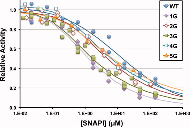
Comparison of the inhibitory profiles for the five GST-SNAPIs with Gly insertions to that of the wildtype GST-SNAP25(141–206) are shown in Figure 1. Three of these GST-SNAPIs, 2G, 4G, and 5G, showed IC50 values half that of the wildtype substrate, while GST-SNAPIs with 1G or 3G insertions were 10 times more effective inhibitors than the wildtype peptide with IC50 values of 1.3 and 1.7 μM, respectively (Table I). To further verify if these Gly insertion mutants are also cleavage resistant, these Gly insertion SNAPIs were incorporated into our previously described GFP fusion proteins containing a C-terminal 33-residue tag sequence for testing susceptibility to cleavage by BoNT/A-LC.23 Gel-shift assay showed that all five Gly insertion mutants were cleavage resistant, with G1, G4, and G5 exhibiting only a small amount of cleavage after prolonged treatment with BoNT/A-LC (Fig. 2), while G2 and G3 remain cleavage free even after 2 days of incubation (data not shown).
Figure 1.

Inhibitory profiles of Gly-insertion GST-SNAPIs for BoNT/A-catalyzed cleavage of YsCsYDE16. Solid circle (WT), solid diamond (1G), open diamond (2G), solid square (3G), open square (4G), and solid triangle (5G).
Figure 2.

BoNT/A-LC cleavage resistance of Gly-insertion GFP-SNAPIs. Shown are images of coomassie blue-stained 12.5% PAGE gels for aliquots taken at the indicated time points from reaction mixtures containing wildtype GFP-SNAP25 (WT) or GFP-SNAPIs (1G, 2G, 3G, 4G, 5G).
Comparative modeling of SNAPI with Gly insertions at the active site of BoNT/A-LC
To evaluate the feasibility of packing the extra Gly residues into the active site cleft of BoNT/A-LC, we performed comparative modeling using MODELLER software31, 32, 33 and the pdb1xtg file as the template. The SNAP25 peptide assumed a similar main chain conformation on the surface of wildtype BoNT/A-LC model as it does on the mutant BoNT/A-LC(E224Q/Y366F) (Supporting Information Fig. S1, Panels A and B). Models for SNAPI (1G–5G) showed increasing flexibility of the main chain conformation for the Gly insertions (Supporting Information Fig. S1, Panels C–G). A striking feature is the large size of the active site crevice, which allows it to tolerate up to 5 Gly insertions. These modeling results are consistent with the observed inhibition by SNAPIs, all of which exhibited IC50 values 2–10 times lower than that of the wildtype SNAPI.
Discussion
Small proteins with high affinity for BoNT/A-LC could be used as inhibitors or as effectors of protein down-regulation inside neuronal cells.12, 13 Protein-based intracellular intervention strategies become practical once specific delivery vehicles become available.13 SNAP25(141–206)-derived pseudosubstrate inhibitors (SNAPIs) offer an easy approach for potential high affinity binding protein for BoNT/A-LC. A substrate recognition mechanism for BoNT/A-LC was proposed to involve an initial binding of the P5 Asp to the S5 pocket followed by positioning of the P4′ Lys and P1′ Arg to their binding pockets.29 The structure of a catalytically inactive E224Q/Y366F mutant of BoNT/A-LC in complex with SNAP25(141–204) revealed important exosites for substrate binding remote from the active-site pocket.17 Although D194, R198 and K201 of the SNAP25 substrate are in the vicinity of the counter-charged residues R177, D370, and E257 on the enzyme (5.45 Å, 5.20 Å, and 4.65 Å, respectively), the distance between the side-chain nitrogen of the P1′ Arg and the side-chain oxygen of D370 was greater than that found in complexes of BoNT/A-LC with other inhibitors (Table II). The inactive mutant of the enzyme is presumably unable to make the conformational change necessary for catalysis to occur. This nonproductive enzyme-substrate complex also suggests that the anticipated induced-fit for the wildtype enzyme leading to enzyme catalysis did not occur. Using our SNAP25-derived FRET substrate YsCsY, it was possible to measure the binding affinity (in terms of IC50 values) of GST-SNAP25(141–206) for the catalytically active BoNT/A-LC against cleavage of the FRET substrate. The IC50 value for wildtype SNAP25 peptide was similar to that observed for the cleavage-resistant R198A, R198T, and A195S mutants, suggesting that the observed IC50 value (14 μM) is for the initial binding not the induced-fit complex.
Table II.
Distances Between Arg198 of SNAPIs or Corresponding Peptide Inhibitors and Asp370 of BoNT/A-LC
| Name | Distance (Å) | Ki (μM) | IC50 (μM) | Reference |
|---|---|---|---|---|
| RRGC | 2.65 | 0.158 | Ref.21 | |
| QRATKM | 2.90 | 133 | Ref.27 | |
| RRATKM | 2.95 | 95 | Ref.27 | |
| I1 peptide | 2.87 | 0.041 | Ref.22 | |
| CRATKML | 3.12a | 1.9b | (a) Ref.19, (b) Ref.20 | |
| SNAP25 | 5.20c | 14d | (c) Ref.17, (d) this study |
a–d denote that the indicated distance information and inhibitory properties are from different reference sources.
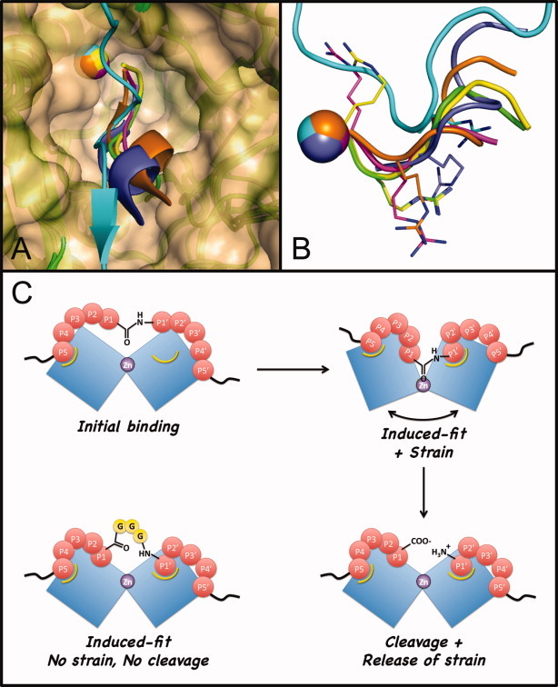
It has been shown through binding of small molecule inhibitors30 and peptide-like molecules22 that the substrate-binding cleft of the BoNT/A LC protease exhibits a remarkable plasticity. Several peptide-like molecules at the substrate-binding cleft not only adapt different backbone conformation [Fig. 3(A)], but also have very different disposition of the common Arg side chain [Fig. 3(B)]. Most remarkably, the D370 loop of BoNT/A-LC that shapes the S1 binding pocket could be repositioned to accommodate the Arg in each of the peptide-like molecules.22 On the basis of the high plasticity of the substrate-binding cleft, we tested the feasibility of creating a new binding pocket with several His mutants. Although some of these His-containing SNAPIs showed IC50 values only two to three times higher than the wildtype SNAP25, no clear pattern was apparent. On the other hand, insertion of up to five Gly residues between the P1-P1′ QR residues at the cleavage site of wildtype SNAP25 peptide resulted in cleavage-resistant SNAPIs with IC50 values 10-fold lower than that of the wildtype SNAP25 peptide (Table I). Since the poly-Gly insert is not likely to produce additional interactions with new binding pockets at the enzyme active site, the enhanced binding could only be from better complementarity between the SNAPI and BoNT/A-LC. We hypothesize that the induced fit in the enzyme-substrate complex preceding peptide bond cleavage could be trapped with these Gly-insertion SNAPIs. Outlined in Figure 3(C) is a proposed model for this inhibition. Upon the initial binding through interactions at remote exosites, wildtype SNAP25 loosely occupies the S5 site and positions the P1′–P5′ residues to the vicinity of their interacting residues in the active-site cleft. This initial binding is followed by a distortion of the enzyme for optimal contacts between the enzyme and substrate, and a build-up of strain in the complex that shapes the scissile bond toward a transition state for cleavage. After peptide-bond cleavage the strain is released and the optimal binding prior to cleavage no longer exists. In the case of SNAPI with Gly insertion, after initial binding, the optimal induced fit for the entire P5 to P1 and P1′–P5′ residues ensues, but no penalty of strain accompanies this binding. The Gly linker in the 1G or 3G mutants provides the best accommodation to the induced conformational change, while 2G may be geometrically less ideal and 4G, 5G may be too flexible and cost some entropy.
Figure 3.

Active-site plasticity of BoNT/A-LC. A: Shown are superposed structures of BoNT/A-LC(E224Q/Y366F) mutant complexed with SNAP25(141-204) (pdb1xtg, cyan) and 5 wildtype BoNT/A-LC complexes with Ac-CRATKML (pdb3boo, purple); I1 peptide (pdb3ds9, orange); QRATKM-NH2 (pdb3dda, green); RRATKM-NH2 (pdb3ddb, yellow); or RRGC-NH2 (pdb3c88, magenta). Zinc ions are shown as spheres. The surface at the BoNT/A-LC active site was generated from the pdb1xtg structure. B: Shown are the main chains of the pseudosubstrates or peptidomimetics from the same overlaying structures as in (A) after a 90° rotation. Side chains of all Arg residues are shown as sticks. The graphic image was generated with Pymol using the corresponding Protein Data Base (PDB) files. C: An induced-fit model for BoNT/A-LC-catalyzed cleavage of SNAP25 and inhibition of BoNT/A-LC by the Gly-insertion SNAPI (3G). Blue blocks represent enzyme surface at the catalytic cleft. Pockets marked with yellow curves are the induced, high affinity sites for P5 Asp and P1′ Arg.
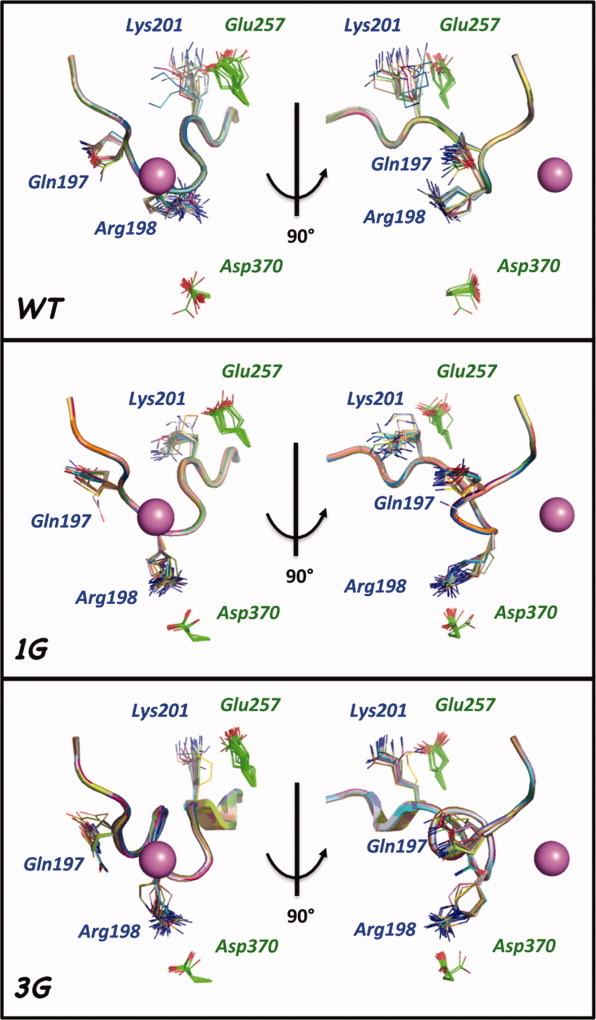
Comparative modeling has been widely used in modeling loop structures.32 Using MODELLER software we observed remarkable feasibility of the BoNT/A-LC active site to accommodate insertion of up to 5 Gly residues between Q197 and R198 of the substrate peptide (Supporting Information Fig. S1). The structures for small peptide inhibitors in complex with BoNT/A-LC suggest that close contact between the R198 equivalent of inhibitor mimetics and D370 of the enzyme is an important factor for tight binding (Table II). Most models generated for SNAPI-1G or SNAPI-3G in complex with BoNT/A-LC using 1XTG as template displayed a shorter distance between K201-E257 than R198-D370 (Supporting Information Table S1). The models of SNAPI-1G and SNAPI-3G with relatively low energy and short R198-D370 distances were used as new reference templates for additional modeling (Supporting Information Fig. S1, Panels H and I) to generate models with converged main chain conformations allowing for optimal interaction between R198 of SNAPI and D370 of BoNT/A-LC. As shown in Figure 4, SNAPI-1G and SNAPI-3G could have stable conformers bringing the side chain of R198 closer to D370 of BoNT/A-LC than that observed with the wildtype SNAP25 peptide. This further supports the hypothesis that SNAPI (1G and 3G) are able to utilize the optimal binding provided by the induced-fit of normal substrate upon binding the enzyme, yet the flexible Gly insertion alleviates the build up of conformational strain associated with this binding interaction.
Figure 4.

Comparative modeling of Gly insertion pseudosubstrates at BoNT/A-LC active-site. Shown are models of wildtype SNAP25 (residues 194–204) (top), the corresponding SNAPI with single Gly insertion (middle) or with three Gly insertions (bottom) complexed at the active site of BoNT/A-LC, generated using MODELLER software and presented with Pymol software (see Methods). Also indicated are the side chains of selected residues: Q197, R198, K201 of SNAP25 (shown in blue) and E257, D370 of BoNT/A (shown in green). The active site Zn ions are shown as purple spheres. Gly insertions were not numbered. Images of models on the left in each panel were vertically rotated 90° to generate the corresponding images on the right.
The discovery that Gly insertion at the SNAP25 cleavage site leads to 10-fold enhanced binding is consistent with a model for induced fit without conformational strain. Modification of this poly-Gly linker with other amino acids may uncover additional pockets for further improvement of the affinity of SNAPI for BoNT/A-LC. This phenomenon also has broader implications for other protease inhibitor designs by creating alternative pseudosubstrates capable of capturing the transient induced-fit state prior to peptide-bond cleavage, particularly for those with specificity defined by extended regions of the substrate. This molecular sculpturing approach could also be used in de novo design of nonantibody, high-affinity molecules.
Materials and Methods
Materials
BoNT/A-LC was prepared as described previously.23 PCR primers were purchased from IDT. PCR Reddy Mix from Abgene was used for all plasmid constructions. Restriction enzymes were purchased from Fermentas. BSA standard was obtained from Pierce.
Preparation of GST-SNAPI proteins
The previously described pGST-SNAP25(141-206) vector23 was used as a template to generate all GST-SNAPI vectors by PCR, using a forward primer in the GST region and two overlapping reverse primers to introduce the mutation and an EcoRI site after the stop codon. The PCR product was fragment-changed into the SacI-EcoRI sites of the parent vector. All His-insertion GST-SNAPI protein constructs were designed as poly-His mutants, but some as indicated were verified as containing Gln after sequencing, presumably due to mutation introduced by PCR. All GST-fusion proteins were isolated and purified from 1 liter of E. coli Rosetta strain cultures, as described previously.23 The protein preparation after glutathione-agarose affinity chromatography was concentrated to 1.5 mL using a Millipore filter concentrator with 30-kDa molecular weight cutoff, and then desalted through a PD-10 column in PBS. The proteins were stored at −20°C until use.
Preparation of GFP-SNAPI proteins
The vectors encoding GST-SNAPI 1G, 2G, 3G, 4G, or 5G, generated as above, were used as templates to generate the corresponding SNAPI PCR products, which were then inserted into the Sac I-Not I sites of the pGFP-SNAP25(141–206) vector23 to generate the corresponding GFP-SNAPI proteins with a 33-amino acid tag at their C-terminus. The resulting GFP-SNAPI proteins were isolated and purified, as described above, and used for gel shift cleavage assay.
Preparation of YsCsYDE16 substrate
The previously described YsCsY FRET substrate,23 YFP-SNAP-CFP-SNAP-YFP (where SNAP denotes the 66-mer substrate peptide corresponding to residues 141–206 of SNAP25), was modified by addition of 16 Asp-Glu repeats (DE16) to the C terminus by PCR amplification of the sequence coding for DE16 from pGFP-DE16-BoNTA-HC,23 followed by addition of it to the 3′-end of the coding region of YsCsY through PCR primer extension method. The resulting modified YsCsYDE16 protein was expressed and purified as described before,23 and exhibited similar fluorescence properties and kinetic behavior as YsCsY (data not shown), but was more easily purified by Ni-chelation affinity and ANX-cation exchange chromatography.
FRET-based BoNT/A-LC activity assay
The FRET-based cleavage assay was carried out in a reaction buffer containing 20 mM HEPES, pH 7.5, 1.25 mM DTT, 0.2% Tween-20, 0.1 mg/mL BSA, and 10 mM ZnCl2, as described before.23 Using a 384-well assay plate, GST-SNAPI fusion proteins were setup in 10-well rows with 3-fold serial dilutions, followed by wells with positive or negative controls. BoNT/A-LC was added to each well, except for the control wells without enzyme. Serial dilution of each SNAPI was carried out directly on the microtiter assay plate using a mechanical liquid handler epMotion 5070 (Eppendorf). Solutions of YsCsYDE16 substrate (2 μM, final concentration) and BoNT/A-LC (100 pM, final concentration) were added by using a microfiller (Biotek). The assay plate was read on a Synergy 4 UV-Vis-Fluorescence plate reader (Biotek) using 400/30 nm filter for excitation and 485/20 nm and 528/20 nm filters for emission. The 528 nm/485 nm fluorescence emission ratio for each well was calculated by using Gen5 software and data was exported as an excel file. The relative YsCsYDE16 substrate cleavage rates in the presence of each of the GST-SNAPI fusion proteins at the indicated serial dilutions were determined from the initial decrease in 528 nm/485 nm ratio comparing with the fluorescence ratio for the control reaction wells without SNAPI. The relative rates for each SNAPI at a series of inhibitor concentrations were fitted to a logistic function for calculating the corresponding IC50 value.
Gel-shift cleavage assay
For gel-shift cleavage assays the corresponding GFP-fusion proteins were used instead of the GST-fusions because of the greater gel separation observed between the uncleaved substrate and the resulting cleaved product (data not shown). Reactions (200 μL total volume), containing GFP-SNAP(141–206) or GFP-SNAPI (1G, 2G, 3G, 4G, 5G) at a final concentration of 6 μM, were initiated and maintained at 30°C upon addition of BoNT/A-LC (300 pM, final concentration) to the reaction buffer, as described above for the FRET assay. At various time points, an aliquot (25 μL) was removed from each reaction, mixed with an equal volume of SDS-PAGE sampling buffer, and immediately boiled for 1 min. The samples were analyzed by 12.5% SDS-PAGE and stained with Coomassie blue for visualization.
Data analysis
The change in 528 nm/485 nm fluorescence ratio for the initial three time points (5-min intervals) was used for calculating initial rates. Relative rates normalized to the control with enzyme only (without GST-SNAPI) were fitted to an empirical logistic function  , where y is the relative rate and x is the concentrations for GST-SNAPI. Least squares curve fitting was calculated by minimizing the sum of squares
  , using the Solver add-in for Excel (Frontline Systems). IC50 values were determined by finding x for y = 0.5 in the solved logistic equation with optimal parameters. The standard deviation for the IC50 value was estimated from the y variance of the curve-fitting and
  , where Δx is the difference in x for
  . For selected proteins, at least two series of serial dilutions were used for determination of Ki (see Table I). And for wildtype SNAPI, SNAPI-1G, and SNAPI-3G the serial dilutions from different protein preparations were repeated at least three times. All protein concentrations were determined based on densitometry of Coomassie blue-stained gels using BSA as the standard.
Comparative modeling for SNAPI pseudosubstrates
Comparative models were produced using MODELLER software (version 9.8)31, 32 to generate the models for each pseudosubstrate interfacing with BoNT/A-LC. Models were presented with Pymol software. Models for BoNT/A-LC complexed with SNAP25(141–204) and each of the pseudosubstrate peptides, where 1–5 Gly residues were incorporated between Q197 and R198 of SNAP25(141–206), were generated using the model-single.py function, based on the alignment with the double mutant BoNT/A-LC-SNAP25 complex (PDB ID:pdb1xtg). To restore the crystal structure of the double mutant to the wildtype protein, the amino acids mutated in the double mutant were with the corresponding wildtype amino acid (E224Q/Y366F), and the active site zinc and water as well as a chloride ion were also incorporated into the model. The CHARMM22 forcefield was used in conjunction with the variable target function method to anneal the peptide to the substrate surface by using increasingly restrained simulated annealing and molecular dynamics. The calculated energetics of the final structure were obtained using the MODELLER scoring function “molpdf”, which is a unitless composite score based on the DOPE (discrete optimized protein energy).33 A control was performed aligning 1XTG with 1XTG to confirm the structure was minimally perturbed through the modeling process (bbRMSD ∼ 0.3 Å). For each complex, 25 models were sampled, the superimposed structures of which are shown in Supporting Information Figure 1. For SNAPI-1G and SNAPI-3G, a low energy model with short distance between R198 of SNAP25 and D370 of BoNT/A-LC was used as an initial structure for remodeling.
Supplementary material
Additional Supporting Information may be found in the online version of this article.
References
- 1.Arnon SS, Schechter R, Inglesby TV, Henderson DA, Bartlett JG, Ascher MS, Eitzen E, Fine AD, Hauer J, Layton M, Lillibridge S, Osterholm MT, O'Toole T, Parker G, Perl TM, Russell PK, Swerdlow DL, Tonat K. Botulinum toxin as a biological weapon: medical and public health management. JAMA. 2001;285:1059–1070. doi: 10.1001/jama.285.8.1059. [DOI] [PubMed] [Google Scholar]
- 2.Schiavo G, Matteoli M, Montecucco C. Neurotoxins affecting neuroexocytosis. Physiol Rev. 2000;80:717–766. doi: 10.1152/physrev.2000.80.2.717. [DOI] [PubMed] [Google Scholar]
- 3.Lacy DB, Stevens RC. Unraveling the structures and modes of action of bacterial toxins. Curr Opin Struct Biol. 1998;8:778–784. doi: 10.1016/s0959-440x(98)80098-5. [DOI] [PubMed] [Google Scholar]
- 4.Lacy DB, Tepp W, Cohen AC, DasGupta BR, Stevens RC. Crystal structure of botulinum neurotoxin type A and implications for toxicity. Nat Struct Biol. 1998;5:898–902. doi: 10.1038/2338. [DOI] [PubMed] [Google Scholar]
- 5.Montecucco C, Molgo J. Botulinal neurotoxins: revival of an old killer. Curr Opin Pharmacol. 2005;5:274–279. doi: 10.1016/j.coph.2004.12.006. [DOI] [PubMed] [Google Scholar]
- 6.Montal M. Botulinum neurotoxin: a marvel of protein design. Ann Rev Biochem. 2010;79:591–617. doi: 10.1146/annurev.biochem.051908.125345. [DOI] [PubMed] [Google Scholar]
- 7.Foran PG, Davletov B, Meunier FA. Getting muscles moving again after botulinum toxin: novel therapeutic challenges. Trends Mol Med. 2003;9:291–299. doi: 10.1016/s1471-4914(03)00113-8. [DOI] [PubMed] [Google Scholar]
- 8.Chertow DS, Tan ET, Maslanka SE, Schulte J, Bresnitz EA, Weisman RS, Bernstein J, Marcus SM, Kumar S, Malecki J, Sobel J, Braden CR. Botulism in 4 adults following cosmetic injections with an unlicensed, highly concentrated botulinum preparation. JAMA. 2006;296:2476–2479. doi: 10.1001/jama.296.20.2476. [DOI] [PubMed] [Google Scholar]
- 9.Hakami RM, Ruthel G, Stahl AM, Bavari S. Gaining ground: assays for therapeutics against botulinum neurotoxin. Trends Microbiol. 2010;18:164–172. doi: 10.1016/j.tim.2010.02.001. [DOI] [PubMed] [Google Scholar]
- 10.Goodnough MC, Oyler G, Fishman PS, Johnson EA, Neale EA, Keller JE, Tepp WH, Clark M, Hartz S, Adler M. Development of a delivery vehicle for intracellular transport of botulinum neurotoxin antagonists. FEBS Lett. 2002;513:163–168. doi: 10.1016/s0014-5793(02)02268-8. [DOI] [PubMed] [Google Scholar]
- 11.Ho M, Chang LH, Pires-Alves M, Thyagarajan B, Bloom JE, Gu Z, Aberle KK, Teymorian SA, Bannai Y, Johnson SC, McArdle JJ, Wilson BA. Recombinant botulinum neurotoxin A heavy chain-based delivery vehicles for neuronal cell targeting. Protein Eng Des Sel. 2011;24:247–253. doi: 10.1093/protein/gzq093. [DOI] [PMC free article] [PubMed] [Google Scholar]
- 12.Tsai YC, Maditz R, Kuo CL, Fishman PS, Shoemaker CB, Oyler GA, Weissman AM. Targeting botulinum neurotoxin persistence by the ubiquitin-proteasome system. Proc Natl Acad Sci USA. 2010;107:16554–16559. doi: 10.1073/pnas.1008302107. [DOI] [PMC free article] [PubMed] [Google Scholar]
- 13.Kuo CL, Oyler GA, Shoemaker CB. Accelerated neuronal cell recovery from botulinum neurotoxin intoxication by targeted ubiquitination. PLoS One. 2011;6:e20352. doi: 10.1371/journal.pone.0020352. [DOI] [PMC free article] [PubMed] [Google Scholar]
- 14.Rubinsztein DC. The roles of intracellular protein-degradation pathways in neurodegeneration. Nature. 2006;443:780–786. doi: 10.1038/nature05291. [DOI] [PubMed] [Google Scholar]
- 15.Chen S, Barbieri JT. Association of botulinum neurotoxin serotype A light chain with plasma membrane-bound SNAP-25. J Biol Chem. 2011;286:15067–15072. doi: 10.1074/jbc.M111.224493. [DOI] [PMC free article] [PubMed] [Google Scholar]
- 16.Washbourne P, Pellizzari R, Baldini G, Wilson MC, Montecucco C. Botulinum neurotoxin types A and E require the SNARE motif in SNAP-25 for proteolysis. FEBS Lett. 1997;4189(1-2):1–5. doi: 10.1016/s0014-5793(97)01328-8. [DOI] [PubMed] [Google Scholar]
- 17.Breidenbach MA, Brunger AT. Substrate recognition strategy for botulinum neurotoxin serotype A. Nature. 2004;432:925–929. doi: 10.1038/nature03123. [DOI] [PubMed] [Google Scholar]
- 18.Brunger AT, Breidenbach MA, Jin R, Fischer A, Santos JS, Montal M. Botulinum neurotoxin heavy chain belt as an intramolecular chaperone for the light chain. PLoS Pathog. 2007;3:1191–1194. doi: 10.1371/journal.ppat.0030113. [DOI] [PMC free article] [PubMed] [Google Scholar]
- 19.Silvaggi NR, Wilson D, Tzipori S, Allen KN. Catalytic features of the botulinum neurotoxin A light chain revealed by high resolution structure of an inhibitory peptide complex. Biochemistry. 2008;47:5736–5745. doi: 10.1021/bi8001067. [DOI] [PubMed] [Google Scholar]
- 20.Schmidt JJ, Stafford RG, Bostian KA. Type A botulinum neurotoxin proteolytic activity: development of competitive inhibitors and implications for substrate specificity at the S1’ binding subsite. FEBS Lett. 1998;435:61–64. doi: 10.1016/s0014-5793(98)01041-2. [DOI] [PubMed] [Google Scholar]
- 21.Hale M, Oyler G, Swaminathan S, Ahmed SA. Basic tetrapeptides as potent intracellular inhibitors of type A botulinum neurotoxin protease activity. J Biol Chem. 2011;286:1802–1811. doi: 10.1074/jbc.M110.146464. [DOI] [PMC free article] [PubMed] [Google Scholar]
- 22.Zuniga JE, Schmidt JJ, Fenn T, Burnett JC, Arac D, Gussio R, Stafford RG, Badie SS, Bavari S, Brunger AT. A potent peptidomimetic inhibitor of botulinum neurotoxin serotype A has a very different conformation than SNAP-25 substrate. Structure. 2008;16:1588–1597. doi: 10.1016/j.str.2008.07.011. [DOI] [PMC free article] [PubMed] [Google Scholar]
- 23.Pires-Alves M, Ho M, Aberle KK, Janda KD, Wilson BA. Tandem fluorescent proteins as enhanced FRET-based substrates for botulinum neurotoxin activity. Toxicon. 2009;53:392–399. doi: 10.1016/j.toxicon.2008.12.016. [DOI] [PMC free article] [PubMed] [Google Scholar]
- 24.O'Sullivan GA, Mohammed N, Foran PG, Lawrence GW, Oliver Dolly J. Rescue of exocytosis in botulinum toxin A-poisoned chromaffin cells by expression of cleavage-resistant SNAP-25. Identification of the minimal essential C-terminal residues. J Biol Chem. 1999;274:36897–36904. doi: 10.1074/jbc.274.52.36897. [DOI] [PubMed] [Google Scholar]
- 25.Chen S, Barbieri JT. Unique substrate recognition by botulinum neurotoxins serotypes A and E. J Biol Chem. 2006;281:10906–10911. doi: 10.1074/jbc.M513032200. [DOI] [PubMed] [Google Scholar]
- 26.Raghunath A, Perez-Branguli F, Smith L, Dolly JO. Adeno-associated virus transfer of a gene encoding SNAP-25 resistant to botulinum toxin A attenuates neuromuscular paralysis associated with botulism. J Neurosci. 2008;28:3683–3688. doi: 10.1523/JNEUROSCI.5690-07.2008. [DOI] [PMC free article] [PubMed] [Google Scholar]
- 27.Kumaran D, Rawat R, Ahmed SA, Swaminathan S. Substrate binding mode and its implication on drug design for botulinum neurotoxin A. PLoS Pathog. 2008;4:e1000165. doi: 10.1371/journal.ppat.1000165. [DOI] [PMC free article] [PubMed] [Google Scholar]
- 28.Kumaran D, Rawat R, Ludivico ML, Ahmed SA, Swaminathan S. Structure- and substrate-based inhibitor design for Clostridium botulinum neurotoxin serotype A. J Biol Chem. 2008;283:18883–18891. doi: 10.1074/jbc.M801240200. [DOI] [PubMed] [Google Scholar]
- 29.Chen S, Kim JJ, Barbieri JT. Mechanism of substrate recognition by botulinum neurotoxin serotype A. J Biol Chem. 2007;282:9621–9627. doi: 10.1074/jbc.M611211200. [DOI] [PubMed] [Google Scholar]
- 30.Silvaggi NR, Boldt GE, Hixon MS, Kennedy JP, Tzipori S, Janda KD, Allen KN. Structures of Clostridium botulinum neurotoxin serotype A light chain complexed with small-molecule inhibitors highlight active-site flexibility. Chem Biol. 2007;14:533–542. doi: 10.1016/j.chembiol.2007.03.014. [DOI] [PubMed] [Google Scholar]
- 31.Sali A, Blundell TL. Comparative protein modeling by satisfaction of spatial restraints. J Mol Biol. 1993;234:779–815. doi: 10.1006/jmbi.1993.1626. [DOI] [PubMed] [Google Scholar]
- 32.Marti-Renom MA, Stuart A, Fiser A, Sanchez R, Melo F, Sali A. Comparative protein structure modeling of genes and genomes. Annu Rev Biophys Biomol Struct. 2000;29:291–325. doi: 10.1146/annurev.biophys.29.1.291. [DOI] [PubMed] [Google Scholar]
- 33.Shen MY, Sali A. Statistical potential for assessment and prediction of protein structures. Prot Sci. 2006;15:2507–2524. doi: 10.1110/ps.062416606. [DOI] [PMC free article] [PubMed] [Google Scholar]
Associated Data
This section collects any data citations, data availability statements, or supplementary materials included in this article.


