Abstract
Objective
The current study longitudinally evaluated concentrations of fibrinogen (Fib), D-Dimer, plasminogen activator type-1 (PAI-1) and tissue type plasminogen activator (T-Pa) before pregnancy and in the first and third trimesters of pregnancy with a focus on the pregnancy transition.
Study design
Twenty healthy, nonsmoking, nulliparous women, aged 29.8 ± 3.0 years, BMI 23.3 ± 3.2 kg/m2 were studied during menstrual cycle day 8 ± 4 and again in early (11 – 15 wks) and late (31 – 34 wks) pregnancy. Seventeen women had singleton conceptions and delivered at term with uncomplicated pregnancies (CTL) and three women developed complicated hypertension (CH) during pregnancy after the third trimester (late pregnancy) evaluation. Data are means ± SEM, Significance was based on p < 0.05.
Results
Fib was the only protein evaluated that increased in early pregnancy relative to the prepregnancy assessment. D-dimer, PAI-1 and T-Pa increased in the third trimester compared with prepregnant and early pregnant values (p < .001). T-PA was significantly higher during late pregnancy in CH subjects compared with CTL (8.1 ± 0.7 ng/ml vs. 5.0 ± 0.2 ng/ml, p = .02). There were no other differences between groups.
Conclusions
Increases in fibrinogen are evident in early pregnancy whereas fibrinolysis, perhaps in response to the procoagulant environment of pregnancy, is increased during late pregnancy. Before development of clinically overt hypertension, T-Pa is increased without concomitant changes in other proteins assessed. This is consistent with altered endothelial function with preeclampsia that may contribute to, or reflect, the vasculopathy accompanying this disorder.
Keywords: pregnancy, preeclampsia, coagulation, fibrinolysis, T-Pa, fibrinogen
Introduction
Pregnancy is accompanied by changes in the expression of coagulation and fibrinolytic proteins that favor a balance towards clot formation. Increases in the activation of the coagulation cascade contribute to a reduction in hemorrhage risk that could otherwise be detrimental to fetal and maternal health. Alterations in both the extrinsic and intrinsic pathways of coagulation have been well-characterized and show that prothrombin, coagulation factors VII, X, XII and VIII are increased during pregnancy [1]. Fibrinogen, the precursor of fibrin, has been shown to increase during pregnancy contributing to a procoagulant environment [1–3].
Proteins in the fibrinolytic system that inhibit fibrinolysis are upregulated thereby further favoring coagulation. Plasminogen activator inhibitor type-1 (PAI-1) is an antifibrinolytic protein implicated in vascular remodeling [4]. PAI-1 is secreted by endothelial cells stimulated by a number of signaling moieties including thrombin [5]. During pregnancy, PAI-1 is a primary inhibitor of tissue type plasminogen activator (T-Pa), a key protein involved in fibrin degradation [6, 7]. T-PA is produced by endothelial cells, secreted locally in response to endothelial injury, and in the presence of fibrin, is a potent activator of plasminogen, a crucial step in fibrin degradation [5]. T-PA increases during pregnancy; however, increases in PAI-1 supersede those in T-PA thereby maintaining a procoagulable environment [8]. Though coagulation processes are predominant over fibrinolysis during pregnancy, D-Dimer, a product of fibrin degradation, increases with progressing pregnancy indicating a functionally active fibrinolytic system [8].
Whereas normal pregnancy is accompanied by increased coagulation, women who develop preeclampsia (PE) have augmented coagulation compared with those with to normal pregnancy [9]. Clinical manifestations of increased coagulation include excess fibrin deposition and microthrombus formation involving both maternal organs and the placenta [10]. The hypercoagulability of PE has been attributed to endothelial dysfunction, decreased antithrombin levels, and increased levels in markers of thrombin activity [9, 11]. Other proteins involved in hemostasis such as factor VIII, soluble fibrin, thrombomodulin and D-dimer are also reported to be altered in PE [12–16].
The objective of the current study was to evaluate concentrations of coagulation and fibrinolytic system proteins longitudinally in nulliparous women prior to pregnancy and through pregnancy with a primary focus on the transition from the nonpregnant state to pregnancy. We evaluated concentrations of fibrinogen, PAI-1, T-Pa, and fibrin fragment D-Dimer before pregnancy and during early and late pregnancy. In addition, we evaluated differences at these time points in women who subsequently developed preeclampsia.
Methods
Thirty four nulligravid women interested in conception were enrolled in this research study through an open advertisement. Women were provided with ovulation detection kits (Quidel Corporation, San Diego, CA) to assist with achieving a successful conception. All subjects were young (18–40) healthy, nonsmokers with regular menstrual cycles at the time of enrollment. None of the women had a history of hypertension, autoimmune disease, diabetes or other disorders known to affect blood pressure. Thirty women subsequently conceived. Eight subjects conceived before baseline prepregnancy studies were performed; one subject had a first trimester miscarriage; one subject was lost to follow-up. The remaining 20 subjects, all of whom conceived singleton pregnancies, had complete prepregnancy assessments and successful pregnancy outcomes comprise the current report. One woman missed her first trimester assessment and was therefore not included in the early pregnancy study day data. Three women developed complicated hypertension (CH) during pregnancy. Two of which had classically defined preeclampsia with 24 hour urine collections demonstrating proteinuria >300 mg/dl and blood pressure >140/90 mmHg. The third woman had new onset third trimester, elevated blood pressure >140/90 mmHg, elevated liver enzymes, elevated uric acid concentration (>5mg/dl) and fetal growth restriction with iatrogenic delivery at 37 weeks. Women were enrolled consecutively over a 33 month period, from May 2004 through February 2007. Prior to each study visit subjects were provided with a 3500-mg sodium-balanced diet for 72 hours. Each subject was asked to abstain from alcohol and caffeine beginning at least 24 hours before the study and to avoid the use of decongestants and nonsteroidal medications beginning at least 48 hours before the study. All prepregnancy assessments were performed during the follicular phase. Assessments during pregnancy were performed between 11 and 15 menstrual weeks. Ovulation detection and early pregnancy ultrasound assessments were used to calculate gestational age. The research protocols were approved by the University of Vermont Human Investigational Committees. All women studied provided written informed consent.
Each periodic assessment was conducted between 8 AM and 10 AM. Subjects were admitted to the University of Vermont General Clinical Research Center on the day of the study after an overnight fast. For subjects’ prepregnancy visit, first-void urine was obtained to confirm nonpregnant state. Following height and weight determination, subjects rested in the supine position for the remainder of the study with a minimum of 30 minutes before blood collection.
Blood samples were collected into EDTA or sodium citrate tubes, depending on the assay, from the antecubital fossa with the use of an indwelling venous saline lock and following a 2.5 mL discard. They were centrifuged within 60 minutes for 15 minutes at 3000 × g to isolate plasma. Plasma was then aliquoted and stored at −70°C until analysis.
Assay Methods
Coagulation
Fibrinogen concentrations in plasma were determined with the use of the Clauss method [17]. Fibrinogen levels are directly correlated with clotting time of a diluted plasma sample in the presence of excess thrombin. Fibrinogen concentrations were quantified with the STAR automated coagulation analyzer (Diagnostica Stago, Parsippany, NJ). Patient fibrinogen results were then compared based on control results. Inter-assay CV ranged from 2 – 6%.
Fibrinolysis
T-PA antigen and PAI-1 antigen were assessed with the use of commercially available ELISAs, consistent with manufacturer’s instructions (TriniLIZE T-Pa Antigen kit and TriniLIZE PAI-1 Antigen kit, Diagnostica Stago). PAI-1 antigen reflects both active and inactive PAI-1, as well as PAI-1 complexed with T-Pa and urokinase plasminogen activator.
PAI-1 activity was assessed as previously described using a modified chromogenic substrate enzymatic assay method developed by Chmielewska and Wiman [18].
D-Dimer was measured with the use of a commercially available immuno-turbidometric assay (Liatest D-Di, Diagnostica Stago) consistent with the manufacturer’s instructions. The interassay CV ranged from 5 to 14%.
Statistical Analyses
For demographic information listed in Table 1, Student’s t-test was used to determine statistical significance. P-value for birth weight percentile reflects a probit transformation of percentiles to z-scores prior to analysis. Two-factor repeated measures analyses of variance were used to compare women with normal pregnancies and to those complicated by hypertension on longitudinal changes in coagulation and fibrinolytic proteins. The two factors in the ANOVA represented pregnancy group (an across-subject factor) and assessment time (a within-subject factor). When appropriate, F-tests for simple effects were used to examine temporal trends each groups subsequent to the overall ANOVA. Pairwise comparisons were performed using Fisher’s LSD. Spearman’s rank correlation (r) was used to examine the correlation between outcome measures within and across time points Analyses were performed using SAS Statistical Software Version 9.2 (SAS Institute, Cary NC). Statistical significance was determined based on α=.05.
Table 1.
Maternal demographic characteristics and pregnancy outcomes
| Characteristic | CTL (n = 17) Mean ± SE |
CH (n = 3)* Mean ± SE |
p-value# |
|---|---|---|---|
| Maternal age (years) | 28.9 ± 0.7 | 31.3 ± 1.2 | .21 |
| BMI (kg/m2) | 23.6 ± 0.8 | 21.5 ± 0.9 | .31 |
| Prepregnancy cycle day | 8.6 ± 1.1 | 7.7 ± 1.2 | .71 |
| First trimester study day† | 93.3 ± 2.6 | 97 ± 7.6 | .60 |
| Third trimester study day | 227.8 ± 2.0 | 212.0 ± 0.6 | .005 |
| Gestational age at delivery (weeks) | 39.7 ± 0.3 | 38 ± 1.0 | .13 |
| Birth weight (g) | 3582 ± 96 | 2514 ± 421 | .001 |
| Birth weight percentile | 55.2 ± 6.0 | 12 ± 9.1 | .003 |
Three women developed complicated hypertension (CH) during pregnancy. Two of which had classically defined preeclampsia, the third woman had elevated blood pressure, elevated liver enzymes, elevated uric acid concentration and fetal growth restriction in the third trimester. The remaining 17 women are designated as uncomplicated pregnancies or controls (CTL).
Significance based on two sample t-tests. P-value for birth weight percentile reflects a probit transformation of percentiles to z-scores prior to analysis.
One control subject missed her first trimester study day. The demographic and outcome data presented in this table include all 17 women.
Results
Subject characteristics
All pregnancies were singletons, and the majority of the subjects were Caucasian, 90% (18/20). Clinical and demographic characteristics are presented in Table 1. There were no significant differences between non-hypertensive (CTL) and women developed complicated hypertension (CH) in age, body mass index (BMI), prepregnancy cycle day, early pregnancy study day and gestational age at delivery (Table 1). There was a significant difference between CTL and CH third trimester study day where CTLs were studied sixteen days later than CH. Birthweight and birthweight percentile of the newborns were significantly lower in CH subjects compared with CTL (p = .001 and p = .01, respectively). Two of the CH newborns were small for gestational age (1st and 5th birthweight percentiles) whereas the third CH newborn was in the 30th percentile. CTL newborn birthweights ranged from the 15th to 95th percentile.
The mean and SE for each of the hemostatic assessments in both of the groups are listed in Table 2 and presented in Figures 1 and 2.
Table 2.
Numerical values and statistical comparisons of fibrinogen, and fibrinolytic system proteins before pregnancy and during pregnancy.
| Prepregnancy | Early Pregnancy† | Late Pregnancy | p-value# | |||||||
|---|---|---|---|---|---|---|---|---|---|---|
| Variable | CTL | CH | CTL | CH | CTL | CH | Assessment Time |
Pregnancy Group |
Time by Group Interaction |
|
|
Coagulation System |
||||||||||
| Fibrinogen | 223 ± 12 | 199 ± 21 | 360 ± 17 | 318 ± 24 | 473 ± 27 | 330 ± 129 | <.001 | .07 | .17 | |
|
Fibrinolytic System |
||||||||||
| D-Dimer | 0.26 ± 0.04 | 0.3 ± 0.08 | 0.49 ± 0.06 | 0.45 ± 0.04 | 0.97 ± 0.10 | 0.96 ± 0.25 | <.001 | .99 | .96 | |
| PAI-1 Antigen |
17.3 ± 5.7 | 20.1 ± 10.5 | 17.7 ± 1.9 | 24.7 ± 3.2 | 66.4 ± 4.9 | 57.7 ± 12.9 | <.001 | .96 | .68 | |
| PAI-1 Activity | 9.3 ± 1.9 | 6.1 ± 1.7 | 9.0 ± 0.8 | 8.9 ± 2.6 | 31.4 ± 3.0 | 30.0 ± 3.1 | <.001 | .65 | .91 | |
| T-PA | 4.2 ± 0.4 | 3.2 ± 0.3 | 3.6 ± 0.2 | 4.1 ± 0.5 | 5.0 ± 0.22 | 8.1 ± 0.69 | <.001 | .10 | .001 | |
Significance levels based on repeated measures analyses of variance
One control subject missed her first trimester study day. Therefore, the first trimester measurements only include 16 women.
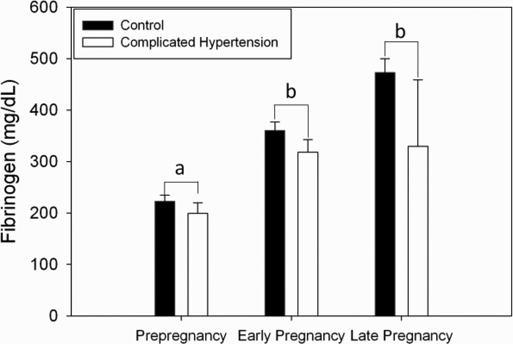
Figure 1.
Fibrinogen increased in early pregnancy compared with prepregnancy values and remained high in the third trimester. Means with a common letter are not significantly different (p < .05). There were no differences between those women who developed complicated hypertension and those with uncomplicated pregnancies.
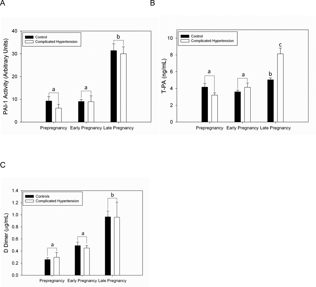
Figure 2.
Fibrinolytic protein profiles before pregnancy and during pregnancy. Means with a common letter are not significantly different (p < .05). A) Plasminogen activator inhibitor type-1 (PAI-1) increased in late pregnancy compared with prepregnancy and early pregnancy measurements. There were no differences at any time point between those women who developed complicated hypertension and those with uncomplicated pregnancies. B) T-Pa is increased during late pregnancy in both groups compared with earlier time points. During the third trimester, T-Pa was significantly higher in those who developed complicated hypertension during pregnancy, before the development of disease (p < .001). There are no differences between groups before pregnancy or in early pregnancy. C) D-Dimer increases in late pregnancy compared with prepregnancy and early pregnancy values. There are no differences between those women who developed complicated hypertension and those with uncomplicated pregnancies.
Coagulation
Plasma levels of fibrinogen increased in early pregnancy compared with prepregnancy levels and remained elevated at the third trimester study day in both CTL and CH groups (Fig 1; p < .001). There was a trend for lower fibrinogen in CH compared to CTL independent of assessment time (p = .07) with no evidence of an interaction between the pregnancy group and time (p = .17).
Fibrinolysis
Plasma PAI-1 antigen was increased in the third trimester in both CTL and CH compared with the earlier study time points (Table 2, p <.001). There were no differences between CTL and CH in plasma PAI-1 levels. We also assessed PAI-1 activity (Fig 2A). The pattern of PAI-1 activity paralleled the pattern of PAI-1 antigen change in pregnancy. There was a significant change over time of T-Pa antigen. There was evidence that the temporal pattern was different between the two groups (group by time interaction, p = .001, Fig 2B) due to significantly increased T-PA antigen in the CH group compared with CTL in the third trimester. These different temporal patterns resulted in significant differences between CTL and CH during late pregnancy, (p < 0.001). It should be noted that these assessments were made prior to the development of clinical disease. Third trimester plasma D-dimer was increased in the third trimester compared with both prepregnancy and early pregnancy study days (Fig 2C, p < .0001). There were no differences between CTL and CH D-dimer levels at any of the time points studied.
Correlation Analyses
We were interested in determining the contribution of prepregnancy coagulation and fibrinolytic proteins to early pregnancy and late pregnancy values. To address this question, we performed correlation analyses on all subjects, examining the relationship between visits for each of the coagulation and fibrinolytic factors. We found no significant correlation between prepregnant fibrinogen and either early or late pregnancy fibrinogen (p = .12 and p = .24, respectively). There was a significant positive correlation between prepregnancy and early pregnancy PAI-1 activity (r = .63, p = .004). However, this relationship was no longer present in late pregnancy (r = .007, p = .98). Prepregnancy T-PA levels tended to be associated with early pregnancy values (r = .40, p = .09), and there was no significant relationship between prepregnancy and late pregnancy T-PA (r = −.07, p = .77).
Discussion
This study was designed to assess the contribution of prepregnancy coagulation and fibrinolytic status to measurements made during pregnancy. We evaluated prepregnancy concentrations of fibrinogen, PAI-1, T-Pa and D-dimer with reassessment of these proteins during early and late pregnancy.
Pregnancy is known to be a procoagulable state; therefore, it is not surprising that we, and others, have observed an increase in fibrinogen beginning in early pregnancy [1–3]. In our group of longitudinally studied women, fibrinogen increased significantly in early pregnancy compared with prepregnancy and remained high during late pregnancy. Others have found very similar increases in fibrinogen from first to third trimester [8], though reports evaluating fibrinogen longitudinally, beginning before pregnancy and longitudinally through pregnancy are lacking. We did not detect any difference in fibrinogen in women who later developed CH, though preeclampsia is known to be a hypercoagulable state, primarily due to increases in thrombin and markers of thrombin activity, [9, 19, 20]. Fibrinogen has been reported to be higher in women with PE, and has been shown to correlate with severity of disease [21, 22]. Our evaluation of fibrinogen was before the onset of clinical disease in women who developed term PE, rather than more severe, early onset, forms of preeclampsia.
PAI-1 has anti-fibrinolytic activity through inhibition of T-PA, thus opposing fibrinolysis [23]. Indeed, clot stability is favored during pregnancy because concentrations of PAI-1 increase during pregnancy [8, 24, 25]. Reported increases in PAI-1 during pregnancy begin as early as the second trimester to the third trimester [8, 25]. We observed a significant increase in PAI-1 at the late pregnancy assessment compared with the prepregnancy and early pregnancy levels. We did not perform a second trimester assessment and PAI-1 may have increased before our late pregnancy study date. Our data further suggest that concentrations of PAI-1 remain relatively stable through the nonpregnant to pregnant transition and are not a predisposing factor to CH. Whereas correlation analyses suggest that prepregnancy values of PAI-1 portend early pregnancy values of PAI-1, third trimester concentrations of PAI-1 are independent of prepregnancy or early pregnancy concentrations of PAI-1. Thus, the predictive value of prepregnancy PAI-1 measurements is negligible. We did not detect any difference in concentrations of PAI-1 at any time point in our small cohort of women with CH compared with normal pregnant women. This is likely to be due to the very small sample size in our CH group as several other studies have found a significant increase in PAI-1 detectable by 23 weeks that persists through delivery [24, 26, 27].
Early studies investigating fibrinolysis during pregnancy reported decreased fibrinolysis reflected by plasminogen activator levels [2, 28, 29]. Later, studies suggested that alterations occurred in concentrations of both fibrinolytic system proteins such as plasminogen activator inhibitors and pro-coagulant proteins including antithrombin and von Willebrand factor during pregnancy [30–32]. One pro-fibrinolytic protein, T-Pa, has been shown to increase during the second trimester and remain elevated until 8 weeks postpartum [8, 25, 26]. Our studies confirm previous observations of increasead T-Pa in late pregnancy. T-PA increased simultaneously with PAI-1 suggesting a compensatory effect. This might be expected because fibrinolysis does not intensify until late pregnancy, T-PA is not increased during the early transition into pregnancy. Nevertheless, in our very small cohort of CH women, we found a significant increase in T-Pa during late pregnancy before the onset of clinical disease. Elevated levels of T-PA have been observed in women during the second trimester, before clinical disease in those who later developed PE [24]. In the face of CH and PE, T-Pa appears to potentially mitigate the hypercoagulable state seen with PE. Thus, increases in T-Pa may, one can speculate, identify women prone to develop CH or PE.
As pregnancy progresses into the 3rd trimester, fibrinolysis increases. This is evidenced by increased T-Pa, unchanged fibrinogen and increasing D-dimer. D-dimer has been reported to increase beginning with the onset of pregnancy and continuing until postpartum [8]. We did not find evidence of increasing D-dimer from the prepregnant to early pregnant state. In our cohort of women, D-dimer did not increase significantly until the third trimester. Additionally, the impending development of CH did not appear to be associated with D-dimer levels. Interestingly, most studies have not identified a difference in D-dimer in PE compared with normal pregnancy, but these studies have not segregated for timing of onset of clinical disease [24, 26, 27, 33, 34]. D-dimer levels may be associated with severity of PE, since D-dimer is significantly increased in PE diagnosed before 34 weeks compared with PE diagnosed at a later gestational age or when compared to normal pregnancy [9].
Our longitudinal study highlights the temporal nature of changes in the coagulation and fibrinolysis cascades beginning before pregnancy and transitioning through pregnancy. Initially, increases in fibrinogen (and likely thrombin generation) occur with normal pregnancy. During late pregnancy increases in T-Pa augment fibrinolysis, however, persistent increases in fibrinogen coupled with simultaneous increases in PAI-1 may shift the balance towards coagulation. In CH, T-Pa is further increased, perhaps as a compensatory response to excessive coagulation and formation of microthrombi characteristic of hypertensive-complicated pregnancy or potentially reflective of endothelial injury.
Acknowledgments
Research was performed at the University of Vermont, Burlington, VT
Grant support: NIH HL 71944 (IMB) M01 RR109 (UVM GCRC)
Abbreviations
- CTL
control
- CH
complicated hypertension
- PAI-1
plasminogen activator inhibitor-1
- T-Pa
tissue type plasminogen activator
Footnotes
Publisher's Disclaimer: This is a PDF file of an unedited manuscript that has been accepted for publication. As a service to our customers we are providing this early version of the manuscript. The manuscript will undergo copyediting, typesetting, and review of the resulting proof before it is published in its final citable form. Please note that during the production process errors may be discovered which could affect the content, and all legal disclaimers that apply to the journal pertain.
Contributor Information
Sarah A. Hale, Email: Sarah.hale@uvm.edu.
Burton Sobel, Email: Burton.sobel@uvm.edu.
Anna Benvenuto, Email: Anna.Benvenuto@vtmednet.org.
Gary J. Badger, Email: Gary.Badger@uvm.edu.
Ira M. Bernstein, Email: Ira.bernstein@uvm.edu.
References
- 1.O'Riordan MN, Higgins JR. Haemostasis in normal and abnormal pregnancy. Best Pract Res Clin Obstet Gynaecol. 2003 Jun;17(3):385–396. doi: 10.1016/s1521-6934(03)00019-1. [DOI] [PubMed] [Google Scholar]
- 2.Bonnar J, McNicol GP, Douglas AS. Fibrinolytic enzyme system and pregnancy. Br Med J. 1969 Aug 16;3(5667):387–389. doi: 10.1136/bmj.3.5667.387. [DOI] [PMC free article] [PubMed] [Google Scholar]
- 3.Stirling Y, Woolf L, North WR, Seghatchian MJ, Meade TW. Haemostasis in normal pregnancy. Thromb Haemost. 1984 Oct 31;52(2):176–182. [PubMed] [Google Scholar]
- 4.Diebold I, Kraicun D, Bonello S, Gorlach A. The 'PAI-1 paradox' in vascular remodeling. Thromb Haemost. 2008 Dec;100(6):984–991. [PubMed] [Google Scholar]
- 5.Norris LA. Blood coagulation. Best Pract Res Clin Obstet Gynaecol. 2003 Jun;17(3):369–383. doi: 10.1016/s1521-6934(03)00014-2. [DOI] [PubMed] [Google Scholar]
- 6.Ranby M, Brandstrom A. Biological control of tissue plasminogen activator-mediated fibrinolysis. Enzyme. 1988;40(2–3):130–143. doi: 10.1159/000469155. [DOI] [PubMed] [Google Scholar]
- 7.Jorgensen M, Philips M, Thorsen S, Selmer J, Zeuthen J. Plasminogen activator inhibitor-1 is the primary inhibitor of tissue-type plasminogen activator in pregnancy plasma. Thromb Haemost. 1987 Oct 28;58(3):872–878. [PubMed] [Google Scholar]
- 8.Choi Pai. Tissue plasminogen activator levels change with plasma fibrinogen concentrations during pregnancy. Annals of Hematology. 2002;81(11):611–615. doi: 10.1007/s00277-002-0549-1. [DOI] [PubMed] [Google Scholar]
- 9.Heilmann L, Rath W, Pollow K. Hemostatic abnormalities in patients with severe preeclampsia. Clin Appl Thromb Hemost. 2007 Jul;13(3):285–291. doi: 10.1177/1076029607299986. [DOI] [PubMed] [Google Scholar]
- 10.Brown MA. The physiology of pre-eclampsia. Clin Exp Pharmacol Physiol. 1995 Nov;22(11):781–791. doi: 10.1111/j.1440-1681.1995.tb01937.x. [DOI] [PubMed] [Google Scholar]
- 11.Kobayashi T, Tokunaga N, Sugimura M, Suzuki K, Kanayama N, Nishiguchi T, et al. Coagulation/fibrinolysis disorder in patients with severe preeclampsia. Semin Thromb Hemost. 1999;25(5):451–454. doi: 10.1055/s-2007-994949. [DOI] [PubMed] [Google Scholar]
- 12.Dusse LM, Carvalho MG, Getliffe K, Voegeli D, Cooper AJ, Lwaleed BA. Increased circulating thrombomodulin levels in pre-eclampsia. Clin Chim Acta. 2008 Jan;387(1–2):168–171. doi: 10.1016/j.cca.2007.08.015. [DOI] [PubMed] [Google Scholar]
- 13.Higgins JR, Walshe JJ, Darling MR, Norris L, Bonnar J. Hemostasis in the uteroplacental and peripheral circulations in normotensive and pre-eclamptic pregnancies. Am J Obstet Gynecol. 1998 Aug;179(2):520–526. doi: 10.1016/s0002-9378(98)70389-8. [DOI] [PubMed] [Google Scholar]
- 14.Howie PW, Prentice CR, McNicol GP. Coagulation, fibrinolysis and platelet function in preeclampsia, essential hypertension and placental insufficiency. J Obstet Gynaecol Br Commonw. 1971 Nov;78(11):992–1003. doi: 10.1111/j.1471-0528.1971.tb00216.x. [DOI] [PubMed] [Google Scholar]
- 15.Redman CW, Denson KW, Beilin LJ, Bolton FG, Stirrat GM. Factor-VIII consumption in preeclampsia. Lancet. 1977 Dec 17;2(8051):1249–1252. doi: 10.1016/s0140-6736(77)92661-7. [DOI] [PubMed] [Google Scholar]
- 16.Schjetlein R, Abdelnoor M, Haugen G, Husby H, Sandset PM, Wisloff F. Hemostatic variables as independent predictors for fetal growth retardation in preeclampsia. Acta Obstet Gynecol Scand. 1999 Mar;78(3):191–197. [PubMed] [Google Scholar]
- 17.Clauss A. Rapid physiological coagulation method in determination of fibrinogen. Acta Haematol. 1957 Apr;17(4):237–246. doi: 10.1159/000205234. [DOI] [PubMed] [Google Scholar]
- 18.Chmielewska J, Wiman B. Determination of tissue plasminogen activator and its "fast" inhibitor in plasma. Clin Chem. 1986 Mar;32(3):482–485. [PubMed] [Google Scholar]
- 19.von Dadelszen P, Magee LA, Roberts JM. Subclassification of preeclampsia. Hypertens Pregnancy. 2003;22(2):143–148. doi: 10.1081/PRG-120021060. [DOI] [PubMed] [Google Scholar]
- 20.Sattar N, Greer IA, Rumley A, Stewart G, Shepherd J, Packard CJ, et al. A longitudinal study of the relationships between haemostatic, lipid, and oestradiol changes during normal human pregnancy. Thromb Haemost. 1999 Jan;81(1):71–75. [PubMed] [Google Scholar]
- 21.Üstün Y, Engin-Üstün Y, Kamacl M. Association of fibrinogen and C-reactive protein with severity of preeclampsia. Eur J Obstet Gynecol Reprod Biol. 2005;121(2):154–158. doi: 10.1016/j.ejogrb.2004.12.009. [DOI] [PubMed] [Google Scholar]
- 22.Williams VK, Griffiths ABM, Carbone S, Hague WM. Fibrinogen Concentration and Factor VIII Activity in Women with Preeclampsia. Hypertens Pregnancy. 2007;26(4):415–421. doi: 10.1080/10641950701548240. [DOI] [PubMed] [Google Scholar]
- 23.Van Meijer MPH. Structure of plasminogen activator inhibitor 1 (PAI-1) and its function in fibrinolysis: an update. Fibrinolysis. 1995;9:263–276. [Google Scholar]
- 24.Sartori MT, Serena A, Saggiorato G, Campei S, Faggian D, Pagnan A, et al. Variations in fibrinolytic parameters and inhibin-A in pregnancy: related hypertensive disorders. J Thromb Haemost. 2008 Feb;6(2):352–358. doi: 10.1111/j.1538-7836.2008.02840.x. [DOI] [PubMed] [Google Scholar]
- 25.Coolman M, de Groot CJ, Steegers EA, Geurts-Moespot A, Thomas CM, Steegers-Theunissen RP, et al. Concentrations of plasminogen activators and their inhibitors in blood preconceptionally, during and after pregnancy. Eur J Obstet Gynecol Reprod Biol. 2006 Sep-Oct;128(1–2):22–28. doi: 10.1016/j.ejogrb.2006.02.004. [DOI] [PubMed] [Google Scholar]
- 26.Catarino C, Rebelo I, Belo L, Rocha S, Castro EB, Patricio B, et al. Relationship between maternal and cord blood hemostatic disturbances in preeclamptic pregnancies. Thromb Res. 2008;123(2):219–224. doi: 10.1016/j.thromres.2008.02.007. [DOI] [PubMed] [Google Scholar]
- 27.Hunt BJ, Missfelder-Lobos H, Parra-Cordero M, Fletcher O, Parmar K, Lefkou E, et al. Pregnancy outcome and fibrinolytic, endothelial and coagulation markers in women undergoing uterine artery Doppler screening at 23 weeks. J Thromb Haemost. 2009 Jun;7(6):955–961. doi: 10.1111/j.1538-7836.2009.03344.x. [DOI] [PubMed] [Google Scholar]
- 28.Menon IS, Peberdy M, Rannie GH, Weightman D, Dewar HA. A comparative study of blood fibrinolytic activity in normal women, pregnant women and women on oral contraceptives. J Obstet Gynaecol Br Commonw. 1970 Aug;77(8):752–756. doi: 10.1111/j.1471-0528.1970.tb03604.x. [DOI] [PubMed] [Google Scholar]
- 29.Shaper AG, Macintosh DM, Evans CM, Kyobe J. Fibrinolysis and plasminogen levels in pregnancy and the puerperium. Lancet. 1965 Oct 9;2(7415):706–708. doi: 10.1016/s0140-6736(65)90451-4. [DOI] [PubMed] [Google Scholar]
- 30.Cadroy Y, Grandjean H, Pichon J, Desprats R, Berrebi A, Fournie A, et al. Evaluation of six markers of haemostatic system in normal pregnancy and pregnancy complicated by hypertension or pre-eclampsia. Br J Obstet Gynaecol. 1993 May;100(5):416–420. doi: 10.1111/j.1471-0528.1993.tb15264.x. [DOI] [PubMed] [Google Scholar]
- 31.Halligan A, Bonnar J, Sheppard B, Darling M, Walshe J. Haemostatic, fibrinolytic and endothelial variables in normal pregnancies and pre-eclampsia. Br J Obstet Gynaecol. 1994 Jun;101(6):488–492. doi: 10.1111/j.1471-0528.1994.tb13147.x. [DOI] [PubMed] [Google Scholar]
- 32.Koh CL, Viegas OA, Yuen R, Chua SE, Ng BL, Ratnam SS. Plasminogen activators and inhibitors in normal late pregnancy, postpartum and in the postnatal period. Int J Gynaecol Obstet. 1992 May;38(1):9–18. doi: 10.1016/0020-7292(92)90723-v. [DOI] [PubMed] [Google Scholar]
- 33.Ho CH, Yang ZL. The predictive value of the hemostasis parameters in the development of preeclampsia. Thromb Haemost. 1992 Feb 3;67(2):214–218. [PubMed] [Google Scholar]
- 34.Koh SC, Anandakumar C, Montan S, Ratnam SS. Plasminogen activators, plasminogen activator inhibitors and markers of intravascular coagulation in pre-eclampsia. Gynecol Obstet Invest. 1993;35(4):214–221. doi: 10.1159/000292703. [DOI] [PubMed] [Google Scholar]


