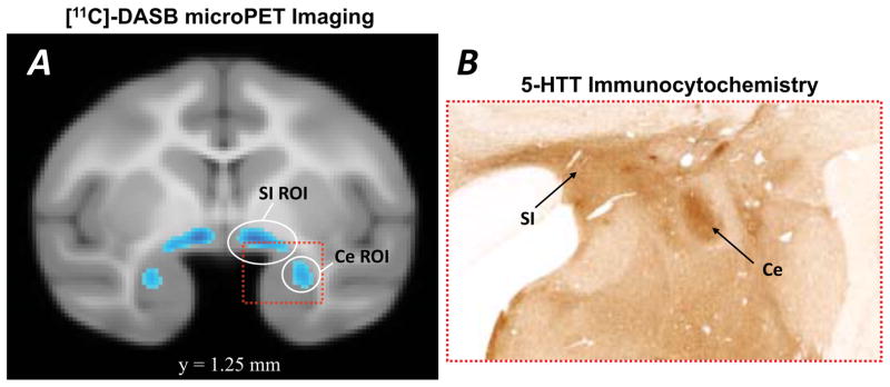
Fig 2. Serotonin transporter (5-HTT) labeling in the central nucleus (Ce) region of rhesus monkey amygdala used to delineate the seed region for monkey fMRI analysis.
(A) In vivo PET image demonstrating 5-HTT binding availability in the Ce and dorsomedially adjacent substantia innominata (SI) region, adapted from data first reported in Christian et al., (2009). The image is thresholded at 250X the background binding level, and the circles indicate the regions of interest (ROIs) used in the present study as seed clusters for analysis of the monkey functional connectivity data. (B) Comparable low-power photomicrograph showing the dense and selective expression of 5-HTT in the lateral division of the Ce (image provided by Dr. Julie Fudge, University of Rochester School of Medicine and reprinted with permission of the publisher).

