Abstract
INTRODUCTION
Acute appendicitis is one of the most frequent causes of surgical abdominal pain presenting to the Emergency Department. The diagnosis is confirmed by a set of clinical signs, blood tests and imaging.
The typical presentation consists of periumbilical pain radiating to the right lower quadrant with peritoneal reaction on palpation (Mac Burney).
PRESENTATION OF CASE
In this article, we report a case of acute appendicitis presenting with a left upper quadrant pain due to intestinal malrotation and we describe the radiologic findings on computed tomography.
DISCUSSION
With an Alvarado score of 4 and a nonconclusive abdominal U/S, the diagnosis of acute appendicitis was a long shot. Persistence of pain and increasing inflammatory parameters in her blood exams pushed the medical team to further investigate and a CT scan revealed intestinal malrotation with acute appendicitis.
CONCLUSION
An examining physician should not be mislead by the atypical presentation of acute appendicitis and should bear in mind the diagnosis to avoid serious complications.
Keywords: Acute appendicitis, Intestinal malrotation, Computed tomography
1. Introduction
Acute appendicitis is one of the most frequent causes of surgical abdominal pain presenting to the Emergency Department.
Typical presentation includes periumbilical pain radiating to the right lower quadrant with peritoneal reaction on palpation, fever and anorexia.
However, several atypical presentations have been reported and included backeache,1 left lower quadrant pain2 especially in case of situs inversus,3 Groin pain from a strangulated femoral hernia containing the appendix.4
Early diagnosis and surgical management decreases perforation rates.
Several scoring systems have been established (modified Alvarado score, Ohmann score and Eskelinen score) but none seems to be 100% specific.
The diagnosis is often confirmed by abdominal ultrasound and computed tomography.
We report a case presenting for a left upper quadrant pain that was diagnosed after computed tomography as an acute appendicitis on intestinal malrotation.
2. Presentation of case
It is about a 15 year-old girl without any particular medical history who presented to the Emergency Department of our institution for a 3 days history of left upper quadrant pain of colic type (7/10) radiating to the epigastric area.
Pain was associated with nausea, low grade fever (38 °C) and several episodes of vomiting. The patient denied any dysmenorrhea or lower urinary tract symptoms.
On Physical examination, she had pain and defense on deep palpation of the Left upper quadrant and the periumbilical region.
Blood exams revealed 14,000 White Blood Cells with 80% Neutrophiles.
A C-reactive protein level of 143 mg/L with normal liver function tests and normal amylase and lipase level.
A urinalysis also came negative.
With a wide range of differential diagnosis, the patient had undergone an abdominal ultrasound that was unable to justify her symptoms.
So she was admitted to the hospital and had an abdominal CT scan the next day with IV contrast.
She had an intestinal malrotation with the caecum at the umbilical level and the inflammatory appendix extending laterally toward the left flank (Figs. 1 and 2).
Fig. 1.

Enhanced CT showing a mesenteric malrotation with a mesenteric vein (arrow) located to the left of the mesenteric artery (arrowhead).
Fig. 2.

Enhanced CT showing a tubular appendicular structure originating from a malpositioned caecum located on the midline at the level of the umbilicus anteriorly and the aorto-iliac bifurcation posteriorly. The appendix extends laterally toward the left flank, with a 10 mm diameter, appendiceal wall enhancement and a peri-appendicular fat stranding.
Patient was transferred to the operating room for laparoscopic appendicectomy.
After scrubbing and insufflation of the abdominal cavity through the umbilicus, we explored the abdomen with a 0 degree optic. We first noticed the ascending colon laying parallel to the transverse with the caecum in the midline.
Another two 5 mm trocars were inserted to the left and the right sides of the umbilicus at midclavicular lines. We did not find a Ladd's band or any other GI malformation, the omentum was found to be inflamed and adherent to the distal part of the caecum.
Dissection was performed easily and the appendix was inflamed and hard especially at the tip.
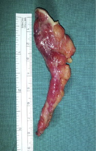
Appendectomy was done and the inflamed appendix was extracted in an endobag and was sent to pathology (Photo 1).
Photo 1.

Photo showing the inflamed appendix.
The patient was discharged 3 days later without any complications.
Pathology report mentioned acute appendicitis with an acute periappendicular reaction.
3. Discussion
Acute appendicitis typically presents as a periumbilical pain radiating to the right lower quadrant. Atypical locations are frequently seen but the left upper quadrant seems to be a very uncommon site.
A Case of Left flank pain was reported in the literature but acute appendicitis was diagnosed after laparoscopy and to our knowledge, never upon imaging.2 Other cases of acute appendicitis on intestinal malrotation presenting for Left lower quadrant pain have been reported.5–8
In our case, an Alvarado score of 4 and a nonconclusive abdominal U/S made the diagnosis of acute appendicitis less likely. But the persistence of pain and the increasing inflammatory parameters in her blood exams pushed the medical team to further investigate and a CT scan revealed intestinal malrotation with acute appendicitis.
Intestinal malrotation is very rare and is generally asymptomatic so an acute appendicitis on intestinal malrotation seems even less likely.9
Scores like the Alvarado score are supposed to help the physician in his assessment of the severity of the case and to guide his decision concerning the treatment.
Such scores lack both specificity and sensitivity and their use in some anatomical variations is highly debatable as it rules out acute appendicitis and leads to potential serious complications.
Conclusion
Atypical presentations of acute appendicitis should not mislead the examining physician and the diagnosis should always be in his differential whenever a patient presents to the ER for abdominal pain.
Conflict of interest statement
None.
Funding
None.
Ethical approval
Written informed consent was obtained from the patient for publication of this case report and accompanying images. A copy of the written consent is available for review by the Editor-in-Chief of this journal on request.
Author contributions
Charbel Tawk: Author of the paper.
Rana Zgheib: contributed in analysis and annotation of the CT scan.
Seba Mehanna: Review of the paper.
References
- 1.Stevenson W.O. A case off appendicitis with most unusual symptoms. Canadian Medical Association Journal. 1938;39(September (3)):263–264. [PMC free article] [PubMed] [Google Scholar]
- 2.Talanow R. An unusual manifestation of acute appendicitis with left flank pain. Radiology Case. 2008;2(July (1)):8–11. doi: 10.3941/jrcr.v2i1.27. [DOI] [PMC free article] [PubMed] [Google Scholar]
- 3.Akbulut S., Caliskan A., Ekin A., Yagmun Y. Left-sided acute appendicitis with situs inversus totalis: review of 63 published cases and report of two cases. Journal of Gastrointestinal Surgery. 2010;14:1422–1428. doi: 10.1007/s11605-010-1210-2. [DOI] [PubMed] [Google Scholar]
- 4.Nguyen E., Komenaka I. Strangulated femoral hernia containing a perforated appendix. Canadian Journal of Surgery. 2004;47:68–69. [PMC free article] [PubMed] [Google Scholar]
- 5.Pinto A., Raimondo D.D., Tuttolomondo A., Fernandez P., Caronia A., Lagalla R. An atypical clinical presentation of acute appendicitis in a young man with midgut malrotation. Radiography. 2007;13:164–168. [Google Scholar]
- 6.Welte F.J., Grosso M. Left-sided appendicitis in a patient with congenital gastrointestinal malrotation: a case report. Journal of Medical Case Reports. 2007;1:92. doi: 10.1186/1752-1947-1-92. [DOI] [PMC free article] [PubMed] [Google Scholar]
- 7.Flesch J., Oswald P., Grebici M., Schmaltz C., Bruant P., Burguet J.L. Mésentère commun complet révélé par une appendicite perforée gauche. Journal de Radiologie. 2010;91:915–916. doi: 10.1016/s0221-0363(10)70136-0. [DOI] [PubMed] [Google Scholar]
- 8.Israelit S., Brook O.R., Nira B.R., Guralnik L., Hershko D. Left-sided perforated acute appendicitis in an adult with midgut malrotation: the role of computed tomography. Emergency Radiology. 2009;16(May (3)):217–218. doi: 10.1007/s10140-008-0746-x. [DOI] [PubMed] [Google Scholar]
- 9.Au A.C.Y., Syed A., Bradpiece H.A. A rare case of intestinal malrotation presenting as appendicitis in late adulthood. Journal of Strength and Conditioning Research. 2010;8:3. doi: 10.1093/jscr/2010.8.3. [DOI] [PMC free article] [PubMed] [Google Scholar]


