Abstract
Uterine perforation is a serious problem which can happen after intrauterine device (IUD) insertion. Migration of the IUD to the pelvic and abdominal cavity or adjacent organs may be seen following perforation of the uterus. Migration of an IUD to a far intra-abdominal site is extremely rare. The patient reported here had undergone an IUD placement 30 years previously and had no problems during this period. The IUD was incidentally found at the left upper quadrant of the abdomen in the mesentery.
Introduction
Intrauterine device (IUD) placement is one of the most frequent methods of contraception.1,2 Uterine perforation due to an IUD is seen in 0.05 to 13 cases out of 1000 IUD placements.1,3,4 Uterine perforation following IUD insertion may be observed during or soon after the procedure or as a delayed event.1,2,4 Delayed rupture can be due to uterine spasms.1,4
Following the uterine rupture, an IUD may potentially migrate to the pelvic or intra-abdominal cavity causing several complications.2 There are not many reports on the the far-migration of an IUD.2,3 The longer the distance from the uterus, the more likely the patient will have severe symptoms.2 Our case describes a patient with a far-migrated intra-abdominal IUD causing no symptoms over the past 30 years and detected incidentally.
Case report
A 68-year-old woman was admitted to the outpatient urology clinic for acute right lumber pain. Her medical history revealed medical treatment for right lower back pain with no previous abdominal or urologic surgery. Thirty years before, three years after her second delivery, she had undergone an IUD placement. There had been no further examinations from that time for the IUD’s location and the IUS had not been replaced or removed. She even got pregnant two years after the IUD insertion and had an abortion. She had no severe chronic or acute pelvic and/or abdominal pain during the past 30 years.
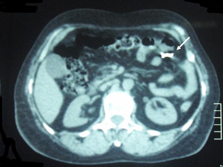
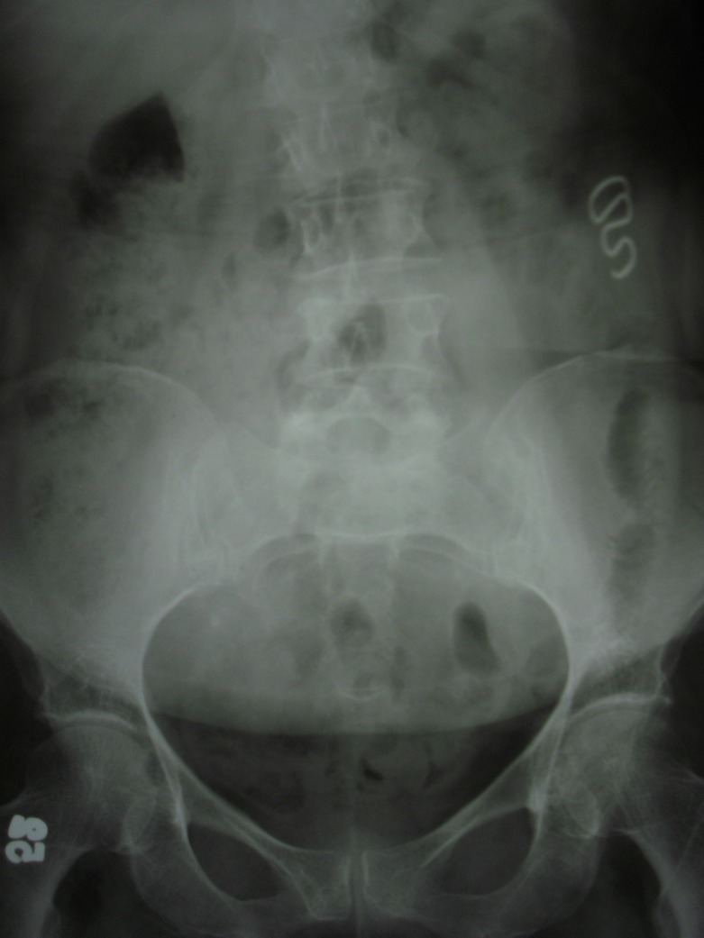
Her physical examination was normal. Routine laboratory investigations, including urinalysis, revealed normal findings. A plain x-ray of the abdomen demonstrated an opaque spiral shaped shadow, resembling an IUD, located in the left upper quadrant of the abdomen (Fig. 1). Computerized tomography (CT) of the pelvis and abdomen confirmed the IUD at the left half of the abdomen in the mesentery (Fig. 2). To remove the intra-abdominally far-migrated IUD, we planned a laparoscopic removal of the apparatus. However, the patient did not want surgery since she had no severe symptoms over the past 30 years following the IUD placement.
Fig. 1.
Plain X-ray of the abdomen demonstrated an opaque spiral shaped shadow resembling an intrauterine device at the left upper quadrant of the abdomen.
Fig. 2.

A computed tomography scan confirmed the intrauterine device (arrow) at the left half of the abdomen in the mesentery.
Discussion
Uterine perforation is one of the most serious complications of an IUD insertion.1–3 Perforation of the uterus due to IUD placement may be seen soon after the procedure or as a delayed event. It has been advocated that IUDs should be placed following proper patient selection by trained clinicians.1 An IUD may potentially perforate through the uterine wall into the gynecologic, urinary or gastrointestinal system organs.2 Migration of an IUD to the pelvic and abdominal cavity or neighbouring organs may result in several complications. More frequent problems include lower urinary tract symptoms, stone formation around the IUD, uterovesical fistula and stricture of the rectosigmoid colon.2,5–8 Rare complications include IUD appendicitis, gangrene of the small intestine and ureterohydronephrosis due to retroperitoneal fibrosis caused by the migration of an IUD through the peritoneum.6,9–11 Our case is an extremely rare example of an IUD migration to a far intra-abdominal site causing no symptoms for 30 years.
IUDs should be examined periodically.3 An ultrasound is a simple, rapid and non-invasive imaging method to assess the position of the IUD.1–3 Our patient had no examinations for the position of the apparatus after the IUD placement 30 years previously. She even got pregnant two years after the IUD insertion and had an abortion. Since she had no symptoms, there was no examination of a potention IUD migration.
A simple question (i.e., “Have you undergone IUD placement previously and has your IUD been removed?”) could have prevented serious problems. Although a far-migrated IUD was not considered during the previous ultrasounds to assess the reason for the patient’s lower back pain, the patient reported here is fortunate since she had no complications or symptoms. As in this patient, a simple x-ray of the abdomen is sufficient for the suspicion of a far-migrated IUD. Additional imaging modalities including ultrasound and CT to determine the exact position of a migrated IUD and related complications.1,2
Far-migrated IUD in the abdominal cavity may cause inflammation resulting in adhesion formation, intestinal obstruction, abdominal pain and bowel perforation.1,3 It is important to treat the patient with a migrated IUD for psychosomatic symptoms.3 It has been mentioned that surgical removal of the IUD located in the abdominal cavity is mandatory, even in asymptomatic patients.2,3,12 In contrast, Markovitch and colleagues believe that asymptomatic patients may be managed conservatively in some circumstances.13 Decision for the surgical excision of a migrated IUD in patients with no symptoms depends on the type of IUD and when the IUD was inserted.12,13 Laparoscopic removal of the intra-abdominal IUD should be the preferred choice of surgical management.1–3
Laparoscopy is a safe and minimally invasive procedure with less complications, shorter operative time and hospitalization compared to laparotomy.1–3,14 Sepsis and intestinal perforation needing repair are contraindications for laparoscopy and laparotomy should be preferred in these cases.1,3
Conclusion
Our patient presented with a very rare far-migrated intra-abdominal IUD. Clinicians should be mindful of asymptomatic patients with previously placed IUDs; periodic follow-up is mandatory. Laparoscopic removal of the intra-abdominal IUD or conservative management could be preferred based on the patient and the characteristics of the IUD.
Footnotes
Competing interests: None declared.
This paper has been peer-reviewed.
References
- 1.Sharifiaghdas F, Beigi FM, Abdi H. Laparoscopic removal of a migrated intrauterine device. Urol J. 2007;4:177–9. [PubMed] [Google Scholar]
- 2.Sun CC, Chang CC, Yu MH. Far-migrated intra-abdominal intrauterine device with abdominal pain. Taiwan J Obstet Gynecol. 2008;47:244–6. doi: 10.1016/S1028-4559(08)60095-9. [DOI] [PubMed] [Google Scholar]
- 3.Mulayim B, Mulayim S, Celik NY. A lost intrauterine device. Guess where we found it and how it happened? Eur J Contracept Reprod Health Care. 2006;11:47–9. doi: 10.1080/13625180500456791. [DOI] [PubMed] [Google Scholar]
- 4.Grimaldi L, De Giorgio F, Andreotta P, et al. Medicolegal aspects of an unusual uterine perforation with multiload-Cu 375R. Am J Forensic Med Pathol. 2005;26:365–6. doi: 10.1097/01.paf.0000188083.15245.a5. [DOI] [PubMed] [Google Scholar]
- 5.Szabo Z, Ficsor E, Nyiradi J, et al. Rare case of the utero-vesical fistula caused by intrauterine contraceptive device. Acta Chir Hung. 1997;36:337–9. [PubMed] [Google Scholar]
- 6.El-Hefnawy AS, El-Nahas AR, Osman Y, et al. Urinary complications of migrated intrauterine contraceptive device. Int Urogynecol J Pelvic Floor Dysfunct. 2008;19:241–5. doi: 10.1007/s00192-007-0413-x. [DOI] [PubMed] [Google Scholar]
- 7.Sepulveda WH, Ciuffardi I, Olivari A, et al. Sonographic diagnosis of bladder perforation by an intrauterine device. A case report. J Reprod Med. 1993;38:911–3. [PubMed] [Google Scholar]
- 8.Nceboz US, Ozcakir HT, Uyar Y, et al. Migration of an intrauterine contraceptive device to the sigmoid colon: A case report. Eur J Contracept Reprod Health Care. 2003;8:229–32. [PubMed] [Google Scholar]
- 9.Chang HM, Chen TW, Hsieh CB, et al. Intrauterine contraceptive device appendicitis: a case report. World J Gastroenterol. 2005;11:5414–5. doi: 10.3748/wjg.v11.i34.5414. [DOI] [PMC free article] [PubMed] [Google Scholar]
- 10.Barranco CJ. IUD gangrene of small intestine. Am J Surg. 1978. p. 717. [DOI] [PubMed]
- 11.Ohana E, Sheiner E, Leron E, et al. Appendix perforation by an intrauterine contraceptive device. Eur J Obstet Gynecol Reprod Biol. 2000;88:129–31. doi: 10.1016/S0301-2115(99)00142-6. [DOI] [PubMed] [Google Scholar]
- 12.Gorsline J, Osborne N. Management of the missing intrauterine contraceptive device: Report of a case. Am J Obstet Gynecol. 1985;153:228–30. doi: 10.1016/0002-9378(85)90122-x. [DOI] [PubMed] [Google Scholar]
- 13.Markovitch O, Klein Z, Gidoni Y, et al. Extrauterine mislocated IUD: is surgical removal mandatory? Contraception. 2002;66:105–8. doi: 10.1016/S0010-7824(02)00327-X. [DOI] [PubMed] [Google Scholar]
- 14.Demir SC, Cetin MT, Ucunsak IF, et al. Removal of intra-abdominal intrauterine device by laparoscopy. Eur J Contracept Reprod Health Care. 2002;7:20–3. [PubMed] [Google Scholar]


