Abstract
Although in theory, the differences in design between fixed-core and mobile-core prostheses should influence motion restoration, in vivo kinematic differences linked with prosthesis design remained unclear. The aim of this study was to investigate the rationale that the mobile-core design seems more likely to restore physiological motion since the translation of the core could help to mimic the kinematic effects of the natural nucleus. In vivo intervertebral motion characteristics of levels implanted with the mobile-core prosthesis were compared with untreated levels of the same population, levels treated by a fixed-core prosthesis, and normal levels (data from literature). Patients had a single-level implantation at L4L5 or L5S1 including 72 levels with a mobile-core prosthesis and 33 levels with a fixed-core prosthesis. Intervertebral mobility characteristics included the range of motion (ROM), the motion distribution between flexion and extension, the prosthesis core translation (CT), and the intervertebral translation (VT). A method adapted to the implanted segments was developed to measure the VT: metal landmarks were used instead of the bony landmarks. The reliability assessment of the VT measurement method showed no difference between three observers (p < 0.001), a high level of agreement (ICC = 0.908) and an interobserver precision of 0.2 mm. Based on this accurate method, this in vivo study demonstrated that the mobile-core prosthesis replicated physiological VT at L4L5 levels but not at L5S1 levels, and that the fixed-core prosthesis did not replicate physiological VT at any level by increasing VT. As the VT decreased when the CT increased (p < 0.001) it was proven that the core mobility minimized the VT. Furthermore, some physiologic mechanical behaviors seemed to be maintained: the VT was higher at implanted the L4L5 level than at the implanted L5S1 level, and the CT appeared lower at the L4L5 level than at the L5S1 level. ROM and motion distribution were not different between the mobile-core prosthesis and the fixed-core prosthesis implanted levels. This study validated in vivo the concept that a mobile-core helps to restore some physiological mechanical characteristics of the VT at the implanted L4L5 level, but also showed that the minimizing effect of core mobility on the VT was not sufficient at the L5S1 level.
Keywords: Total disc replacement, Lumbar spine, Segmental motion, Intervertebral translation, Prosthesis design
Introduction
For the treatment of lumbar degenerative disc disease, total disc arthroplasty appears as an alternative to conventional fusion techniques.
In the long-term, lumbar disc prosthesis treatment is thought to have the advantage of slowing degenerative changes in adjacent levels by preserving motion and consequently leading to lower incidence of adjacent surgery for arthritic progression. Furthermore, within the field of motion preservation, the disc prosthesis must also replicate kinematics at the implanted level to avoid any increase in load on the facet joints [16] which may generate arthritic progression. This eventual arthritic progression at the implanted level is significant as it could affect long-term clinical results [5, 8, 14, 19].
In physiology, the intervertebral mobility at a given spine segment includes a coupled motion of rotation and translation in both the sagittal and frontal planes. A change of dorsoventral intervertebral translation in relation to the change in sagittal rotation angle is small in the healthy spine [6, 7]. The small amount of translation between adjacent vertebrae in physiology is the consequence of the migration of the nucleus, which allows a moving axis of rotation during the flexion–extension motion. Thus, the migration of the nucleus minimizes the horizontal sagittal intervertebral translation (VT), also called olisthesis. The abnormal translations between vertebrae in the sagittal plane (i.e. spondylolisthesis and retrolisthesis or excessive translations) are clinically important signs of dysfunction or instability [1, 10].
However, it is reported [13, 21] that effects of disc prosthesis implantation with respect to restoration of segmental motion are inconsistent. Thus, disc prosthesis implantation could disturb intervertebral motion, including the postoperative decrease of sagittal range of motion (ROM) and a change to the flexion–extension distribution with loss of motion predominating in flexion [13]. Furthermore, disc prosthesis placement may increase the VT [21] and consequently may increase any load on the facet joints. This report [21] involved different types of lumbar prosthesis designs with different amounts of excess VT occurring with sagittal rotation.
The different types of prosthesis designs can be roughly classified into two families [11, 16]: mobile-core and fixed-core. In the mobile-core design, the core is free to translate in the transverse plane during flexion–extension and lateral bending allowing a moving axis of rotation. This allows the adjacent vertebra to rotate without necessary accompanying translation. The fixed-core design allows motion through a ball-and-socket configuration where the amount of VT occurring with rotation depends on the radius of the core’s curvature. The translation of the adjacent vertebrae is thereby coupled with the rotation; the center of rotation is fixed and corresponds to the center of the ball.
However, from a biomechanical point of view, the mobile-core design seems more likely to restore physiological motion given that the translation of the core could help to mimic the kinematic effects of the migration of the natural nucleus and consequently could avoid or minimize any excess of VT. This theoretical concept was never validated in vivo according to our knowledge.
The goal of this study was to investigate in vivo the hypothesis that the multidirectional plane translation of the Mobidisc® prosthesis’ mobile-core design (LDR Médical, Troyes, France) may contribute to the replication of physiologic characteristics of intervertebral displacement including the VT. A new method of measurement of the VT adapted from Frobin’s method was developed by the authors to optimize the accuracy of the measures.
In vivo intervertebral motion characteristics of levels implanted with the mobile-core prosthesis were compared with (1) untreated levels in the same population, (2) levels treated with a fixed-core prosthesis (ProDisc-L®, Synthes Spine), and (3) normal patient data from literature based on asymptomatic subjects [6].
Materials and methods
Study population
One group included 72 consecutive patients receiving a Mobidisc® disc prosthesis in a prospective multicenter study. The patients selected had a prosthesis with a sagittal rotation mobility superior to 3°. From the 72 patients, 9 with a motionless prosthesis (motion < 3°) were excluded from the analysis. All the patients had a single-level implantation (24 at L4L5 and 39 at L5S1). The mean duration of follow-up was 30 months in a population with a mean age of 41.
A second group retrospectively included 33 patients receiving a ProDisc-L® in three different centers. This group was not composed of consecutive cases but selected cases through the availability of dynamic X-ray. All the patients had a single-level implantation (15 at L4L5 and 18 at L5S1). The mean follow-up was 25 months and the mean age 44.
Data collection
The radiographs required for measurement comprised a lateral view taken standing, as well as views in maximum active extension and flexion. The X-ray film was digitalized and analyzed using image analysis software (Surgiview® Corporation, Paris, France). The images were then calibrated with subsequent results being reported in millimeter. Normal values were obtained from literature [6] on radiographs of 61 symptom-free people (between 16 and 57 years of age).
Kinematics assessment
The vertebral translation
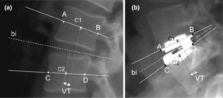
The vertebral translation (VT) measurement method was different for implanted levels and untreated levels. For untreated levels, the method described by Frobin [6] was applied by pointing the corners of the vertebral bodies followed by an automatic detection (Surgiview® Corporation, Paris, France) of the contours of each vertebra which were then manually better defined. Finally, the center point of each vertebra was automatically defined as well as the midplane angle of the cranial and caudal vertebra and their bisectrices. The dorsoventral displacement (VT) was defined by Frobin [6] as the distance between the projections of the center points of the vertebrae on the bisectrix divided by the inferior vertebral endplate length of the cranial vertebra (Fig. 1).
Fig. 1.
The methods of measuring the vertebral translation (VT). a The Frobin’s method uses the bony landmarks to determine the bisectrix (bi) and the projections of the vertebrae’s center points (C1 and C2) on it. b The adapted method uses metal landmarks and substitutes the center points of the vertebrae with the center points of the prosthesis endplates
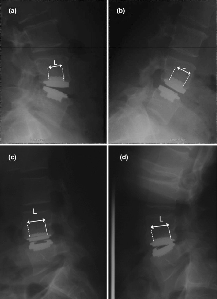
For implanted levels, the planes of the caudal and cranial prosthesis endplates, as well as their bisectrix substituted the midplane angles of the vertebra. The center points of the vertebra were substituted by the center points of the prosthesis endplates. The VT was similarly defined as the distance between the projections of the prosthesis endplates’ center points on their bisectrix divided by the inferior vertebral endplate of the cranial vertebra (Fig. 2). Between flexion X-ray and extension X-ray, the orthogonal incidence was maintained and, therefore, the length of the keels was the same. If they had not the same that would have meant the dynamic X-rays had not been included. This adapted method allowed for more accurate measurements as it was easier to define the limits of the prosthesis endplates (made in metal with angular limits) compared to the corners of the vertebral bodies. The VT is usually expressed [6] in percentage of the superior vertebra caudal endplate per degree of rotation. As X-rays were calibrated, and therefore the size of the endplate known, results in the present study could be expressed in mm. As regards to the sign of the displacement, when going from flexion to extension, if the vertebrae moved into a posterior direction (e.g. a retrolisthesis direction) displacement was counted negatively as in the reference study [6]. There were three observers who made VT measurements on 36 sets of radiographs to approximate the inter-observer precision.
Fig. 2.
Radiographs in extension and in flexion of a fixed-core prosthesis (a) (b) and of a mobile-core prosthesis (c) (d). The length of the keel (L) has to be the same between flexion-film and extension-film to show that the orthogonal incidence was maintained between the two films
The core translation
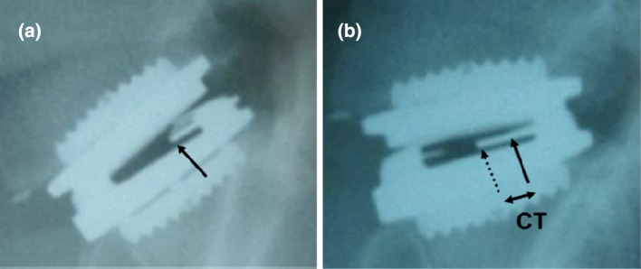
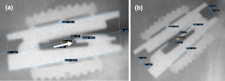
A core translation (CT) measurement of a mobile-core prosthesis was possible because of the presence of a tantalum marker inserted in the core (Fig. 3).
Fig. 3.
Radiographs a in flexion and b in extension of a mobile-core prosthesis. Arrows localize the anterior part of the marker inserted in the core: the full one in flexion and the dotted one in extension. The difference of location between the two radiographs corresponds to the core translation (CT)
The segmental rotational range of motion
ROM was defined as the difference of angles taken in maximum flexion and in maximum extension. The flexion–extension distribution was also evaluated and corresponded to the different disc lordosis in maximum flexion, in maximum extension, and in neutral standing. Also taken into account was the distribution between flexion and extension on either side of the angle obtained in a neutral static position for each level. In case of an eventual reduction in ROM, this analysis allowed us to assess if it predominated in flexion, in extension, or both. Leivseth [13] extracted normal data regarding ROM from the Frobin study [6].
Statistical analysis
For comparisons between implanted and untreated levels, as well as for comparisons with normal data, the Wilcoxon test and Student test were used. The level of significance was set to α = 0.05. To evaluate reproducibility in the VT measurement, inter-observer reliability of the VT measurement method was assessed by calculating the intraclass correlation coefficient (ICC) Type 2 [2]. To approximate the inter-observer precision, the difference between the measurements of the observers was determined for each measurement occasion. The deviation of these differences was then calculated. Precision was determined as the mean of the absolute differences between measurements [15].
Results
Vertebral translation
In order to be able to make comparisons, normal values from the Frobin report [6], which are only available as a percentage of the superior vertebra caudal endplate length per degree of rotation, were expressed on the basis of the mean value of the endplate length of the vertebrae implanted with a prosthesis in our study: 33 mm. Furthermore, results were adjusted to 10° of ROM to be able to compare the amount of VT between each group for the same ROM.
The seven cases of mobile-core prosthesis with a core without any mobility during flexion–extension were separated, the goal being to investigate the effect of the core mobility on the VT.
The assessment of the reliability of the VT measurement method developed in this study showed that the level of agreement was rather high with the ICC being 0.908 (p < 0.001). The inter-observer precision corresponding to the mean absolute difference between the observers was 0.2 mm.
The VT results are listed in Table 1 for L3L4, L4L5 and L5S1 segments with and without prostheses (mobile-core and fixed-core). At L3L4 levels, the mean value of VT between the untreated levels and the normal database was not different. At L4L5 levels, no significant differences were found between untreated and implanted levels with a mobile-core prosthesis (p = 0.58) and a fixed-core prosthesis (p = 0.08). But the amount of VT was significantly higher for the fixed-core prosthesis compared to the mobile-core prosthesis (p < 0.001). The VT of L5S1 untreated levels (below L4L5 implanted levels) was expressed with a high range of standard deviation compared to other levels because of a higher degree of error due to the difficulty of accurately identifying the ventral corner of the S1 endplate (the sacral endplate often had a dome shape). At L5S1 levels, the sign of the displacement was positive for untreated levels, which corresponds to normal levels with a positive sign [6], and negative for treated levels either with mobile-core or fixed-core prostheses. The differences were significant between L5S1 normal levels versus all the other L5S1 levels (untreated, mobile-core prosthesis and fixed-core prosthesis). The difference was also significant between untreated L5S1 levels and L5S1 levels treated with mobile-core prostheses (p < 0.001). In addition, the amount of VT was significantly lower at L5S1 levels treated with mobile-core prostheses compared to L5S1 levels treated with fixed-core prostheses (p < 0.001). Comparing L4L5 treated levels to L5S1 treated levels, the differences were significant in the case of mobile-core prostheses (p = 0.002) and insignificant with fixed-core prostheses. The mean VT of L5S1 levels treated with mobile-core prostheses was lower than the L4L5 levels treated with mobile-core prostheses. The mean VT of levels treated with mobile-core prostheses in which the core had no mobility (n = 7) was higher compared to the treated levels where the core was mobile (−1.6 ± 0.35 mm and−1.1 ± 0.8 mm, respectively).
Table 1.
Vertebral translation at normal levels (data from literature), untreated levels (below or above treated levels), mobile-core implanted levels and fixed-core implanted levels
| Normal levels | Untreated levels | Mobile-core implanted levels | Fixed-core implanted levels | Differences between each group* | ||
|---|---|---|---|---|---|---|
| L3L4 | −1.22 ± 0.60 n = 58 |
−1.29 ± 1.02 n = 46 |
N versus U | NS | ||
| L4L5 | −1.03 ± 0.48 | −1.20 ± 1.28 | −1.10 ± 0.59 | −1.74 ± 0.71 | N versus U | NS |
| n = 49 | n = 29 | n = 21 | n = 15 | N versus M | NS | |
| N versus F | p < 0.001 | |||||
| U versus M | NS | |||||
| U versus F | NS p = 0.08 | |||||
| M versus F | p < 0.001 | |||||
| L5S1 | +0.03 ± 0.97 | +1.07 ± 1.61 | −0.79 ± 0.44 | −1.61 ± 0.4 | N versus U | p = 0.02 |
| n = 37 | n = 17 | n = 35 | n = 18 | N versus M | p < 0.001 | |
| N versus F | p < 0.001 | |||||
| U versus M | p < 0.001 | |||||
| U versus F | p < 0.001 | |||||
| M versus F | p < 0.001 | |||||
| L4L5 versus L5S1 | ML4L5 versus ML5S1 | p = 0.002 | ||||
| FL4L5 versus FL5S1 | NS |
n number of cases available, N normal levels, U untreated levels, M mobile-core implanted levels, F fixed-core implanted levels, NS not significant (p > 0.05)
* The Wilcoxon test was used to compare U and M and F. The Student test was used to compare N to all the other levels. Vertebral translation is expressed in millimeter for 10° of range of motion. Means ± standard deviation are shown
Core translation
In 7 cases, in over 63 prostheses (mobile-core design), the core was not mobile. The mean CT, taking into account all the different sagittal rotations, was 1.13 ± 0.88 mm and 1.07 ± 0.75 mm at L5S1 and L4L5 levels, respectively, with no significant difference. However, the CT, adjusted to 10° of sagittal rotation, averaged 0.97 ± 0.92 mm and 1.27 ± 0.76 mm at L5S1 and L4L5 levels, respectively, with a significant difference between these two levels (p = 0.05).
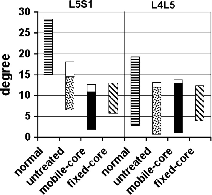
Sagittal range of motion (Fig. 4)
Fig. 4.
Range of motion at L4L5 and L5S1 levels for normal levels (data from literature), untreated levels (adjacent to treated levels), levels implanted with a mobile-core prosthesis and levels implanted with a fixed-core prosthesis. For untreated levels and implanted levels with a mobile-core prosthesis, the range of motion in flexion and in extension are separated
ROM at levels implanted with a mobile-core was higher (but not significantly) at both L4L5 and L5S1 levels compared to levels implanted with a fixed-core prosthesis: 10.3° ± 5 and 6.9° ± 3.5 at L4L5 levels and 8.9° ± 3.9 and 6.4° ± 3.8 at L5S1. The comparison of ROM between treated levels (either with a mobile-core prosthesis or a fixed-core prosthesis) and untreated levels revealed no significant difference. At levels L4L5, both implanted prostheses and untreated groups started the flexion–extension motion at virtually identical angles. At level L5S1, the motion distribution was slightly different between implanted and untreated levels with some more lordosis in untreated L5S1 levels. In comparison to data found in the literature [6] on a symptom-free volunteer group (called “normal” in our study), both implanted and untreated levels at both L4L5 and L5S1 levels had lower ROM, and the motion deficit laid in extension especially at the L5S1 levels.
The ROM of the fixed-core prosthesis compared to the ROM of the mobile-core prosthesis was lower, but the difference was not significant. Any motion distribution was basically similar between both protheses.
Relation between vertebral translation and range of motion
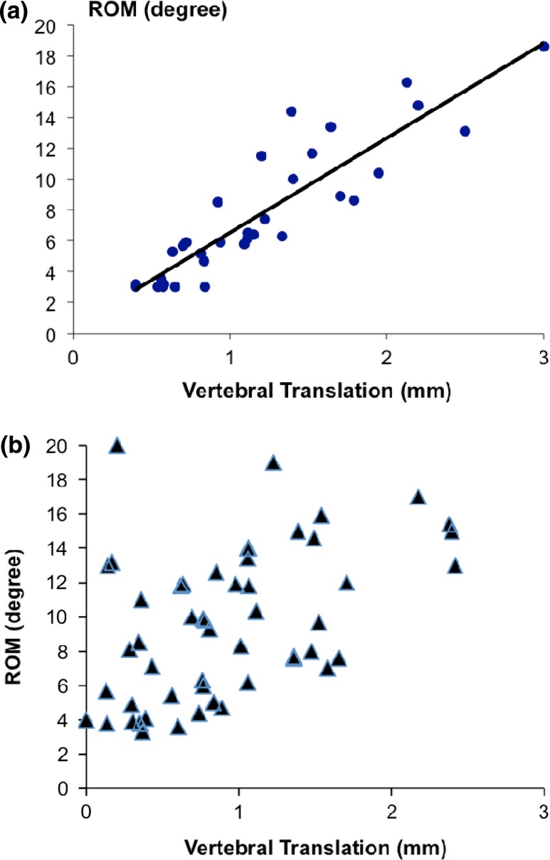
With the mobile-core prosthesis, no correlation was found between the VT and ROM (Fig. 5). Yet, with the fixed-core prosthesis, a strong correlation existed—the VT increased when the ROM increased (p < 0.001) (Fig. 5).
Fig. 5.
Relation between vertebral translation (in mm) and range of motion (in degree) at levels implanted a with a fixed-core prosthesis. (p < 0.001) and b with a mobile-core prosthesis
Relation between vertebral translation and core translation
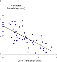
At the L4L5 levels, a higher VT was observed combined with a lower CT, but at the L5S1 levels, a lower VT combined with a higher CT was observed. Without taking into account the sagittal rotations, no correlation was found between the VT and CT. However, when the analysis was adjusted to 10° of ROM, a strong correlation between VT and CT appeared—the VT decreased as the CT increased (p < 0.0001) (Fig. 6).
Fig. 6.
Relation between vertebral translation (in mm) and core translation (in mm) at levels implanted with a mobile-core prosthesis. Analysis was adjusted to 10° of range of motion. (p < 0.0001)
Discussion
This in vivo study demonstrates that the mobile-core prosthesis replicated physiological VT at L4L5 levels, but not at L5S1 levels, and that the fixed-core prosthesis did not replicate physiological VT at any level.
VT measurement method
The method developed in this study to measure the VT was adapted from the well-accepted Frobin’s method using the same principal. But the adapted method provided a higher accuracy rate (0.2 mm of inter-observer precision) and strong reliability (ICC = 0.908) as the landmarks based on the metal angular limits of the prosthesis were more accurate in comparison with the bony corners of the vertebral body. Other authors [3, 15] developed a motion measurement method with landmarks using the spikes or the fins of lumbar disc prostheses instead of the bony landmarks, and also showed better reliability. Johnson [9] and Leivseth [12] reached the same accuracy for VT measurements (0.2 mm) with a very precise technique: radiostereometric analysis. Ordway [18] also applied this technique to measure motion following intervertebral disc replacement with an accuracy rate of 100 micrometers in translation.
The other parameters necessary to Frobin’s method were thus substituted. The plane of the prosthesis’ endplates replaced the vertebral body midplanes; since they are parallel they have identical bisectrices. The center points of the vertebral bodies were substituted by the center points of the two prosthesis endplates. Frobin’s method adaptations appeared coherent as prosthesis endplates and vertebral bodies are attached and consequently have the same translation during flexion–extension motion.
VT results
Untreated levels
The absence of difference between L3L4 and L4L5 normal levels as physiological reference levels [6], compared to L3L4 and L4L5 untreated levels of the present study, validated our measurements, and suggested that untreated levels can be used as reference levels.
Mobile-core prosthesis
Based on our method adapted from Frobin’s method at the treated levels, the results showed that mobile-core prostheses pleasantly replicated physiological VT at L4L5 levels since they made no difference with either normal or untreated L4L5 levels. Cunningham [4], in an in vitro human cadaveric model, also found that VT occurring at the level implanted with a mobile-core prosthesis was not significantly different when being compared to the intact spine. However, at L5S1 levels, VT obtained with mobile-core prostheses was different than untreated and normal L5S1 levels. In addition, untreated L5S1 level VT was unexpectedly different from normal L5S1 level VT because, in the present study, untreated L5S1 levels corresponded to non-degenerative adjacent levels below treated L4L5 levels. One could make the hypothesis that the abnormal high VT of untreated L5S1 levels could be a kinematic consequence of the presence of the prosthesis at the above-mentioned L4L5 level. But, the current study cannot provide any explanation for this difference between untreated and normal levels.
However, in physiology the kinematics between L4L5 and L5S1 levels are different, and L5S1 levels seem to have a more complicated mobility. For example, some authors [6] described that the physiological movement at L5S1 levels can be paradoxal with the possibility that during the flexion–extension motion, going from flexion to extension, the L5 vertebra moves into an anterior (ventral) direction. Tournier [21] also stressed in this study that L5S1 levels have a more complicated biomechanical functioning given the mean center of rotation location was not in a well-defined fixed position compared to L4L5 levels. Thus, we can speculate that L5S1 levels could be biomechanically less adapted than L4L5 levels to receive disc prostheses with the current prosthesis designs.
Nevertheless, the mean VT absolute value was significantly lower at the L5S1 implanted levels than at the L4L5 implanted levels, as in physiology (L5S1 VT is inferior to L4L5 VT [6]).
Fixed-core prosthesis
Unlike the mobile-core prosthesis, the fixed-core prosthesis did not seem to replicate physiological VT at L4L5 levels; VT with a fixed-core prosthesis was higher at both L4L5 and L5S1 levels compared to normal and untreated levels and also to levels implanted with a mobile-core prosthesis. Ordway [18] also reported higher VT than physiology in a study using an accurate method to measure intervertebral mobility with a fixed-core prosthesis (radiostereometry). Furthermore, in the present study, no differences were found between L4L5 levels and L5S1 levels implanted with a fixed-core prosthesis whereas in physiology L5S1 VT is inferior to L4L5 VT. In the end, no difference of VT was found between a fixed-core prosthesis and a mobile-core prosthesis in which the core was immobile, logically behaving as a fixed-core designed prosthesis.
Tournier [21] results studying the VT of fixed-core and mobile-core prostheses contradicted what the present study found. First, Tournier [21] reported a significant increase of VT at the L4L5 levels after both a mobile-core and a fixed-core total disc arthroplasty, but not at L5S1 levels where the range of VT was significantly higher for the mobile-core prosthesis compared to the fixed-core. One explanation for these opposing results could be that the prosthesis belongs to the same families (e.g. fixed-core and mobile-core), but was not the same as those of the present study with a mobile-core in regards to their biomechanical characteristics. Classification into only two families may also be too simple [11], not to mention their method of VT measurement was less accurate because using Frobin’s bony landmarks is inherently less accurate compared to the use of metal landmarks in the present method.
Secondly, Tournier [21] separately measured the VT in flexion, and then in extension, without doing the synthesis of both measurements. That may generate more uncertainty, as the amount of VT for some authors [23] could be variable throughout the displacement and even more variable in its distribution between flexion and extension. Thus, Wu [23], measuring the continuous VT via a videofluoroscopic technique during cervical flexion–extension, showed that translation displayed a relatively inconsistent trend on movement direction throughout the ROM. However, it is true that that translational data could only be assessed at the end of the movement in a static study [6, 20] under the condition of doing the synthesis of the measurements in flexion and in extension.
CT
Of the 63 consecutive patients who received a mobile-core prosthesis (with a ROM > 3°), the majority had cores that were mobile. However, seven prostheses were found to have cores that were not mobile but more mechanical in behavior much like a fixed-core prosthesis regards to the VT, demonstrating that core mobility minimized VT (e.g. the olisthesis). The range of CT at L4L5 levels was significantly lower than at L5S1 levels. This difference could be assimilated by analogy to the difference of centrodes (the path taken by an instantaneous center or rotation during a full ROM). In physiology, the centrode of an L4L5 disc is shorter than the centrode of an L5S1 disc [17]. The analogy with physiology could be forced by the fact that the L4L5 mobile-core prosthesis implanted levels combined a shorter CT and a longer VT compared to L5S1 mobile-core implanted levels, which could be assimilated to the shorter centrode and the longer VT of the natural L4L5.
ROM
The implantation of either a mobile-core prosthesis or a fixed-core prosthesis restored ROM, though not statistically different at either L4L5 or L5S1 when compared to the ROM of untreated levels in the population of the present study (mean age 41 and 44). But, when compared to normal data, both implanted and untreated levels of the present study did exhibit a lower ROM. Regarding the untreated segments in patients with disc prostheses at adjacent levels, Leivseth [12] found (in a population of a median age of 45) the ROM to be equivalent to the ROM of our study, and also found ROM lower than normal. The normal ROM refers to the data compiled by Frobin et al. [6] in an asymptomatic volunteer population between 16 and 57 years of age. In the present study, as in the study of Leivseth [12], the patients were not totally asymptomatic meaning persisting clinical symptoms and apprehension of pain could lead to a lower ROM when performing the flexion–extension movement. What’s more, ROM decreases as people age [22], and the mean age of volunteers in Frobin’s study (which was not reported) could have been different from the mean age in the present study. Technical differences between the studies to obtain both lateral views, one in maximum extension and one in maximum flexion, could also influence the collected ROM (i.e. half the patients in the Frobin study performed the flexion–extension movement passively whereas in the present study it was obtained actively).
However, results concerning ROM have to be interpreted cautiously since the fixed-core prosthesis group was not composed of consecutive cases but of selected ones through the availability of a dynamic X-ray, which represents a bias. Another limitation to the present study is that the analysis was limited to implants (with fixed-core and mobile-core), which were mobile with the exception of a minority of patients.
Finally, motion distribution was basically similar between mobile-core prostheses and fixed-core prostheses compared to the untreated levels. That seems to imply that the difference between the designs of both prostheses in this study does not influence the motion distribution.
Motion distribution of implanted levels compared to normal level motion distribution revealed a deficit in extension, especially at the L5S1 levels. As aforementioned, when compared to normal levels, the lower rate of ROM in implanted and untreated levels could be linked to the population. It brings up the question of age and low back pain, which are supposed to decrease the ROM especially in extension. Therefore, it has to be recognized that the L5S1 disc has a more complicated biomechanical functioning and consequently may be more complicated to mimic given the tendency of the deficit in extension to indeed be stronger at L5S1 levels. Motion distribution of L5S1 implanted levels also showed a deficit in extension when compared to L5S1 untreated levels. However, at L4L5 levels, the motion distribution between untreated levels and implanted levels was mainly similar.
Adjacent untreated levels in the same population seem to be a better adapted reference since this population generally still continues to feel some low back pain even after surgery, and still fails to perform maximum flexion and extension during the dynamic X-ray procedure. However, the reference population called “normal” is composed of healthy, symptom-free volunteers [6] who feel no apprehension of the dynamic X-ray procedure which could explain higher ROM and especially a higher range of extension. To reinforce the argument of untreated adjacent levels being better reference levels, it appeared that the untreated levels between the current study and Leivseth’s study [12] exhibited comparable ROM, motion distribution and VT at L4L5 levels.
Relation between VT and ROM
The fixed-core prosthesis VT in the present study appeared proportional to the ROM (Fig. 5) as expected because with the fixed-core prosthesis, being a pure ball-and-socket joint, translation of the adjacent vertebrae is coupled and proportional with rotation. Translation occurs concurrently with rotation given by the radius of the inner core. The current study illustrated and quantified in vivo this biomechanical principal.
However, with the mobile-core prosthesis, the absence of correlation between VT and ROM (Fig. 5) could be related to the mobility of the core. This absence of correlation is also in favor of a moving axis of rotation during motion as in physiology.
Relation between VT and CT
In comparing L4L5 levels to L5S1 levels, the opposite combination of VT and CT (e.g. a higher VT and a lower CT at L4L5 levels) (Fig. 7) already suggested a correlation between VT and CT. However, the strong correlation between VT and CT appeared when the analysis was adjusted to 10° of rotation (Fig. 6) and demonstrated the influence of the core mobility on the intervertebral mobility. As VT decreased while CT increased it was proven that the core mobility minimized the olisthesis (Fig. 8). This study also confirmed in vivo that core mobility allowed a moving axis of rotation during intervertebral motion, as expected.
Fig. 7.
Radiographs a in flexion and b in extension of a mobile-core prosthesis (case example 1) with a strong core translation and a small or absent vertebral translation. Arrow indicates the tantalum marker inside the core made of polyethylene
Fig. 8.
Radiographs a in flexion and b in extension of a mobile-core prosthesis (case example 2) with a small or absent core translation, and a strong vertebral translation. Arrow indicates the tantalum marker inside the core made of polyethylene
Conclusion
With a mobile-core prosthesis, the different characteristics of segmental rotational–translation motion, including the VT and ROM and the flexion–extension distribution, were all similar between L4L5 implanted levels and L4L5 untreated levels within a 40-year-old population. Furthermore, some physiologic mechanical behaviors seemed to be maintained: VT was higher at implanted L4L5 levels than at implanted L5S1 levels and CT appeared lower at L4L5 levels than at L5S1 levels.
Thus, we can speculate that the presence of a mobile-core helps to restore L4L5 segmental kinematics close to the natural physiology of a 40-year-old population taking into account that untreated levels are preferable reference levels (compared to normal levels) to assess the kinematic effect of the implantation of a total disc arthroplasty.
This study, according to our knowledge, is the first study which validates in vivo the concept that the mobile-core lumbar disc prosthesis replicates normal VT at L4L5 levels. On the contrary, at L5S1 levels, a disc prosthesis even with a mobile-core, appeared to fail to replicate a physiological rotational-motion. The observed “minimizing” effect of core mobility on the VT appeared insufficient at L5S1 levels.
A prosthesis with a fixed-core increased the VT during flexion–extension motion in comparison to the physiology and mobile-core prosthesis at both L4L5 and L5S1 levels. This study could not provide any information on the eventual relation between the amount of VT and the loading rate on the facet joints, and it did not take into account the influence of prosthesis positioning on segmental mobility.
Conflict of interest
None.
References
- 1.Axelsson P, Karlsson BS. Intervertebral mobility in the progressive degenerative process. A radiosterometric analysis. Eur Spine J. 2004;13:567–572. doi: 10.1007/s00586-004-0713-5. [DOI] [PMC free article] [PubMed] [Google Scholar]
- 2.Bonnet DG. Sample size requirents for estimating intraclass correlations with desired precision. Stat Med. 2002;21:1331–1335. doi: 10.1002/sim.1108. [DOI] [PubMed] [Google Scholar]
- 3.Cakir B, Richter M, Puhl W, Schmidt R. Reliability of motion measurements after total disc replacement: the spike and the fin method. Eur Spine J. 2006;15:165–173. doi: 10.1007/s00586-005-0942-2. [DOI] [PMC free article] [PubMed] [Google Scholar]
- 4.Cunningham BW, Gordon JD, Dmitriev AE, Hu N, Mcafee PC. Biomechanical evaluation of total disc replacement arthroplasty: an in vitro human cadaveric model. Spine. 2003;28:S110–S117. doi: 10.1097/01.BRS.0000092209.27573.90. [DOI] [PubMed] [Google Scholar]
- 5.David T. Long-term results of one-level lumbar arthroplasty: minimum 10-year fellow-up of the Charité artificial disc in 106 patients. Spine. 2007;32:661–666. doi: 10.1097/01.brs.0000257554.67505.45. [DOI] [PubMed] [Google Scholar]
- 6.Frobin W, Brinckmann P, Leivseth G, et al. Precision measurement of segmental motion from flexion–extension radiographs of the lumbar spine. Clin Biomech. 1996;11:457–465. doi: 10.1016/S0268-0033(96)00039-3. [DOI] [PubMed] [Google Scholar]
- 7.Frobin W, Brinckmann P, Biggemann M, et al. Precision measurement of disc height, vertebral height and sagittal plane displacement from lateral radiographic views of the lumbar spine. Clin Biomech. 1997;12(suppl 1):1–63. doi: 10.1016/S0268-0033(96)00067-8. [DOI] [PubMed] [Google Scholar]
- 8.Huang RC, Tropiano P, Marnay T, et al. Range of motion and adjacent level degeneration after lumbar total disc replacement. Spine J. 2006;6:242–247. doi: 10.1016/j.spinee.2005.04.013. [DOI] [PubMed] [Google Scholar]
- 9.Johson R, Axelson P, Strömqvist B. Mobility provocation of lumbar fusion evaluated by radiostereometric analysis. Acta Orthop Scand. 1996;67(suppl):45–46. [Google Scholar]
- 10.Kirkaldy-Willis WH, Farfan HF. Instability of the lumbar spine. Clin Orthop. 1982;165:110–123. [PubMed] [Google Scholar]
- 11.Lavaste F (2007) Biomechanics of total disc replacements. In: Vital JM (ed) Alternative à l’arthrodèse lombaire et lombosacrée. Ed Elsevier Masson SAS, Paris, pp 134–145
- 12.Leivseth G, Brinckmann P, Frobin W, Johnsson R, Stromqvist B. Assessment of sagittal plane segmental motion in the lumbar spine—a comparison between distortion-compensated and stereophotogrammetric roentgen analysis. Spine. 1998;23:2648–2655. doi: 10.1097/00007632-199812010-00021. [DOI] [PubMed] [Google Scholar]
- 13.Leivseth G, Braaten S, Frobin W, Brinckmann P. Mobility of lumbar segments instrumented with a ProDisc II prosthesis—a two-year follow-up study. Spine. 2006;15:1726–1733. doi: 10.1097/01.brs.0000224213.45330.68. [DOI] [PubMed] [Google Scholar]
- 14.Lemaire JP, Carrier H, Sariali el H, et al. Clinical and radiological outcomes with the Charité artificial disc: a 10-year minimum follow-up. J Spinal Disord Tech. 2005;18:353–359. doi: 10.1097/01.bsd.0000172361.07479.6b. [DOI] [PubMed] [Google Scholar]
- 15.Lim MR, Girardi FP, Zhang K, Huang RC, Peterson MG, Cammisa FP. Measurement of total disc replacement radiographic range of motion: a comparison of two techniques. J Spinal Disord Tech. 2005;18:252–256. [PubMed] [Google Scholar]
- 16.Moumene M, Geisler FH. Comparison of biomechanical function at ideal and varied surgical placement for two lumbar artificial disc implant designs: Mobile-core versus fixed-core. Spine. 2007;17:1840–1851. doi: 10.1097/BRS.0b013e31811ec29c. [DOI] [PubMed] [Google Scholar]
- 17.Ogston NG, King GJ, Gertzbein SD, Tile M, Kapasouri A, Rubenstein JD. Centrode pattern in the lumbar spine. Baseline studies in normal subjects. Spine. 1986;11:591–595. doi: 10.1097/00007632-198607000-00010. [DOI] [PubMed] [Google Scholar]
- 18.Ordway NR, Fayyazi AH, Abjornson C, et al. Twelve-month follow-up of lumbar spine range of motion following intervertebral disc replacement using radiostereometric analysis. SAS J. 2008;2:9–15. doi: 10.1016/S1935-9810(08)70012-4. [DOI] [PMC free article] [PubMed] [Google Scholar]
- 19.Park CK, Ryu KS, Jee WH. Degenerative changes of discs and facet joints in lumbar total disc replacement using ProDisc II—Minimum two-Year follow-up. Spine. 2008;33:1755–1761. doi: 10.1097/BRS.0b013e31817b8fed. [DOI] [PubMed] [Google Scholar]
- 20.Pickett GE, Rouleau JP, Dugall N. Kinematic analysis of the cervical spine following implantation of an artificial cervical disc. Spine. 2005;30:1949–1954. doi: 10.1097/01.brs.0000176320.82079.ce. [DOI] [PubMed] [Google Scholar]
- 21.Tournier C, Aunoble S, Le Huec JC, Lemaire JP, Tropiano P, Lafage V, Skalli W. Total disc arthroplasty: consequences for sagittal balance and lumbar spine mouvement. Eur Spine J. 2007;16:411–421. doi: 10.1007/s00586-006-0208-7. [DOI] [PMC free article] [PubMed] [Google Scholar]
- 22.Wong KWN, Leong JCY, Chan MK, Luk KDK, Lu WW. The flexion–extension profile of lumbar spine in 100 healthy volunteers. Spine. 2004;29:1636–1641. doi: 10.1097/01.BRS.0000132320.39297.6C. [DOI] [PubMed] [Google Scholar]
- 23.Wu SK, Kuo LC, Lan HCH, Tsai SW, Chen CL, Su FC. The quantitative measurement of the intervertebral angulation and translation during cervical flexion and extension. Eur Spine J. 2007;16:1435–1444. doi: 10.1007/s00586-007-0372-4. [DOI] [PMC free article] [PubMed] [Google Scholar]


