

Abstract
In the title molecule, C9H7F3O4S, the S and the methyl C atoms of the methylsulfonyl group deviate from the benzene ring plane by 0.185 (2) and −1.394 (3) Å, respectively. In the crystal, O—H⋯O hydrogen bonds link the molecules into chains along [201]. Weak C—H⋯O interactions further link these chains into layers parallel to the ac plane.
Related literature
For details of the synthesis, see: Cain et al. (1998 ▶). 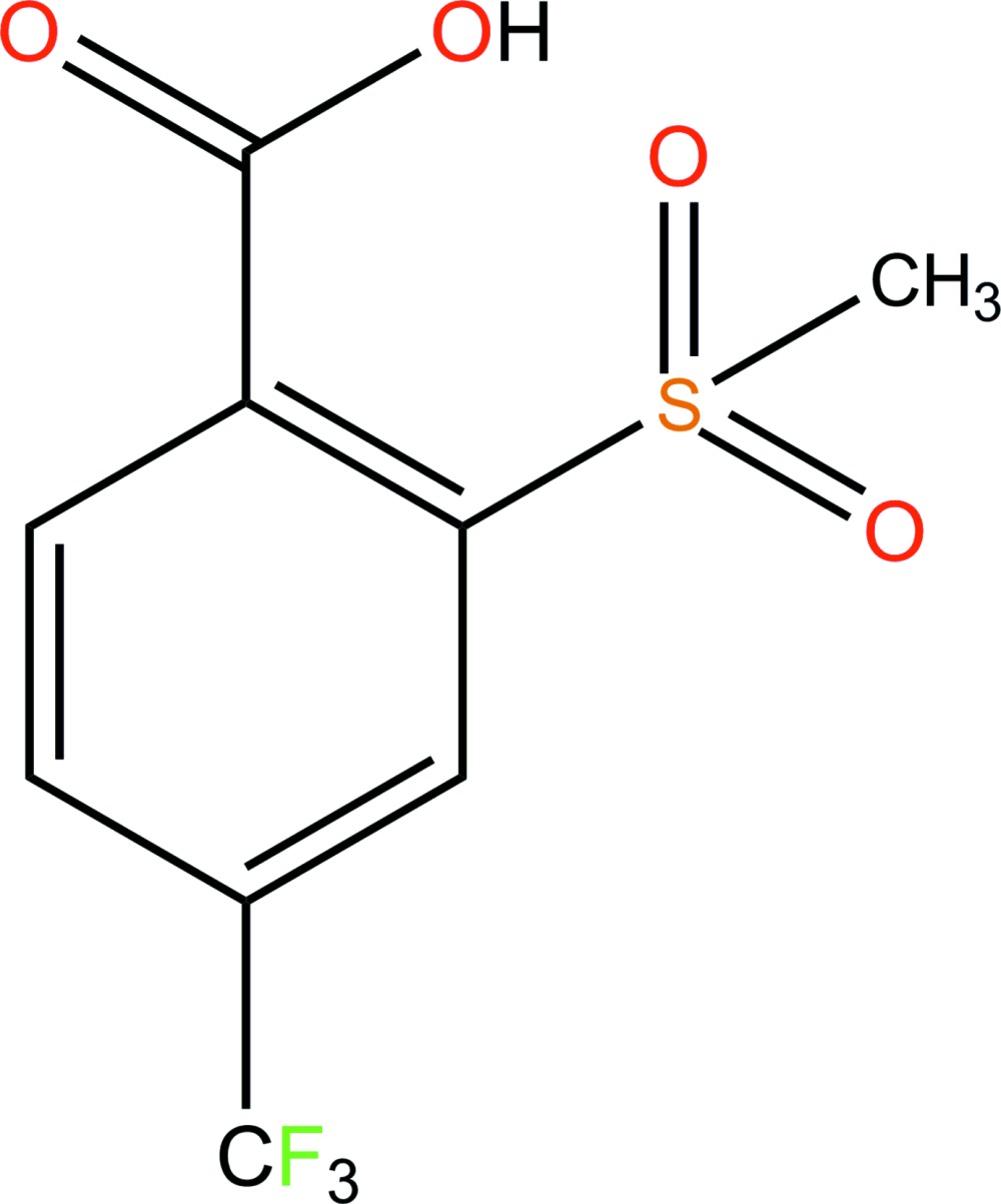
Experimental
Crystal data
C9H7F3O4S
M r = 268.21
Monoclinic,

a = 5.0804 (10) Å
b = 17.345 (4) Å
c = 11.576 (2) Å
β = 95.41 (3)°
V = 1015.6 (4) Å3
Z = 4
Mo Kα radiation
μ = 0.36 mm−1
T = 293 K
0.39 × 0.32 × 0.22 mm
Data collection
Rigaku R-AXIS RAPID diffractometer
Absorption correction: multi-scan (ABSCOR; Higashi, 1995 ▶) T min = 0.871, T max = 0.926
9132 measured reflections
2250 independent reflections
1879 reflections with I > 2σ(I)
R int = 0.029
Refinement
R[F 2 > 2σ(F 2)] = 0.044
wR(F 2) = 0.123
S = 1.11
2250 reflections
158 parameters
1 restraint
H atoms treated by a mixture of independent and constrained refinement
Δρmax = 0.39 e Å−3
Δρmin = −0.38 e Å−3
Data collection: RAPID-AUTO (Rigaku, 1998 ▶); cell refinement: RAPID-AUTO; data reduction: CrystalClear (Rigaku/MSC, 2002 ▶); program(s) used to solve structure: SHELXS97 (Sheldrick, 2008 ▶); program(s) used to refine structure: SHELXL97 (Sheldrick, 2008 ▶); molecular graphics: SHELXTL (Sheldrick, 2008 ▶); software used to prepare material for publication: SHELXL97.
Supplementary Material
Crystal structure: contains datablock(s) I, global. DOI: 10.1107/S160053681202243X/cv5300sup1.cif
Structure factors: contains datablock(s) I. DOI: 10.1107/S160053681202243X/cv5300Isup2.hkl
Supplementary material file. DOI: 10.1107/S160053681202243X/cv5300Isup3.cml
Additional supplementary materials: crystallographic information; 3D view; checkCIF report
Table 1. Hydrogen-bond geometry (Å, °).
| D—H⋯A | D—H | H⋯A | D⋯A | D—H⋯A |
|---|---|---|---|---|
| O2—H2⋯O4i | 0.82 (1) | 1.92 (1) | 2.725 (3) | 169 (4) |
| C9—H9B⋯O3ii | 0.96 | 2.35 | 3.208 (3) | 148 |
Symmetry codes: (i)  ; (ii)
  .
.
Acknowledgments
The authors thank the Project of Innovation Service Platform of Heilongjiang Province (grant No. PG09J001) and Heilongjiang University for support.
supplementary crystallographic information
Comment
The title compound, (I), is a intermediate in the synthesis of sulfonylurea herbicides developed and produced by E. I. du Pont de Nemours and Company. Herein, we report its crystal structure.
In (I) (Fig. 1), the S and the methyl C atoms of the methylsulfonyl group deviate from the benzene ring plane at 0.185 (2) and -1.394 (3) Å, respectively. Intermoleculear O—H···O hydrogen bonds (Table 1) link the molecules into chains along [201] (Fig. 2), and weak C—H···O interactions (Table 1) link further these chains into layers parallel to ac plane.
Experimental
The title compound was prepared by the reaction of 2-(methylsulphenyl)-4-trifluoromethylbenzoic acid and hydrogen peroxide in acetic acid at 10 ° (Cain et al., 1998). A colourless crystal suitable for single-crystal X-ray diffraction was obtained by the recrystallization from dichloromethane.
Refinement
C-bound H atoms were placed in calculated positions and treated as riding on their parent atoms, with C—H = 0.93 – 0.96 Å , and with Uiso(H) = 1.2 – 1.5 Ueq(C). O-bound H atoms were located in a differece Fourier map and were refined with restraint as O—H = 0.82 (1) Å, and Uiso(H) = 1.5Ueq(O).
Figures
Fig. 1.

The molecular structure of (I) showing displacement ellipsoids at the 50% probability level for non-H atoms.
Fig. 2.

A portion of the crystal packing showing hydrogen-bonded (dashed lines) chains.
Crystal data
| C9H7F3O4S | F(000) = 544 |
| Mr = 268.21 | Dx = 1.754 Mg m−3 |
| Monoclinic, P21/c | Mo Kα radiation, λ = 0.71073 Å |
| Hall symbol: -P 2ybc | Cell parameters from 8167 reflections |
| a = 5.0804 (10) Å | θ = 3.0–27.5° |
| b = 17.345 (4) Å | µ = 0.36 mm−1 |
| c = 11.576 (2) Å | T = 293 K |
| β = 95.41 (3)° | Block, colorless |
| V = 1015.6 (4) Å3 | 0.39 × 0.32 × 0.22 mm |
| Z = 4 |
Data collection
| Rigaku R-AXIS RAPID diffractometer | 2250 independent reflections |
| Radiation source: fine-focus sealed tube | 1879 reflections with I > 2σ(I) |
| Graphite monochromator | Rint = 0.029 |
| ω scan | θmax = 27.5°, θmin = 3.5° |
| Absorption correction: multi-scan (ABSCOR; Higashi, 1995) | h = −6→6 |
| Tmin = 0.871, Tmax = 0.926 | k = −22→22 |
| 9132 measured reflections | l = −14→14 |
Refinement
| Refinement on F2 | Primary atom site location: structure-invariant direct methods |
| Least-squares matrix: full | Secondary atom site location: difference Fourier map |
| R[F2 > 2σ(F2)] = 0.044 | Hydrogen site location: inferred from neighbouring sites |
| wR(F2) = 0.123 | H atoms treated by a mixture of independent and constrained refinement |
| S = 1.11 | w = 1/[σ2(Fo2) + (0.0555P)2 + 0.7827P] where P = (Fo2 + 2Fc2)/3 |
| 2250 reflections | (Δ/σ)max = 0.001 |
| 158 parameters | Δρmax = 0.39 e Å−3 |
| 1 restraint | Δρmin = −0.38 e Å−3 |
Special details
| Geometry. All esds (except the esd in the dihedral angle between two l.s. planes) are estimated using the full covariance matrix. The cell esds are taken into account individually in the estimation of esds in distances, angles and torsion angles; correlations between esds in cell parameters are only used when they are defined by crystal symmetry. An approximate (isotropic) treatment of cell esds is used for estimating esds involving l.s. planes. |
| Refinement. Refinement of F2 against ALL reflections. The weighted R-factor wR and goodness of fit S are based on F2, conventional R-factors R are based on F, with F set to zero for negative F2. The threshold expression of F2 > 2sigma(F2) is used only for calculating R-factors(gt) etc. and is not relevant to the choice of reflections for refinement. R-factors based on F2 are statistically about twice as large as those based on F, and R- factors based on ALL data will be even larger. |
Fractional atomic coordinates and isotropic or equivalent isotropic displacement parameters (Å2)
| x | y | z | Uiso*/Ueq | ||
| C1 | 0.2822 (5) | 0.85011 (13) | 0.09229 (19) | 0.0319 (5) | |
| C2 | 0.4291 (5) | 0.80387 (12) | 0.17524 (19) | 0.0284 (5) | |
| C3 | 0.6265 (5) | 0.83602 (13) | 0.25122 (19) | 0.0304 (5) | |
| H3 | 0.7242 | 0.8051 | 0.3051 | 0.037* | |
| C4 | 0.6780 (5) | 0.91494 (13) | 0.2467 (2) | 0.0337 (5) | |
| C5 | 0.5314 (5) | 0.96140 (14) | 0.1677 (2) | 0.0392 (6) | |
| H5 | 0.5636 | 1.0141 | 0.1658 | 0.047* | |
| C6 | 0.3360 (5) | 0.92880 (14) | 0.0911 (2) | 0.0396 (6) | |
| H6 | 0.2386 | 0.9602 | 0.0378 | 0.048* | |
| C7 | 0.0778 (5) | 0.81875 (14) | 0.0022 (2) | 0.0353 (5) | |
| C8 | 0.8793 (5) | 0.94984 (14) | 0.3342 (2) | 0.0389 (6) | |
| C9 | 0.4951 (5) | 0.65356 (15) | 0.0879 (2) | 0.0436 (6) | |
| H9A | 0.4715 | 0.5992 | 0.0984 | 0.065* | |
| H9B | 0.6803 | 0.6649 | 0.0889 | 0.065* | |
| H9C | 0.4062 | 0.6691 | 0.0147 | 0.065* | |
| F1 | 1.0869 (4) | 0.90478 (10) | 0.35927 (19) | 0.0655 (5) | |
| F2 | 0.7743 (4) | 0.96498 (13) | 0.43239 (16) | 0.0743 (6) | |
| F3 | 0.9734 (4) | 1.01622 (10) | 0.29917 (18) | 0.0648 (5) | |
| O1 | 0.0598 (4) | 0.75293 (11) | −0.02770 (17) | 0.0511 (5) | |
| O2 | −0.0822 (4) | 0.87346 (13) | −0.0433 (2) | 0.0594 (6) | |
| H2 | −0.187 (6) | 0.853 (2) | −0.092 (3) | 0.089* | |
| O3 | 0.0826 (3) | 0.69334 (11) | 0.19817 (16) | 0.0409 (4) | |
| O4 | 0.5177 (4) | 0.68310 (10) | 0.30716 (16) | 0.0420 (4) | |
| S1 | 0.36264 (11) | 0.70384 (3) | 0.20026 (5) | 0.02965 (18) |
Atomic displacement parameters (Å2)
| U11 | U22 | U33 | U12 | U13 | U23 | |
| C1 | 0.0313 (12) | 0.0354 (12) | 0.0275 (10) | −0.0014 (9) | −0.0052 (9) | −0.0003 (9) |
| C2 | 0.0292 (11) | 0.0281 (10) | 0.0270 (10) | −0.0019 (8) | −0.0027 (9) | −0.0010 (8) |
| C3 | 0.0292 (11) | 0.0307 (11) | 0.0298 (10) | −0.0012 (9) | −0.0056 (9) | −0.0011 (9) |
| C4 | 0.0343 (13) | 0.0323 (11) | 0.0340 (11) | −0.0041 (9) | 0.0002 (10) | −0.0039 (9) |
| C5 | 0.0465 (15) | 0.0295 (11) | 0.0402 (13) | −0.0049 (10) | −0.0025 (11) | 0.0017 (9) |
| C6 | 0.0461 (15) | 0.0348 (12) | 0.0358 (12) | −0.0008 (10) | −0.0079 (11) | 0.0054 (10) |
| C7 | 0.0363 (13) | 0.0409 (13) | 0.0264 (11) | −0.0014 (10) | −0.0080 (10) | 0.0021 (9) |
| C8 | 0.0381 (14) | 0.0344 (12) | 0.0426 (13) | −0.0063 (10) | −0.0053 (11) | −0.0041 (10) |
| C9 | 0.0411 (14) | 0.0387 (13) | 0.0500 (15) | 0.0002 (11) | −0.0014 (12) | −0.0120 (11) |
| F1 | 0.0520 (11) | 0.0479 (10) | 0.0892 (14) | 0.0019 (8) | −0.0329 (10) | −0.0072 (9) |
| F2 | 0.0700 (14) | 0.1067 (17) | 0.0459 (10) | −0.0298 (12) | 0.0039 (9) | −0.0328 (10) |
| F3 | 0.0673 (12) | 0.0446 (9) | 0.0773 (12) | −0.0257 (8) | −0.0208 (10) | 0.0066 (9) |
| O1 | 0.0628 (14) | 0.0392 (10) | 0.0453 (10) | −0.0044 (9) | −0.0255 (10) | −0.0005 (8) |
| O2 | 0.0596 (14) | 0.0481 (12) | 0.0622 (13) | 0.0101 (10) | −0.0377 (11) | −0.0121 (10) |
| O3 | 0.0279 (9) | 0.0490 (10) | 0.0441 (10) | −0.0075 (7) | −0.0047 (8) | 0.0036 (8) |
| O4 | 0.0404 (10) | 0.0411 (10) | 0.0410 (10) | −0.0038 (7) | −0.0135 (8) | 0.0091 (7) |
| S1 | 0.0264 (3) | 0.0294 (3) | 0.0313 (3) | −0.0036 (2) | −0.0070 (2) | 0.0017 (2) |
Geometric parameters (Å, º)
| C1—C6 | 1.392 (3) | C7—O1 | 1.194 (3) |
| C1—C2 | 1.410 (3) | C7—O2 | 1.326 (3) |
| C1—C7 | 1.502 (3) | C8—F1 | 1.322 (3) |
| C2—C3 | 1.387 (3) | C8—F3 | 1.325 (3) |
| C2—S1 | 1.796 (2) | C8—F2 | 1.326 (3) |
| C3—C4 | 1.396 (3) | C9—S1 | 1.753 (3) |
| C3—H3 | 0.9300 | C9—H9A | 0.9600 |
| C4—C5 | 1.383 (3) | C9—H9B | 0.9600 |
| C4—C8 | 1.498 (3) | C9—H9C | 0.9600 |
| C5—C6 | 1.387 (4) | O2—H2 | 0.816 (10) |
| C5—H5 | 0.9300 | O3—S1 | 1.4325 (18) |
| C6—H6 | 0.9300 | O4—S1 | 1.4484 (18) |
| C6—C1—C2 | 118.2 (2) | O2—C7—C1 | 112.0 (2) |
| C6—C1—C7 | 118.1 (2) | F1—C8—F3 | 106.1 (2) |
| C2—C1—C7 | 123.6 (2) | F1—C8—F2 | 107.8 (2) |
| C3—C2—C1 | 120.5 (2) | F3—C8—F2 | 106.0 (2) |
| C3—C2—S1 | 114.94 (17) | F1—C8—C4 | 112.9 (2) |
| C1—C2—S1 | 124.32 (17) | F3—C8—C4 | 112.8 (2) |
| C2—C3—C4 | 119.8 (2) | F2—C8—C4 | 110.8 (2) |
| C2—C3—H3 | 120.1 | S1—C9—H9A | 109.5 |
| C4—C3—H3 | 120.1 | S1—C9—H9B | 109.5 |
| C5—C4—C3 | 120.3 (2) | H9A—C9—H9B | 109.5 |
| C5—C4—C8 | 120.2 (2) | S1—C9—H9C | 109.5 |
| C3—C4—C8 | 119.3 (2) | H9A—C9—H9C | 109.5 |
| C4—C5—C6 | 119.6 (2) | H9B—C9—H9C | 109.5 |
| C4—C5—H5 | 120.2 | C7—O2—H2 | 107 (3) |
| C6—C5—H5 | 120.2 | O3—S1—O4 | 116.25 (11) |
| C5—C6—C1 | 121.5 (2) | O3—S1—C9 | 112.01 (13) |
| C5—C6—H6 | 119.3 | O4—S1—C9 | 107.14 (13) |
| C1—C6—H6 | 119.3 | O3—S1—C2 | 108.76 (11) |
| O1—C7—O2 | 122.8 (2) | O4—S1—C2 | 106.40 (11) |
| O1—C7—C1 | 125.2 (2) | C9—S1—C2 | 105.64 (12) |
Hydrogen-bond geometry (Å, º)
| D—H···A | D—H | H···A | D···A | D—H···A |
| O2—H2···O4i | 0.82 (1) | 1.92 (1) | 2.725 (3) | 169 (4) |
| C9—H9B···O3ii | 0.96 | 2.35 | 3.208 (3) | 148 |
Symmetry codes: (i) x−1, −y+3/2, z−1/2; (ii) x+1, y, z.
Footnotes
Supplementary data and figures for this paper are available from the IUCr electronic archives (Reference: CV5300).
References
- Cain, P. A., Cramp, S. M., Lambert, C., Wallis, D. I., Yarwood, T. D., Little, G. M., Morris, J., Musil, T., Pettit, S. N. & Smith, P. H. G. (1998). US Patent No. 5804532.
- Higashi, T. (1995). ABSCOR Rigaku Corporation, Tokyo, Japan.
- Rigaku (1998). RAPID-AUTO Rigaku Corporation, Tokyo, Japan.
- Rigaku/MSC (2002). CrystalClear Rigaku/MSC Inc., The Woodlands, Texas, USA.
- Sheldrick, G. M. (2008). Acta Cryst. A64, 112–122. [DOI] [PubMed]
Associated Data
This section collects any data citations, data availability statements, or supplementary materials included in this article.
Supplementary Materials
Crystal structure: contains datablock(s) I, global. DOI: 10.1107/S160053681202243X/cv5300sup1.cif
Structure factors: contains datablock(s) I. DOI: 10.1107/S160053681202243X/cv5300Isup2.hkl
Supplementary material file. DOI: 10.1107/S160053681202243X/cv5300Isup3.cml
Additional supplementary materials: crystallographic information; 3D view; checkCIF report