Abstract
Canonical Wnt signalling plays an important role in development, tissue homeostasis, and cancer. At the cellular level, canonical Wnt signalling acts by regulating cell fate, cell growth, and cell proliferation. With regard to proliferation, there is increasing evidence for a complex interaction between canonical Wnt signalling and the cell cycle. Mitogenic Wnt signalling regulates cell proliferation by promoting G1 phase. In mitosis, components of the Wnt signalling cascade function directly in spindle formation. Moreover, Wnt signalling is strongly activated in mitosis, suggesting that ‘mitotic Wnt signalling’ plays an important role to orchestrate a cell division program. Here, we review the complex interplay between Wnt signalling and the cell cycle.
Keywords: c-myc, cyclin Y, GSK3, mitogenic-Wnt signalling, mitotic Wnt signalling
Introduction
The relationship between canonical Wnt signalling and cell proliferation stood at the very beginning of Wnt discovery in mammals 30 years ago. The pioneering work from Nusse and Varmus (1982) identified Int1 (Wnt1a) as the locus associated with mouse mammary tumour virus-driven tumorigenesis. Another milestone was the discovery that cyclin D1 is induced by Wnt signalling, and thereby triggers G1-phase progression and tumour cell proliferation (Shtutman et al, 1999; Tetsu and McCormick, 1999).
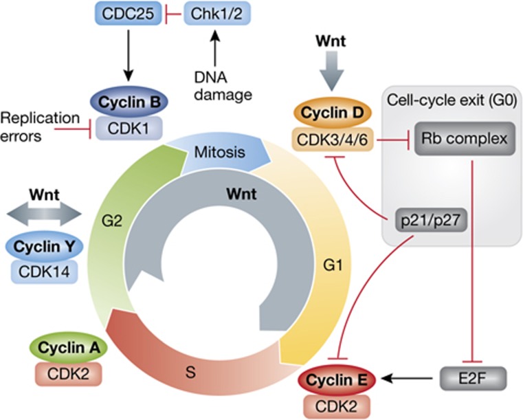
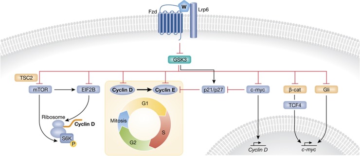
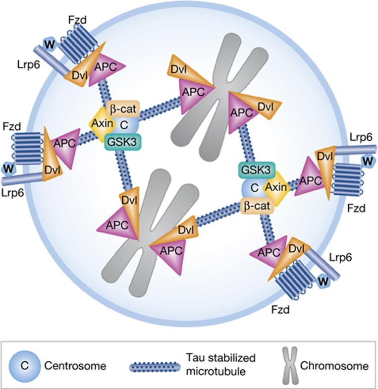
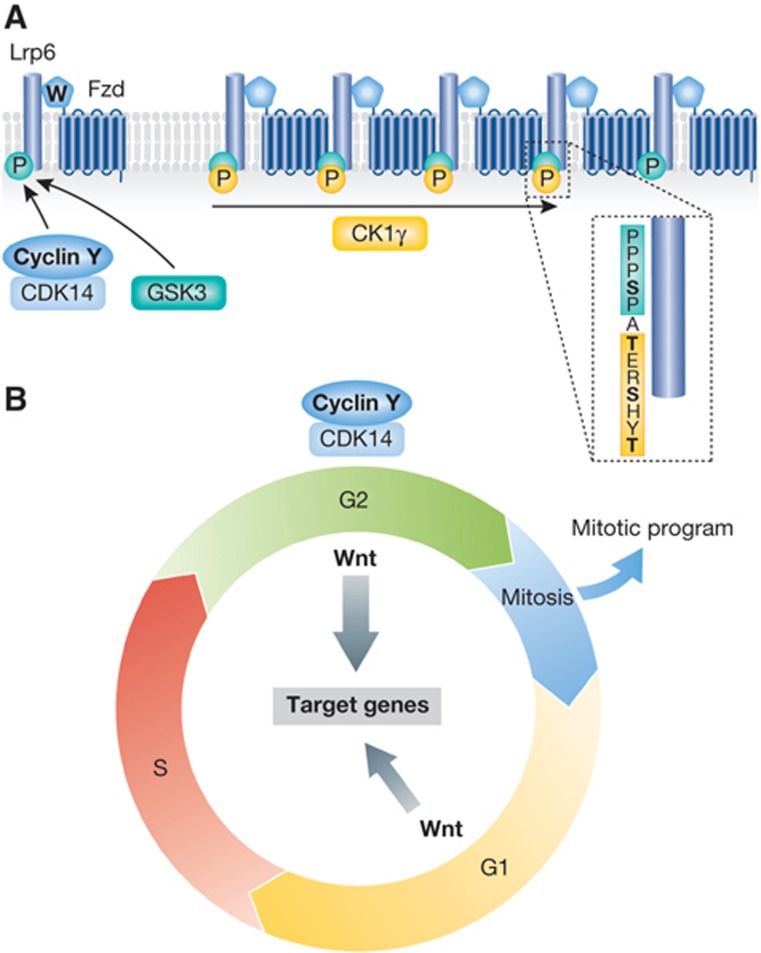
We now know that there is a complex interplay between cell cycle and Wnt signalling (Figure 1). Wnt signalling regulates G1 progression not only via cyclin D1 but also at multiple levels (Figure 2). Furthermore, Wnt signalling also plays an important role in mitosis (Figures 3 and 4). Consequently, misregulation of Wnt signalling can result in aberrant proliferation and chromosome instability, hallmarks of cancer.
Figure 1.
Overview of Wnt in cell-cycle regulation. Cell-cycle progression is controlled by cyclins and their CDKs. In G1, cyclin D initiates Rb complex phosphorylation, which derepresses E2F to induce cyclin E transcription. Components of this complex as well as p21 and p27 oppose these effects and can result in cell-cycle exit to G0. After DNA replication in S phase, different quality controls ensure the integrity of the DNA, while a cyclin B/CDK1 complex orchestrates progression into mitosis. Chromosome abnormalities and DNA damage are reported to this complex via different pathways to delay or stop cell division. Canonical Wnt signalling can regulate the cell cycle at the indicated levels, further described in the text.
Figure 2.
Regulation of G1- to S-phase progression by Wnt signalling. GSK3 inhibition by Wnt signalling acts as a central node for G1 control. GSK3 inhibits or activates the indicated proteins, all of which can contribute to G1- to S-phase progression. GSK3 target proteins known to be regulated by Wnt ligands are indicated in ochre.
Figure 3.
Mitotic spindle regulation by Wnt signalling components. APC and Dvl regulate the attachment of the mitotic spindle to the kinetochores, and together with Fzd and LRP6 modulate spindle orientation. GSK3, β-catenin, and Axin2 are required at the centrosome to ensure a proper distribution of the chromosomes during division. Wnt/GSK3 signalling promotes microtubule assembly by tau stabilization. Inhibition of Wnt signalling or mutations in the indicated components compromise the mitotic spindle and can result in chromosome instability.
Figure 4.
Mitotic Wnt signalling. (A) LRP6 competence depends on PPPSP sites phosphorylation (green) by GSK3 or the G2/M cyclinY/CDK14 complex, which primes CK1γ to further phosphorylate LRP6 upon Wnt stimulation (yellow). The architecture of the Lrp6 phosphorylation motif A is highlighted as an example, further described in Box 1. (B) Phosphorylation of the Wnt coreceptor LRP6 by cyclin Y/CDK14 results in maximal Wnt signalling at G2/M. Whether activation of LRP6 impacts the mitotic program through Wnt signalling components is still unknown.
Here, we review the relation between canonical Wnt signalling and the cell cycle in development and disease. We discuss the role of mitogenic Wnt signalling in G1 progression, self-renewal and proliferation of stem cells, as well as the function of ‘mitotic Wnt signalling’. Excellent reviews dealing with other aspects of Wnt signalling, including the mechanism of canonical Wnt signalling and its role in stem-cell biology and disease are available (Bienz and Clevers, 2000; Reya and Clevers, 2005; Clevers, 2006; MacDonald et al, 2009; van Amerongen and Nusse, 2009; Niehrs and Acebron, 2010; Kikuchi et al, 2011; Metcalfe and Bienz, 2011).
Wnt signalling and G1 phase
In order to divide without losing mass and genetic information, cells have to grow and replicate their DNA before division. Cells compartmentalize these processes in consecutive steps, which compose the cell cycle (Figure 1). In G1 and G2 phases, cell growth and transcription occur. DNA is replicated in S phase, while chromosomes are condensed and segregated after G2, during mitosis. Progression through the cell cycle is regulated by checkpoints that sense defects related to the different phases, in particular errors associated with genomic integrity. These checkpoints are modulated by cyclins and their cyclin-dependent kinases (CDKs), which integrate this information and switch between cell-cycle progression and arrest (Malumbres and Barbacid, 2009; Figure 1). Halting at a checkpoint allows cells to repair defects, such as errors in DNA replication or chromosome segregation. If damage is too severe, then cells may undergo apoptosis. On the other hand, mitogenic signals promote progression at checkpoints and thereby cell proliferation. Misregulation of checkpoints can compromise cell homeostasis and lead to both unscheduled proliferation and accumulation of DNA damage (Malumbres and Barbacid, 2009).
Entering S phase and DNA replication is a key decision that typically forces cells to divide, and hence this decision has to be regulated during G1. Indeed, most signalling pathways that regulate cell proliferation exert their effects in G1 (Massague, 2004). Thus, the balance of signals during this phase needs to be tightly coordinated and not surprisingly misregulations of such pathways are frequently associated with disease, notably cancer (Massague, 2004). Two important checkpoints for the G1 to S progression are regulated by cyclin D, cyclin E and their CDKs (Figure 1). Cyclin D accumulates and regulates many G1 events (Baldin et al, 1993; Malumbres and Barbacid, 2009), including phosphorylation and inhibition of the Retinoblastoma (Rb) complex, which increases cyclin E levels, whose accumulation acts as G1/S checkpoint (Dulic et al, 1992). S phase is then initiated after cyclin E degradation. Under growth inhibitory conditions such as serum starvation or TGFβ signalling, p21 and p27 accumulate and inhibit cyclin D and E, thereby leading to cell-cycle exit and entry into quiescence (G0) (Toyoshima and Hunter, 1994; Robson et al, 1999). Misregulation of cyclins and their CDKs induces unscheduled proliferation and is commonly associated with cancer (Massague, 2004; Malumbres and Barbacid, 2009).
Canonical Wnt signalling (Box 1) plays a key mitogenic role in promoting G1 progression by inhibiting GSK3, which directly regulates β-catenin-dependent c-myc transcription, cell-cycle effectors, and growth regulators.
Canonical Wnt signalling overview.
The essence of canonical Wnt signalling is a cascade of events, which leads to the inhibition of glycogen synthase kinase 3 (GSK3), which regulates many substrates, notably β-catenin. Phosphorylation by GSK3 triggers degradation of many of its substrates, including β-catenin via the proteasome (Aberle et al, 1997) and Wnt signalling stabilizes such proteins (Taelman et al, 2010). In canonical Wnt signaling, Wnt ligands form a ternary complex with Frizzled (Fzd) and LRP6 coreceptors (Wodarz and Nusse, 1998; Tamai et al, 2000; Wehrli et al, 2000). Upon Wnt ligand binding, the signalling complex forms clusters with Dishevelled (Dvl) polymers, to form endocytic LRP6 signalosomes (Bilic et al, 2007; Schwarz-Romond et al, 2007; Cruciat et al, 2010). LRP6 signalosomes promote phosphorylation of LRP6, which is required for signal transduction. So-called PPPSP motifs in LRP6 are phosphorylated in a Wnt-dependent as well as independent manner by a variety of kinases including GSK3 (Zeng et al, 2005) and cyclinY/CDK14 (Davidson et al, 2009). Phosphorylation of PPPSP motifs primes CK1γ to phosphorylate the adjacent Ser/Thr cluster upon Wnt stimulation (Davidson et al, 2005; Zeng et al, 2005). LRP6 phosphorylation by CK1γ triggers recruitment of GSK3 and other components of the β-catenin destruction complex to the signalosomes, notably APC and Axin (Bilic et al, 2007). Thereby, β-catenin is derepressed, accumulates and enters the nucleus to initiate transcription of target genes (listed in http://www.stanford.edu/~rnusse/wntwindow.html). LRP6 signalosomes mature into multivesicular bodies, where GSK3 is sequestered and its inhibition is sustained (Taelman et al, 2010).
β-Catenin-dependent c-myc regulation
Possibly the most important mechanism whereby Wnt signalling promotes G1 progression, is by inducing c-myc, which is a direct target gene of β-catenin (He et al, 1998). C-myc encodes a transcription factor that has a dual role in G1 progression, upregulating cyclin D (Daksis et al, 1994) and repressing p21 and p27 (Figure 2; Gartel et al, 2001; Yang et al, 2001; van de Wetering et al, 2002). Aberrant cyclin D1 and c-myc upregulation takes place when β-catenin is hyperactivated (Tetsu and McCormick, 1999; van de Wetering et al, 2002). This occurs in most colorectal cancers, where mutations of adenomatous polyposis coli (APC), a component of the β-catenin destruction complex, lead to a permissive and fast G1 transition and neoplastic transformation (Arber et al, 1997; Morin et al, 1997; Tetsu and McCormick, 1999).
Stabilization of cell-cycle effectors
Among the substrates which are destabilized by GSK3 phosphorylation are also direct regulators of G1- to S-phase progression, notably cyclin D1, cyclin E1 and c-myc (Diehl et al, 1998; Welcker et al, 2003, 2004; Figure 2). Mutations that impair phosphorylation and proteolysis of these G1–S regulators are often associated with neoplasia (Welcker et al, 2004). Thus, Wnt signalling-mediated GSK3 inhibition not only induces transcription of G1 regulators such as c-myc and cyclin D1 via β-catenin, but may also directly increase their protein stability. Another example is Wnt crosstalk with the sonic hedgehog pathway via GSK3, which phosphorylates Gli/Ci (Borycki et al, 2000; Price and Kalderon, 2002). By inhibiting GSK3, Wnt signalling stabilizes the transcription factor Gli (Borycki et al, 2000), which promotes expression of the G1 regulator N-Myc (akin to c-myc) (Mill et al, 2005).
Regulation of cell growth
When cells divide, they need to increase their protein levels, otherwise they will become smaller. Most cellular growth occurs in G1 and different mitogenic growth signals converge in this phase (Massague, 2004). They synchronize cell-cycle progression and cell mass in order to keep cell size constant. GSK3 is a key inhibitor of cell growth, and this inhibition is released by growth factors like IGF acting through AKT kinase, which blocks GSK3, resulting in an increased glucose uptake and TOR pathway activation (Sutherland et al, 1993). Notably, the TOR pathway is a key regulator of G1 growth by activating protein synthesis.
Similarly to IGF/AKT, Wnt/GSK3 signalling also activates the TOR pathway to stimulate protein translation (Inoki et al, 2006). Wnt activation has a dual role in the TOR pathway because GSK3 directly destabilizes EIF2B, a TOR pathway effector (Welsh et al, 1998), and activates TSC2, an inhibitor of the TOR pathway (Inoki et al, 2006; Figure 2). Interestingly, this TOR activation is yet another mode whereby Wnt signalling increases cyclin D1 protein, because cyclin D1 mRNA translation highly depends on the TOR pathway (Inoki et al, 2006). Thereby Wnt signalling coordinates growth and cell-cycle progression in G1 with GSK3 as its nexus, by directly regulating GSK3 substrates, independent of β-catenin-mediated transcription.
The tight link between Wnt signalling and G1 progression has important consequences in the context of development. Wnt-dependent c-myc/Cyclin D accumulation regulates cell proliferation and thereby patterning during embryogenesis. For example, during spinal cord development Wnts form a dorsal to ventral proliferation gradient (Megason and McMahon, 2002). Another example are developing long bones where Wnt signalling promotes chondrocyte proliferation and longitudinal growth (Yang et al, 2003). In adults, mitogenic Wnt signalling promotes cell turnover during tissue homeostasis or following injury (Stoick-Cooper et al, 2007b; Blanpain and Fuchs, 2009; Haegebarth and Clevers, 2009; Minear et al, 2010; Rabbani et al, 2011). The balance between quiescence and proliferation must be fine-tuned in order to avoid either a fatal loss of the tissue regeneration or neoplasia (Flores et al, 2004; Massague, 2004; Chen et al, 2009; Haegebarth and Clevers, 2009). The dissection of the various modes whereby Wnt signalling impacts G1 may help designing specific cancer therapies.
Wnt signalling and mitosis
The major role whereby Wnt signalling is commonly thought to impact the cell cycle is by transcriptional and translational upregulation of G1 effectors. However, it has become clear that another important phase where Wnt signalling plays a key role is mitosis. In mitosis, cells divide not only chromosomes but also all cellular constituents into daughter cells. Mitosis is therefore predominantly a phase of subcellular mechanics, while transcription and translation are mostly inhibited (Prescott and Bender, 1962; Gottesfeld and Forbes, 1997). Mitotic entry is initiated at late G2 by the cyclin B/CDK1 complex (Takizawa and Morgan, 2000), which is a checkpoint that is only activated when sufficient cell growth has occurred and DNA has been replicated (Malumbres and Barbacid, 2009; Figure 1). A protein network triggered by cyclin B/CDK1 induces nuclear envelope breakdown, chromosome condensation, and centrosome separation (Minshull et al, 1989; Takizawa and Morgan, 2000; Jackman et al, 2003). Microtubules (MTs) project from opposing centrosomes and attach to kinetochores to form the mitotic spindle. When the chromosomes are properly aligned, the Anaphase-Promoting Complex (APC/C; not to be confused with APC) degrades cyclin B and leads to chromosome segregation into daughter cells (Irniger et al, 1995; Zachariae et al, 1998). DNA replication errors trigger the inhibition of the cyclin B/CDK1 complex, for example, via Chk1/2 (Sanchez et al, 1997), and impair entry in mitosis, unless repaired (Figure 1). Once mitosis is initiated several positive feedback loops, notably via CDC25 (Sanchez et al, 1997; Takizawa and Morgan, 2000), reinforce commitment to the mitotic spindle formation. DNA integrity is then monitored by the spindle checkpoint, which inhibits APC/C until the sister chromatids are aligned without abnormalities, to avoid chromosome instability (Michel et al, 2001; Reddy et al, 2007).
Wnt signalling components regulate mitosis
Components of the Wnt signalling pathway modulate different aspects of the mitotic program, including MT dynamics, spindle formation, and centrosome division.
MT dynamics are regulated by Wnt signalling, and not only in mitosis (Salinas, 2007). In mitosis, long-term GSK3 inhibition by LiCl can abolish MT growth, as do stabilizing mutations in β-catenin or Axin2 ablation, while short-time GSK3 inhibition accelerates the reorganization of MTs (Huang et al, 2007). This may reduce phosphorylation of MT-associated proteins, such as the BICD–dynein complex and tau (Hanger et al, 1992; Fumoto et al, 2006), which stabilize MTs and the mitotic spindle (Wakefield et al, 2003; Fumoto et al, 2006). Consequently, GSK3 inhibitors can induce chromosome instability (Tighe et al, 2007).
Axin2 and APC localize to the mitotic spindle and are required for chromosome segregation (Fodde et al, 2001; Hadjihannas et al, 2006; Figure 3). APC associates with the MT plus ends and connects them to kinetochores, which is essential for chromosome organization and segregation. Mutations in APC that are associated with chromosome instability in colon cancer (Fodde et al, 2001; Hadjihannas et al, 2006) impair the linkage between MTs and kinetochores (Fodde et al, 2001) and upregulate Axin2, compromising the mitotic spindle checkpoint (Hadjihannas et al, 2006). Similarly, Dvl cooperates with Plk1 in establishing the spindle orientation and this depends on Fzd and LRP6 coreceptors (Kikuchi et al, 2010; Figure 3).
Centrosomes, which align the mitotic spindle, are another hot spot for Wnt components. Axin2, GSK3, and β-catenin accumulate at the centrosomes, where they regulate MT growth. For example, β-catenin depletion or LiCl treatment cause disruption of radial MTs (Huang et al, 2007). Moreover, stabilizing mutations in β-catenin or Axin2 ablation lead to premature splitting of the centrosome or multiple centrosomes (Huang et al, 2007; Bahmanyar et al, 2008; Hadjihannas et al, 2010). Finally, GSK3 may also play a role in the curious phenomenon whereby proteins destined for proteasomal degradation localize preferentially to only one of the two centrosomes during mitosis. This leads to asymmetric protein distribution, even in otherwise ‘non-polarized’ cells (Fuentealba et al, 2008). Thus, Wnt pathway components organize various centrosomal functions in mitosis.
While it is well established that multiple Wnt signalling components play an important role during mitosis, there is little evidence that these processes are actually regulated by Wnt signalling. The function of Dvl and APC at the mitotic spindle requires LRP6 and Fzd (Kikuchi et al, 2010) but an involvement of Wnt ligand signalling has not been demonstrated and is an important question that needs to be addressed.
Mitotic Wnt signalling
An unexpected finding was that Wnt signalling not only impacts the cell cycle, but that the reverse is also true, specifically that G2/M is a privileged phase for Wnt signalling. This is because the competence of the Wnt coreceptor LRP6 to respond to Wnt ligands is maximal during G2/M. The competence of LRP6 for Wnt signalling depends on a priming phosphorylation of its PPPSP sites (Box 1). The cyclin-dependent kinase 14 (CDK14/PFTK1) phosphorylates the LRP6 PPPSP motifs and associates with and is regulated by the G2/M cyclin Y. As a consequence, LRP6 phosphorylation peaks during G2/M and thus the coreceptor is maximally primed to respond to incoming Wnt signals (Davidson et al, 2009; Figure 4A and B). This explains previous observations that cytoplasmic β-catenin levels oscillate with the cell cycle, peaking in mitosis (Orford et al, 1999; Olmeda et al, 2003). Interestingly, IGF signalling also peaks at G2/M (Shtivelman et al, 2002), suggesting a coordinated cell-cycle modulation of mitogenic signals, notably since both pathways converge in GSK3 regulation.
Recently, it has been confirmed that expression of the Wnt/β-catenin target genes Lgr5 and Axin2 peaks at G2/M. Intriguingly, other Wnt targets like c-myc peaked at G1/S (Hadjihannas et al, 2012). Differences in peak target gene expression may be due to crosstalk with other signalling pathways as well as to a negative feedback loop promoted by Axin2 (Davidson et al, 2009; Hadjihannas et al, 2012).
Wnt signalling and cell-cycle regulation in stem cells
Wnt signalling has a prominent role in stem cell biology, including self-renewal, pluripotency, and differentiation of both embryonic stem (ES) and somatic stem cells (reviewed in Reya and Clevers, 2005; Clevers, 2006; Nusse et al, 2008; Sokol, 2011).
Wnt regulation of ES cells
Pluripotency of ES cells is maintained by a core network consisting of Oct4, Sox2, and Nanog (Pan and Thomson, 2007). In mouse ES cells, Wnt signalling plays a key role in the maintenance of pluripotency and transcriptional regulation of this core network (Kielman et al, 2002; Sato et al, 2004; Hao et al, 2006; Ogawa et al, 2006; Miyabayashi et al, 2007; ten Berge et al, 2011). In a dual mode, β-catenin forms a complex with Tcf1 to upregulate Oct4 transcription (Sato et al, 2004; ten Berge et al, 2011) and blocks Tcf3 (Cole et al, 2008; Kelly et al, 2011; Wray et al, 2011), which is an inhibitor of the core network (Cole et al, 2008). The effect of Wnt/β-catenin signalling on maintaining core network expression is fully accounted for by Tcf3 repression and Tcf1-dependent transcription (Niwa, 2011; Yi et al, 2011). Although β-catenin is required for the self-renewal promoted by Wnt/GSK3 signalling (Kelly et al, 2011; Niwa, 2011; Wray et al, 2011; Yi et al, 2011), it is not essential for mouse ES cell self-renewal under specific culture conditions (Lyashenko et al, 2011; Wray et al, 2011).
Despite the prominent role of canonical Wnt signalling in ES cells self-renewal, surprisingly little is known about whether Wnt signalling regulates ES cell cycle directly. ES cells have a fast cell cycle with a short G1 phase (Becker et al, 2006) and progress in the absence of cyclin D (Burdon et al, 2002), suggesting that mitogenic Wnt signalling would not impact ES cell-cycle progression via cyclin D1 upregulation. On the other hand, c-myc promotes ES cell self-renewal (Cartwright et al, 2005; Ying et al, 2008), although its upregulation is not essential for ES cell maintenance under specific culture conditions (Cartwright et al, 2005; Ying et al, 2008). In ES cells, c-myc forms a regulatory network together with E2F (Chen et al, 2008), which upregulates cyclin E transcription (Geng et al, 1996; Stead et al, 2002). Whether Wnt signalling promotes cell-cycle progression in ES cells by c-myc upregulation is a question that remains unsolved.
Wnt regulation of adult stem cells
A hallmark of adult stem cells is their low proliferation, notably in contrast to ES cells, which prevents their exhaustion (Cheng et al, 2000; Orford and Scadden, 2008). Quiescence of adult stem cells is maintained by p21 (Cheng et al, 2000), which keeps cells arrested in G0. This is the case in intestinal crypt stem cells, where Wnt signalling promotes cell-cycle entry by inducing c-myc and cyclin D1 expression and downregulating p21 (Korinek et al, 1998; Kuhnert et al, 2004).
In haematopoietic stem cells (HSCs), Wnt signalling increases both self-renewal and differentiation by c-myc induction (Reya et al, 2003). Although the essential role of β-catenin in this process was controversial (Cobas et al, 2004), recent data suggest that high and low Wnt levels promote differentiation and HSCs self-renewal, respectively (Luis et al, 2011).
In hair follicles, Wnt signalling is crucial for both self-renewal and activation of bulge stem cells (Reya and Clevers, 2005; Greco et al, 2009). Although the molecular mechanisms underlying these effects are not yet clear, both Wnt/β-catenin and c-myc transcriptional programs overlap at the hair follicle (Choi et al, 2008) and both promote proliferation of bulge stem cells (Chan et al, 1999; Andl et al, 2002; Greco et al, 2009). This suggests that Wnt may exert its activities through c-myc by promoting cell-cycle entry. Moreover, different components of the Wnt pathway including GSK3, β-catenin, lef1, and TCF3/4 play distinct roles to coordinate bulge stem-cell differentiation and transit through the follicle (DasGupta and Fuchs, 1999; Nguyen et al, 2009; Wu et al, 2011).
These examples highlight that Wnt signalling in adult stem cells promotes cell-cycle re-entry by upregulation of c-myc/cyclin D1 and thereby functions as switch between quiescence and division/differentiation. C-myc has a preeminent role in cell-cycle re-entry and as a common driver of tumorigenesis (Pelengaris et al, 2002). Aberrant c-myc activation by Wnt signalling in adult stem cells initiates unscheduled proliferation and neoplasia, associated with cancer in the intestine, haematopoietic lineage, and hair follicle (He et al, 1998; van de Wetering et al, 2002; Pelengaris et al, 2002; Weng et al, 2006). Importantly, c-myc ablation fully rescues APC loss-driven tumorigenesis (Sansom et al, 2007), highlighting its importance in mediating the cell proliferation effects of Wnt signalling during tissue regeneration.
Wnt signalling in G0: quiescent cells
Wnt signalling has been mostly characterized in dividing cells. However, many Wnts are also expressed in differentiated adult tissues, suggesting that Wnt signalling plays important roles in postmitotic cells. Such is the case in neurons, where Wnt signalling regulates axon guidance and neurite outgrowth. In the distal end of axons, Wnt signalling regulates APC association with MTs and thereby induces MT remodelling (Salinas and Zou, 2008). Moreover, Wnt3a and Wnt7a enhance neurite outgrowth via tau derepression (Hall et al, 2000; Endo et al, 2008). Wnt signalling directly inhibits tau phosphorylation by GSK3 and is therefore β-catenin independent (Hanger et al, 1992; Scali et al, 2006). Tau phosphorylation is critical for MT dynamics in axons (Drechsel et al, 1992), and its misregulation is a hallmark of several neurodegenerative disorders, notably Alzheimer (Grundke-Iqbal et al, 1986; Bramblett et al, 1993).
In adult liver, APC is essential for zonation, the phenomenon that different metabolic processes are compartmentalized in specialized liver areas (Benhamouche et al, 2006). APC is differentially expressed within the liver and restricts Wnt/β-catenin signalling to regions where its protein levels are low. In these zones, Wnt signalling activates ammonia metabolism-associated genes, and thereby promotes hepatocyte specialization (Benhamouche et al, 2006). Notably, the role of Wnt/β-catenin signalling in liver zonation does not require upregulation of cell-cycle effectors (Burke et al, 2009).
In postmitotic cardiomyocytes, components of Wnt signalling regulate cell growth and are notably involved in cardiac hypertrophy, an enlargement which can lead to heart failure (ter Horst et al, 2012). Inhibition of GSK3, overexpression of Dvl, and stabilization of β-catenin all lead to aberrant cell growth and heart hypertrophy (Haq et al, 2003; Malekar et al, 2010). This occurs through many different modes that include TOR activation, NFAT upregulation, and transcription of β-catenin target genes (Blankesteijn et al, 2008; ter Horst et al, 2012). While there is evidence for a role of Fzd2 in promoting hypertrophy (Blankesteijn et al, 2008), it remains unclear whether Wnt ligands or only downstream components are involved in cardiac hypertrophy (Haq et al, 2003).
Concluding remarks
Three decades ago, it was discovered that Wnt signalling is associated with deregulated proliferation and neoplasia. Since then key roles of the canonical pathway in cell-cycle regulation have been well established and the underlying mechanisms are emerging. But these studies have also raised new questions that need to be addressed.
What are the targets of β-catenin-independent canonical Wnt signalling?
While canonical Wnt signalling is often equated with β-catenin-dependent transcriptional regulation of target genes, such as c-myc and cyclin D1, it is becoming increasingly clear that this is only one of the modes whereby the pathway regulates the cell cycle. The other major mode is by inhibiting GSK3, a key kinase with pleiotropic effects and a multitude of substrates, which are typically negatively regulated by GSK3. Recent data from De Robertis and colleagues indicate that a significant part of the proteome is stabilized by Wnt signalling halting GSK3-driven proteolysis (Taelman et al, 2010). Bioinformatic analysis of putative GSK3 substrates revealed that many of them function in mitosis, cell cycle, chromatin, and ribosome biogenesis (Taelman et al, 2010), suggesting a largely unexplored layer of cell-cycle regulation by Wnt signalling.
What is the physiological significance of ‘mitotic Wnt signalling’?
The fact that Wnt signalling peaks in mitotic cells and conversely that Wnt components play such an important role in mitosis suggests that Wnt may orchestrate a mitotic program. What role does Wnt-dependent signalling play in regulating mitotic Wnt components? Dvl, APC, Axin2, and β-catenin all play important roles in spindle mechanics. Yet, whether their functions are Wnt regulated is unresolved.
What is the role of Wnt signalling in quiescent cells?
While most work on Wnt signalling has focussed on dividing cells, the examples of neurons, cardiomyocytes, and hepatocytes indicate important cellular roles in postmitotic tissues. Yet, we still know little about Wnt signalling in quiescent cells. Are there common themes? Individual components of the Wnt pathway have both Wnt-dependent and Wnt-independent functions. Is the requirement for Wnt components in quiescent cells reflecting actual Wnt ligand signalling, or do these components function Wnt independently? This could be addressed, for example, by overexpression of Wnt antagonists such as Dkk1 or studying LRP6 knockout mice.
What is the basis for tissue-specific involvement of Wnt signalling in cancer?
Deregulation of Wnt signalling has been well documented as the basis for a limited number of tumours. Yet, Wnt signalling occurs much more widespread, but without apparent association with neoplasia. What determines this specificity? Crosstalk with other mitogenic pathways likely plays a role and hence it will be important to define these.
How can we exploit Wnt cell-cycle regulation in regenerative medicine and cancer therapy?
Wnt signalling is a prime target in cancer treatment (Barker and Clevers, 2006) and interestingly not just pathway inhibition but also activation may cure certain cancer types (Biechele et al, 2012). Given the many roles of Wnt signaling in adult tissues, it appears crucial to identify specific targets to manipulate the pathway in cancer in order to achieve the desired therapy without deleterious side effects. Similarly, in regenerative medicine, specific modulation of Wnt signaling holds great promises (Stoick-Cooper et al, 2007a) but again specificity and control are essential, notably in light of the tumorigenic effects of the pathway.
Acknowledgments
This work was supported by the Deutsche Forschungsgemeinschaft.
Footnotes
The authors declare that they have no conflict of interest.
References
- Aberle H, Bauer A, Stappert J, Kispert A, Kemler R (1997) beta-Catenin is a target for the ubiquitin-proteasome pathway. EMBO J 16: 3797–3804 [DOI] [PMC free article] [PubMed] [Google Scholar]
- Andl T, Reddy ST, Gaddapara T, Millar SE (2002) WNT signals are required for the initiation of hair follicle development. Dev Cell 2: 643–653 [DOI] [PubMed] [Google Scholar]
- Arber N, Doki Y, Han EK, Sgambato A, Zhou P, Kim NH, Delohery T, Klein MG, Holt PR, Weinstein IB (1997) Antisense to cyclin D1 inhibits the growth and tumorigenicity of human colon cancer cells. Cancer Res 57: 1569–1574 [PubMed] [Google Scholar]
- Bahmanyar S, Kaplan DD, Deluca JG, Giddings TH Jr, O’Toole ET, Winey M, Salmon ED, Casey PJ, Nelson WJ, Barth AI (2008) beta-Catenin is a Nek2 substrate involved in centrosome separation. Genes Dev 22: 91–105 [DOI] [PMC free article] [PubMed] [Google Scholar]
- Baldin V, Lukas J, Marcote MJ, Pagano M, Draetta G (1993) Cyclin D1 is a nuclear protein required for cell cycle progression in G1. Genes Dev 7: 812–821 [DOI] [PubMed] [Google Scholar]
- Barker N, Clevers H (2006) Mining the Wnt pathway for cancer therapeutics. Nat Rev Drug Discov 5: 997–1014 [DOI] [PubMed] [Google Scholar]
- Becker KA, Ghule PN, Therrien JA, Lian JB, Stein JL, van Wijnen AJ, Stein GS (2006) Self-renewal of human embryonic stem cells is supported by a shortened G1 cell cycle phase. J Cell Physiol 209: 883–893 [DOI] [PubMed] [Google Scholar]
- Benhamouche S, Decaens T, Godard C, Chambrey R, Rickman DS, Moinard C, Vasseur-Cognet M, Kuo CJ, Kahn A, Perret C, Colnot S (2006) Apc tumor suppressor gene is the ‘zonation-keeper’ of mouse liver. Dev Cell 10: 759–770 [DOI] [PubMed] [Google Scholar]
- Biechele TL, Kulikauskas RM, Toroni RA, Lucero OM, Swift RD, James RG, Robin NC, Dawson DW, Moon RT, Chien AJ (2012) Wnt/beta-catenin signaling and AXIN1 regulate apoptosis triggered by inhibition of the mutant kinase BRAFV600E in human melanoma. Sci Signal 5 ra3 [DOI] [PMC free article] [PubMed] [Google Scholar]
- Bienz M, Clevers H (2000) Linking colorectal cancer to Wnt signaling. Cell 103: 311–320 [DOI] [PubMed] [Google Scholar]
- Bilic J, Huang YL, Davidson G, Zimmermann T, Cruciat CM, Bienz M, Niehrs C (2007) Wnt induces LRP6 signalosomes and promotes dishevelled-dependent LRP6 phosphorylation. Science 316: 1619–1622 [DOI] [PubMed] [Google Scholar]
- Blankesteijn WM, van de Schans VA, ter Horst P, Smits JF (2008) The Wnt/frizzled/GSK-3 beta pathway: a novel therapeutic target for cardiac hypertrophy. Trends Pharmacol Sci 29: 175–180 [DOI] [PubMed] [Google Scholar]
- Blanpain C, Fuchs E (2009) Epidermal homeostasis: a balancing act of stem cells in the skin. Nat Rev Mol Cell Biol 10: 207–217 [DOI] [PMC free article] [PubMed] [Google Scholar]
- Borycki A, Brown AM, Emerson CP Jr (2000) Shh and Wnt signaling pathways converge to control Gli gene activation in avian somites. Development 127: 2075–2087 [DOI] [PubMed] [Google Scholar]
- Bramblett GT, Goedert M, Jakes R, Merrick SE, Trojanowski JQ, Lee VM (1993) Abnormal tau phosphorylation at Ser396 in Alzheimer's disease recapitulates development and contributes to reduced microtubule binding. Neuron 10: 1089–1099 [DOI] [PubMed] [Google Scholar]
- Burdon T, Smith A, Savatier P (2002) Signalling, cell cycle and pluripotency in embryonic stem cells. Trends Cell Biol 12: 432–438 [DOI] [PubMed] [Google Scholar]
- Burke ZD, Reed KR, Phesse TJ, Sansom OJ, Clarke AR, Tosh D (2009) Liver zonation occurs through a beta-catenin-dependent, c-Myc-independent mechanism. Gastroenterology 136: 2316–2324.e1–3 [DOI] [PubMed] [Google Scholar]
- Cartwright P, McLean C, Sheppard A, Rivett D, Jones K, Dalton S (2005) LIF/STAT3 controls ES cell self-renewal and pluripotency by a Myc-dependent mechanism. Development 132: 885–896 [DOI] [PubMed] [Google Scholar]
- Chan EF, Gat U, McNiff JM, Fuchs E (1999) A common human skin tumour is caused by activating mutations in beta-catenin. Nat Genet 21: 410–413 [DOI] [PubMed] [Google Scholar]
- Chen B, Dodge ME, Tang W, Lu J, Ma Z, Fan CW, Wei S, Hao W, Kilgore J, Williams NS, Roth MG, Amatruda JF, Chen C, Lum L (2009) Small molecule-mediated disruption of Wnt-dependent signaling in tissue regeneration and cancer. Nat Chem Biol 5: 100–107 [DOI] [PMC free article] [PubMed] [Google Scholar]
- Chen X, Xu H, Yuan P, Fang F, Huss M, Vega VB, Wong E, Orlov YL, Zhang W, Jiang J, Loh YH, Yeo HC, Yeo ZX, Narang V, Govindarajan KR, Leong B, Shahab A, Ruan Y, Bourque G, Sung WK et al. (2008) Integration of external signaling pathways with the core transcriptional network in embryonic stem cells. Cell 133: 1106–1117 [DOI] [PubMed] [Google Scholar]
- Cheng T, Rodrigues N, Shen H, Yang Y, Dombkowski D, Sykes M, Scadden DT (2000) Hematopoietic stem cell quiescence maintained by p21cip1/waf1. Science 287: 1804–1808 [DOI] [PubMed] [Google Scholar]
- Choi J, Southworth LK, Sarin KY, Venteicher AS, Ma W, Chang W, Cheung P, Jun S, Artandi MK, Shah N, Kim SK, Artandi SE (2008) TERT promotes epithelial proliferation through transcriptional control of a Myc- and Wnt-related developmental program. PLoS Genet 4: e10. [DOI] [PMC free article] [PubMed] [Google Scholar]
- Clevers H (2006) Wnt/beta-catenin signaling in development and disease. Cell 127: 469–480 [DOI] [PubMed] [Google Scholar]
- Cobas M, Wilson A, Ernst B, Mancini SJ, MacDonald HR, Kemler R, Radtke F (2004) Beta-catenin is dispensable for hematopoiesis and lymphopoiesis. J Exp Med 199: 221–229 [DOI] [PMC free article] [PubMed] [Google Scholar]
- Cole MF, Johnstone SE, Newman JJ, Kagey MH, Young RA (2008) Tcf3 is an integral component of the core regulatory circuitry of embryonic stem cells. Genes Dev 22: 746–755 [DOI] [PMC free article] [PubMed] [Google Scholar]
- Cruciat CM, Ohkawara B, Acebron SP, Karaulanov E, Reinhard C, Ingelfinger D, Boutros M, Niehrs C (2010) Requirement of prorenin receptor and vacuolar H+-ATPase-mediated acidification for Wnt signaling. Science 327: 459–463 [DOI] [PubMed] [Google Scholar]
- Daksis JI, Lu RY, Facchini LM, Marhin WW, Penn LJ (1994) Myc induces cyclin D1 expression in the absence of de novo protein synthesis and links mitogen-stimulated signal transduction to the cell cycle. Oncogene 9: 3635–3645 [PubMed] [Google Scholar]
- DasGupta R, Fuchs E (1999) Multiple roles for activated LEF/TCF transcription complexes during hair follicle development and differentiation. Development 126: 4557–4568 [DOI] [PubMed] [Google Scholar]
- Davidson G, Shen J, Huang YL, Su Y, Karaulanov E, Bartscherer K, Hassler C, Stannek P, Boutros M, Niehrs C (2009) Cell cycle control of wnt receptor activation. Dev Cell 17: 788–799 [DOI] [PubMed] [Google Scholar]
- Davidson G, Wu W, Shen J, Bilic J, Fenger U, Stannek P, Glinka A, Niehrs C (2005) Casein kinase 1 gamma couples Wnt receptor activation to cytoplasmic signal transduction. Nature 438: 867–872 [DOI] [PubMed] [Google Scholar]
- Diehl JA, Cheng M, Roussel MF, Sherr CJ (1998) Glycogen synthase kinase-3beta regulates cyclin D1 proteolysis and subcellular localization. Genes Dev 12: 3499–3511 [DOI] [PMC free article] [PubMed] [Google Scholar]
- Drechsel DN, Hyman AA, Cobb MH, Kirschner MW (1992) Modulation of the dynamic instability of tubulin assembly by the microtubule-associated protein tau. Mol Biol Cell 3: 1141–1154 [DOI] [PMC free article] [PubMed] [Google Scholar]
- Dulic V, Lees E, Reed SI (1992) Association of human cyclin E with a periodic G1-S phase protein kinase. Science 257: 1958–1961 [DOI] [PubMed] [Google Scholar]
- Endo Y, Beauchamp E, Woods D, Taylor WG, Toretsky JA, Uren A, Rubin JS (2008) Wnt-3a and Dickkopf-1 stimulate neurite outgrowth in Ewing tumor cells via a Frizzled3- and c-Jun N-terminal kinase-dependent mechanism. Mol Cell Biol 28: 2368–2379 [DOI] [PMC free article] [PubMed] [Google Scholar]
- Flores I, Murphy DJ, Swigart LB, Knies U, Evan GI (2004) Defining the temporal requirements for Myc in the progression and maintenance of skin neoplasia. Oncogene 23: 5923–5930 [DOI] [PubMed] [Google Scholar]
- Fodde R, Kuipers J, Rosenberg C, Smits R, Kielman M, Gaspar C, van Es JH, Breukel C, Wiegant J, Giles RH, Clevers H (2001) Mutations in the APC tumour suppressor gene cause chromosomal instability. Nat Cell Biol 3: 433–438 [DOI] [PubMed] [Google Scholar]
- Fuentealba LC, Eivers E, Geissert D, Taelman V, De Robertis EM (2008) Asymmetric mitosis: unequal segregation of proteins destined for degradation. Proc Natl Acad Sci USA 105: 7732–7737 [DOI] [PMC free article] [PubMed] [Google Scholar]
- Fumoto K, Hoogenraad CC, Kikuchi A (2006) GSK-3beta-regulated interaction of BICD with dynein is involved in microtubule anchorage at centrosome. EMBO J 25: 5670–5682 [DOI] [PMC free article] [PubMed] [Google Scholar]
- Gartel AL, Ye X, Goufman E, Shianov P, Hay N, Najmabadi F, Tyner AL (2001) Myc represses the p21(WAF1/CIP1) promoter and interacts with Sp1/Sp3. Proc Natl Acad Sci USA 98: 4510–4515 [DOI] [PMC free article] [PubMed] [Google Scholar]
- Geng Y, Eaton EN, Picon M, Roberts JM, Lundberg AS, Gifford A, Sardet C, Weinberg RA (1996) Regulation of cyclin E transcription by E2Fs and retinoblastoma protein. Oncogene 12: 1173–1180 [PubMed] [Google Scholar]
- Gottesfeld JM, Forbes DJ (1997) Mitotic repression of the transcriptional machinery. Trends Biochem Sci 22: 197–202 [DOI] [PubMed] [Google Scholar]
- Greco V, Chen T, Rendl M, Schober M, Pasolli HA, Stokes N, Dela Cruz-Racelis J, Fuchs E (2009) A two-step mechanism for stem cell activation during hair regeneration. Cell Stem Cell 4: 155–169 [DOI] [PMC free article] [PubMed] [Google Scholar]
- Grundke-Iqbal I, Iqbal K, Tung YC, Quinlan M, Wisniewski HM, Binder LI (1986) Abnormal phosphorylation of the microtubule-associated protein tau (tau) in Alzheimer cytoskeletal pathology. Proc Natl Acad Sci USA 83: 4913–4917 [DOI] [PMC free article] [PubMed] [Google Scholar]
- Hadjihannas MV, Bernkopf DB, Bruckner M, Behrens J (2012) Cell cycle control of Wnt/beta-catenin signalling by conductin/axin2 through CDC20. EMBO Rep 13: 347–354 [DOI] [PMC free article] [PubMed] [Google Scholar]
- Hadjihannas MV, Bruckner M, Behrens J (2010) Conductin/axin2 and Wnt signalling regulates centrosome cohesion. EMBO Rep 11: 317–324 [DOI] [PMC free article] [PubMed] [Google Scholar]
- Hadjihannas MV, Bruckner M, Jerchow B, Birchmeier W, Dietmaier W, Behrens J (2006) Aberrant Wnt/beta-catenin signaling can induce chromosomal instability in colon cancer. Proc Natl Acad Sci USA 103: 10747–10752 [DOI] [PMC free article] [PubMed] [Google Scholar]
- Haegebarth A, Clevers H (2009) Wnt signaling, lgr5, and stem cells in the intestine and skin. Am J Pathol 174: 715–721 [DOI] [PMC free article] [PubMed] [Google Scholar]
- Hall AC, Lucas FR, Salinas PC (2000) Axonal remodeling and synaptic differentiation in the cerebellum is regulated by WNT-7a signaling. Cell 100: 525–535 [DOI] [PubMed] [Google Scholar]
- Hanger DP, Hughes K, Woodgett JR, Brion JP, Anderton BH (1992) Glycogen synthase kinase-3 induces Alzheimer's disease-like phosphorylation of tau: generation of paired helical filament epitopes and neuronal localisation of the kinase. Neurosci Lett 147: 58–62 [DOI] [PubMed] [Google Scholar]
- Hao J, Li TG, Qi X, Zhao DF, Zhao GQ (2006) WNT/beta-catenin pathway up-regulates Stat3 and converges on LIF to prevent differentiation of mouse embryonic stem cells. Dev Biol 290: 81–91 [DOI] [PubMed] [Google Scholar]
- Haq S, Michael A, Andreucci M, Bhattacharya K, Dotto P, Walters B, Woodgett J, Kilter H, Force T (2003) Stabilization of beta-catenin by a Wnt-independent mechanism regulates cardiomyocyte growth. Proc Natl Acad Sci USA 100: 4610–4615 [DOI] [PMC free article] [PubMed] [Google Scholar]
- He TC, Sparks AB, Rago C, Hermeking H, Zawel L, da Costa LT, Morin PJ, Vogelstein B, Kinzler KW (1998) Identification of c-MYC as a target of the APC pathway. Science 281: 1509–1512 [DOI] [PubMed] [Google Scholar]
- Huang P, Senga T, Hamaguchi M (2007) A novel role of phospho-beta-catenin in microtubule regrowth at centrosome. Oncogene 26: 4357–4371 [DOI] [PubMed] [Google Scholar]
- Inoki K, Ouyang H, Zhu T, Lindvall C, Wang Y, Zhang X, Yang Q, Bennett C, Harada Y, Stankunas K, Wang CY, He X, MacDougald OA, You M, Williams BO, Guan KL (2006) TSC2 integrates Wnt and energy signals via a coordinated phosphorylation by AMPK and GSK3 to regulate cell growth. Cell 126: 955–968 [DOI] [PubMed] [Google Scholar]
- Irniger S, Piatti S, Michaelis C, Nasmyth K (1995) Genes involved in sister chromatid separation are needed for B-type cyclin proteolysis in budding yeast. Cell 81: 269–278 [DOI] [PubMed] [Google Scholar]
- Jackman M, Lindon C, Nigg EA, Pines J (2003) Active cyclin B1-Cdk1 first appears on centrosomes in prophase. Nat Cell Biol 5: 143–148 [DOI] [PubMed] [Google Scholar]
- Kelly KF, Ng DY, Jayakumaran G, Wood GA, Koide H, Doble BW (2011) beta-catenin enhances Oct-4 activity and reinforces pluripotency through a TCF-independent mechanism. Cell Stem Cell 8: 214–227 [DOI] [PMC free article] [PubMed] [Google Scholar]
- Kielman MF, Rindapaa M, Gaspar C, van Poppel N, Breukel C, van Leeuwen S, Taketo MM, Roberts S, Smits R, Fodde R (2002) Apc modulates embryonic stem-cell differentiation by controlling the dosage of beta-catenin signaling. Nat Genet 32: 594–605 [DOI] [PubMed] [Google Scholar]
- Kikuchi A, Yamamoto H, Sato A, Matsumoto S (2011) New insights into the mechanism of Wnt signaling pathway activation. Int Rev Cell Mol Biol 291: 21–71 [DOI] [PubMed] [Google Scholar]
- Kikuchi K, Niikura Y, Kitagawa K, Kikuchi A (2010) Dishevelled, a Wnt signalling component, is involved in mitotic progression in cooperation with Plk1. EMBO J 29: 3470–3483 [DOI] [PMC free article] [PubMed] [Google Scholar]
- Korinek V, Barker N, Moerer P, van Donselaar E, Huls G, Peters PJ, Clevers H (1998) Depletion of epithelial stem-cell compartments in the small intestine of mice lacking Tcf-4. Nat Genet 19: 379–383 [DOI] [PubMed] [Google Scholar]
- Kuhnert F, Davis CR, Wang HT, Chu P, Lee M, Yuan J, Nusse R, Kuo CJ (2004) Essential requirement for Wnt signaling in proliferation of adult small intestine and colon revealed by adenoviral expression of Dickkopf-1. Proc Natl Acad Sci USA 101: 266–271 [DOI] [PMC free article] [PubMed] [Google Scholar]
- Luis TC, Naber BA, Roozen PP, Brugman MH, de Haas EF, Ghazvini M, Fibbe WE, van Dongen JJ, Fodde R, Staal FJ (2011) Canonical wnt signaling regulates hematopoiesis in a dosage-dependent fashion. Cell Stem Cell 9: 345–356 [DOI] [PubMed] [Google Scholar]
- Lyashenko N, Winter M, Migliorini D, Biechele T, Moon RT, Hartmann C (2011) Differential requirement for the dual functions of beta-catenin in embryonic stem cell self-renewal and germ layer formation. Nat Cell Biol 13: 753–761 [DOI] [PMC free article] [PubMed] [Google Scholar]
- MacDonald BT, Tamai K, He X (2009) Wnt/beta-catenin signaling: components, mechanisms, and diseases. Dev Cell 17: 9–26 [DOI] [PMC free article] [PubMed] [Google Scholar]
- Malekar P, Hagenmueller M, Anyanwu A, Buss S, Streit MR, Weiss CS, Wolf D, Riffel J, Bauer A, Katus HA, Hardt SE (2010) Wnt signaling is critical for maladaptive cardiac hypertrophy and accelerates myocardial remodeling. Hypertension 55: 939–945 [DOI] [PubMed] [Google Scholar]
- Malumbres M, Barbacid M (2009) Cell cycle, CDKs and cancer: a changing paradigm. Nat Rev Cancer 9: 153–166 [DOI] [PubMed] [Google Scholar]
- Massague J (2004) G1 cell-cycle control and cancer. Nature 432: 298–306 [DOI] [PubMed] [Google Scholar]
- Megason SG, McMahon AP (2002) A mitogen gradient of dorsal midline Wnts organizes growth in the CNS. Development 129: 2087–2098 [DOI] [PubMed] [Google Scholar]
- Metcalfe C, Bienz M (2011) Inhibition of GSK3 by Wnt signalling--two contrasting models. J Cell Sci 124: 3537–3544 [DOI] [PubMed] [Google Scholar]
- Michel LS, Liberal V, Chatterjee A, Kirchwegger R, Pasche B, Gerald W, Dobles M, Sorger PK, Murty VV, Benezra R (2001) MAD2 haplo-insufficiency causes premature anaphase and chromosome instability in mammalian cells. Nature 409: 355–359 [DOI] [PubMed] [Google Scholar]
- Mill P, Mo R, Hu MC, Dagnino L, Rosenblum ND, Hui CC (2005) Shh controls epithelial proliferation via independent pathways that converge on N-Myc. Dev Cell 9: 293–303 [DOI] [PubMed] [Google Scholar]
- Minear S, Leucht P, Jiang J, Liu B, Zeng A, Fuerer C, Nusse R, Helms JA (2010) Wnt proteins promote bone regeneration. Sci Transl Med 2: 29ra30. [DOI] [PubMed] [Google Scholar]
- Minshull J, Blow JJ, Hunt T (1989) Translation of cyclin mRNA is necessary for extracts of activated xenopus eggs to enter mitosis. Cell 56: 947–956 [DOI] [PubMed] [Google Scholar]
- Miyabayashi T, Teo JL, Yamamoto M, McMillan M, Nguyen C, Kahn M (2007) Wnt/beta-catenin/CBP signaling maintains long-term murine embryonic stem cell pluripotency. Proc Natl Acad Sci USA 104: 5668–5673 [DOI] [PMC free article] [PubMed] [Google Scholar]
- Morin PJ, Sparks AB, Korinek V, Barker N, Clevers H, Vogelstein B, Kinzler KW (1997) Activation of beta-catenin-Tcf signaling in colon cancer by mutations in beta-catenin or APC. Science 275: 1787–1790 [DOI] [PubMed] [Google Scholar]
- Nguyen H, Merrill BJ, Polak L, Nikolova M, Rendl M, Shaver TM, Pasolli HA, Fuchs E (2009) Tcf3 and Tcf4 are essential for long-term homeostasis of skin epithelia. Nat Genet 41: 1068–1075 [DOI] [PMC free article] [PubMed] [Google Scholar]
- Niehrs C, Acebron SP (2010) Wnt signaling: multivesicular bodies hold GSK3 captive. Cell 143: 1044–1046 [DOI] [PubMed] [Google Scholar]
- Niwa H (2011) Wnt: what’s needed to maintain pluripotency? Nat Cell Biol 13: 1024–1026 [DOI] [PubMed] [Google Scholar]
- Nusse R, Fuerer C, Ching W, Harnish K, Logan C, Zeng A, ten Berge D, Kalani Y (2008) Wnt signaling and stem cell control. Cold Spring Harb Symp Quant Biol 73: 59–66 [DOI] [PubMed] [Google Scholar]
- Nusse R, Varmus HE (1982) Many tumors induced by the mouse mammary tumor virus contain a provirus integrated in the same region of the host genome. Cell 31: 99–109 [DOI] [PubMed] [Google Scholar]
- Ogawa K, Nishinakamura R, Iwamatsu Y, Shimosato D, Niwa H (2006) Synergistic action of Wnt and LIF in maintaining pluripotency of mouse ES cells. Biochem Biophys Res Commun 343: 159–166 [DOI] [PubMed] [Google Scholar]
- Olmeda D, Castel S, Vilaro S, Cano A (2003) Beta-catenin regulation during the cell cycle: implications in G2/M and apoptosis. Mol Biol Cell 14: 2844–2860 [DOI] [PMC free article] [PubMed] [Google Scholar]
- Orford K, Orford CC, Byers SW (1999) Exogenous expression of beta-catenin regulates contact inhibition, anchorage-independent growth, anoikis, and radiation-induced cell cycle arrest. J Cell Biol 146: 855–868 [DOI] [PMC free article] [PubMed] [Google Scholar]
- Orford KW, Scadden DT (2008) Deconstructing stem cell self-renewal: genetic insights into cell-cycle regulation. Nat Rev Genet 9: 115–128 [DOI] [PubMed] [Google Scholar]
- Pan G, Thomson JA (2007) Nanog and transcriptional networks in embryonic stem cell pluripotency. Cell Res 17: 42–49 [DOI] [PubMed] [Google Scholar]
- Pelengaris S, Khan M, Evan G (2002) c-MYC: more than just a matter of life and death. Nat Rev Cancer 2: 764–776 [DOI] [PubMed] [Google Scholar]
- Prescott DM, Bender MA (1962) Synthesis of RNA and protein during mitosis in mammalian tissue culture cells. Exp Cell Res 26: 260–268 [DOI] [PubMed] [Google Scholar]
- Price MA, Kalderon D (2002) Proteolysis of the Hedgehog signaling effector Cubitus interruptus requires phosphorylation by Glycogen Synthase Kinase 3 and Casein Kinase 1. Cell 108: 823–835 [DOI] [PubMed] [Google Scholar]
- Rabbani P, Takeo M, Chou W, Myung P, Bosenberg M, Chin L, Taketo MM, Ito M (2011) Coordinated activation of Wnt in epithelial and melanocyte stem cells initiates pigmented hair regeneration. Cell 145: 941–955 [DOI] [PMC free article] [PubMed] [Google Scholar]
- Reddy SK, Rape M, Margansky WA, Kirschner MW (2007) Ubiquitination by the anaphase-promoting complex drives spindle checkpoint inactivation. Nature 446: 921–925 [DOI] [PubMed] [Google Scholar]
- Reya T, Clevers H (2005) Wnt signalling in stem cells and cancer. Nature 434: 843–850 [DOI] [PubMed] [Google Scholar]
- Reya T, Duncan AW, Ailles L, Domen J, Scherer DC, Willert K, Hintz L, Nusse R, Weissman IL (2003) A role for Wnt signalling in self-renewal of haematopoietic stem cells. Nature 423: 409–414 [DOI] [PubMed] [Google Scholar]
- Robson CN, Gnanapragasam V, Byrne RL, Collins AT, Neal DE (1999) Transforming growth factor-beta1 up-regulates p15, p21 and p27 and blocks cell cycling in G1 in human prostate epithelium. J Endocrinol 160: 257–266 [DOI] [PubMed] [Google Scholar]
- Salinas PC (2007) Modulation of the microtubule cytoskeleton: a role for a divergent canonical Wnt pathway. Trends Cell Biol 17: 333–342 [DOI] [PubMed] [Google Scholar]
- Salinas PC, Zou Y (2008) Wnt signaling in neural circuit assembly. Annu Rev Neurosci 31: 339–358 [DOI] [PubMed] [Google Scholar]
- Sanchez Y, Wong C, Thoma RS, Richman R, Wu Z, Piwnica-Worms H, Elledge SJ (1997) Conservation of the Chk1 checkpoint pathway in mammals: linkage of DNA damage to Cdk regulation through Cdc25. Science 277: 1497–1501 [DOI] [PubMed] [Google Scholar]
- Sansom OJ, Meniel VS, Muncan V, Phesse TJ, Wilkins JA, Reed KR, Vass JK, Athineos D, Clevers H, Clarke AR (2007) Myc deletion rescues Apc deficiency in the small intestine. Nature 446: 676–679 [DOI] [PubMed] [Google Scholar]
- Sato N, Meijer L, Skaltsounis L, Greengard P, Brivanlou AH (2004) Maintenance of pluripotency in human and mouse embryonic stem cells through activation of Wnt signaling by a pharmacological GSK-3-specific inhibitor. Nat Med 10: 55–63 [DOI] [PubMed] [Google Scholar]
- Scali C, Caraci F, Gianfriddo M, Diodato E, Roncarati R, Pollio G, Gaviraghi G, Copani A, Nicoletti F, Terstappen GC, Caricasole A (2006) Inhibition of Wnt signaling, modulation of Tau phosphorylation and induction of neuronal cell death by DKK1. Neurobiol Dis 24: 254–265 [DOI] [PubMed] [Google Scholar]
- Schwarz-Romond T, Fiedler M, Shibata N, Butler PJ, Kikuchi A, Higuchi Y, Bienz M (2007) The DIX domain of Dishevelled confers Wnt signaling by dynamic polymerization. Nat Struct Mol Biol 14: 484–492 [DOI] [PubMed] [Google Scholar]
- Shtivelman E, Sussman J, Stokoe D (2002) A role for PI 3-kinase and PKB activity in the G2/M phase of the cell cycle. Curr Biol 12: 919–924 [DOI] [PubMed] [Google Scholar]
- Shtutman M, Zhurinsky J, Simcha I, Albanese C, D’Amico M, Pestell R, Ben-Ze’ev A (1999) The cyclin D1 gene is a target of the beta-catenin/LEF-1 pathway. Proc Natl Acad Sci USA 96: 5522–5527 [DOI] [PMC free article] [PubMed] [Google Scholar]
- Sokol SY (2011) Maintaining embryonic stem cell pluripotency with Wnt signaling. Development 138: 4341–4350 [DOI] [PMC free article] [PubMed] [Google Scholar]
- Stead E, White J, Faast R, Conn S, Goldstone S, Rathjen J, Dhingra U, Rathjen P, Walker D, Dalton S (2002) Pluripotent cell division cycles are driven by ectopic Cdk2, cyclin A/E and E2F activities. Oncogene 21: 8320–8333 [DOI] [PubMed] [Google Scholar]
- Stoick-Cooper CL, Moon RT, Weidinger G (2007a) Advances in signaling in vertebrate regeneration as a prelude to regenerative medicine. Genes Dev 21: 1292–1315 [DOI] [PubMed] [Google Scholar]
- Stoick-Cooper CL, Weidinger G, Riehle KJ, Hubbert C, Major MB, Fausto N, Moon RT (2007b) Distinct Wnt signaling pathways have opposing roles in appendage regeneration. Development 134: 479–489 [DOI] [PubMed] [Google Scholar]
- Sutherland C, Leighton IA, Cohen P (1993) Inactivation of glycogen synthase kinase-3 beta by phosphorylation: new kinase connections in insulin and growth-factor signalling. Biochem J 296: Part 115–19 [DOI] [PMC free article] [PubMed] [Google Scholar]
- Taelman VF, Dobrowolski R, Plouhinec JL, Fuentealba LC, Vorwald PP, Gumper I, Sabatini DD, De Robertis EM (2010) Wnt signaling requires sequestration of glycogen synthase kinase 3 inside multivesicular endosomes. Cell 143: 1136–1148 [DOI] [PMC free article] [PubMed] [Google Scholar]
- Takizawa CG, Morgan DO (2000) Control of mitosis by changes in the subcellular location of cyclin-B1-Cdk1 and Cdc25C. Curr Opin Cell Biol 12: 658–665 [DOI] [PubMed] [Google Scholar]
- Tamai K, Semenov M, Kato Y, Spokony R, Liu C, Katsuyama Y, Hess F, Saint-Jeannet JP, He X (2000) LDL-receptor-related proteins in Wnt signal transduction. Nature 407: 530–535 [DOI] [PubMed] [Google Scholar]
- ten Berge D, Kurek D, Blauwkamp T, Koole W, Maas A, Eroglu E, Siu RK, Nusse R (2011) Embryonic stem cells require Wnt proteins to prevent differentiation to epiblast stem cells. Nat Cell Biol 13: 1070–1075 [DOI] [PMC free article] [PubMed] [Google Scholar]
- ter Horst P, Smits JF, Blankesteijn WM (2012) The Wnt/Frizzled pathway as a therapeutic target for cardiac hypertrophy: where do we stand? Acta Physiol 204: 110–117 [DOI] [PubMed] [Google Scholar]
- Tetsu O, McCormick F (1999) Beta-catenin regulates expression of cyclin D1 in colon carcinoma cells. Nature 398: 422–426 [DOI] [PubMed] [Google Scholar]
- Tighe A, Ray-Sinha A, Staples OD, Taylor SS (2007) GSK-3 inhibitors induce chromosome instability. BMC Cell Biol 8: 34. [DOI] [PMC free article] [PubMed] [Google Scholar]
- Toyoshima H, Hunter T (1994) p27, a novel inhibitor of G1 cyclin-Cdk protein kinase activity, is related to p21. Cell 78: 67–74 [DOI] [PubMed] [Google Scholar]
- van Amerongen R, Nusse R (2009) Towards an integrated view of Wnt signaling in development. Development 136: 3205–3214 [DOI] [PubMed] [Google Scholar]
- van de Wetering M, Sancho E, Verweij C, de Lau W, Oving I, Hurlstone A, van der Horn K, Batlle E, Coudreuse D, Haramis AP, Tjon-Pon-Fong M, Moerer P, van den Born M, Soete G, Pals S, Eilers M, Medema R, Clevers H (2002) The beta-catenin/TCF-4 complex imposes a crypt progenitor phenotype on colorectal cancer cells. Cell 111: 241–250 [DOI] [PubMed] [Google Scholar]
- Wakefield JG, Stephens DJ, Tavare JM (2003) A role for glycogen synthase kinase-3 in mitotic spindle dynamics and chromosome alignment. J Cell Sci 116: 637–646 [DOI] [PubMed] [Google Scholar]
- Wehrli M, Dougan ST, Caldwell K, O’Keefe L, Schwartz S, Vaizel-Ohayon D, Schejter E, Tomlinson A, DiNardo S (2000) Arrow encodes an LDL-receptor-related protein essential for Wingless signalling. Nature 407: 527–530 [DOI] [PubMed] [Google Scholar]
- Welcker M, Orian A, Jin J, Grim JE, Harper JW, Eisenman RN, Clurman BE (2004) The Fbw7 tumor suppressor regulates glycogen synthase kinase 3 phosphorylation-dependent c-Myc protein degradation. Proc Natl Acad Sci USA 101: 9085–9090 [DOI] [PMC free article] [PubMed] [Google Scholar]
- Welcker M, Singer J, Loeb KR, Grim J, Bloecher A, Gurien-West M, Clurman BE, Roberts JM (2003) Multisite phosphorylation by Cdk2 and GSK3 controls cyclin E degradation. Mol Cell 12: 381–392 [DOI] [PubMed] [Google Scholar]
- Welsh GI, Miller CM, Loughlin AJ, Price NT, Proud CG (1998) Regulation of eukaryotic initiation factor eIF2B: glycogen synthase kinase-3 phosphorylates a conserved serine which undergoes dephosphorylation in response to insulin. FEBS Lett 421: 125–130 [DOI] [PubMed] [Google Scholar]
- Weng AP, Millholland JM, Yashiro-Ohtani Y, Arcangeli ML, Lau A, Wai C, Del Bianco C, Rodriguez CG, Sai H, Tobias J, Li Y, Wolfe MS, Shachaf C, Felsher D, Blacklow SC, Pear WS, Aster JC (2006) c-Myc is an important direct target of Notch1 in T-cell acute lymphoblastic leukemia/lymphoma. Genes Dev 20: 2096–2109 [DOI] [PMC free article] [PubMed] [Google Scholar]
- Wodarz A, Nusse R (1998) Mechanisms of Wnt signaling in development. Annu Rev Cell Dev Biol 14: 59–88 [DOI] [PubMed] [Google Scholar]
- Wray J, Kalkan T, Gomez-Lopez S, Eckardt D, Cook A, Kemler R, Smith A (2011) Inhibition of glycogen synthase kinase-3 alleviates Tcf3 repression of the pluripotency network and increases embryonic stem cell resistance to differentiation. Nat Cell Biol 13: 838–845 [DOI] [PMC free article] [PubMed] [Google Scholar]
- Wu X, Shen QT, Oristian DS, Lu CP, Zheng Q, Wang HW, Fuchs E (2011) Skin stem cells orchestrate directional migration by regulating microtubule-ACF7 connections through GSK3beta. Cell 144: 341–352 [DOI] [PMC free article] [PubMed] [Google Scholar]
- Yang W, Shen J, Wu M, Arsura M, FitzGerald M, Suldan Z, Kim DW, Hofmann CS, Pianetti S, Romieu-Mourez R, Freedman LP, Sonenshein GE (2001) Repression of transcription of the p27(Kip1) cyclin-dependent kinase inhibitor gene by c-Myc. Oncogene 20: 1688–1702 [DOI] [PubMed] [Google Scholar]
- Yang Y, Topol L, Lee H, Wu J (2003) Wnt5a and Wnt5b exhibit distinct activities in coordinating chondrocyte proliferation and differentiation. Development 130: 1003–1015 [DOI] [PubMed] [Google Scholar]
- Yi F, Pereira L, Hoffman JA, Shy BR, Yuen CM, Liu DR, Merrill BJ (2011) Opposing effects of Tcf3 and Tcf1 control Wnt stimulation of embryonic stem cell self-renewal. Nat Cell Biol 13: 762–770 [DOI] [PMC free article] [PubMed] [Google Scholar]
- Ying QL, Wray J, Nichols J, Batlle-Morera L, Doble B, Woodgett J, Cohen P, Smith A (2008) The ground state of embryonic stem cell self-renewal. Nature 453: 519–523 [DOI] [PMC free article] [PubMed] [Google Scholar]
- Zachariae W, Schwab M, Nasmyth K, Seufert W (1998) Control of cyclin ubiquitination by CDK-regulated binding of Hct1 to the anaphase promoting complex. Science 282: 1721–1724 [DOI] [PubMed] [Google Scholar]
- Zeng X, Tamai K, Doble B, Li S, Huang H, Habas R, Okamura H, Woodgett J, He X (2005) A dual-kinase mechanism for Wnt co-receptor phosphorylation and activation. Nature 438: 873–877 [DOI] [PMC free article] [PubMed] [Google Scholar]


