Figure 3.
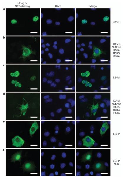
Characterization of the hairy/enhancer-of-split related with YRPW motif 1 (HEY1) nuclear localization signal. COS-1 cells were transfected with expression vectors for Flag-tagged HEY1 (a), the variant L94M (c), the triple-point mutants HEY1-K51A/ R53G/R51A (b) and L94M-K51A/R53G/R51A (d), enhanced green fluorescent protein (EGFP) (e) or EGFP-nuclear localization signal (NLS) (f) and assayed by indirect immunofluorescence with anti-Flag antibody, or direct EGFP fluorescence, as described in Materials and methods. The first column shows the indirect immunofluorescence with anti-Flag antibody or the direct EGFP fluorescence (green), the second column shows 4,6-diamidino-2-phenylindole (DAPI) staining of DNA (blue) and the third column shows the merge image indicating the degree of colocalization of green and blue fluorescence. Bars, 20 μm.

