

Abstract
This case report describes extraction of a fractured left maxillary lateral incisor tooth, followed by immediate placement of a dental implant in the prepared socket and temporization by a bonded restoration. The tooth was atraumatically extracted, the socket was prepared to the required depth and a Biohorizon Implant was inserted followed a week later by temporization by a bonded restoration. An impression was made 4 months after implant insertion, and a definitive restoration was placed. The atraumatic operating technique and the immediate insertion of the Implant resulted in the preservation of the hard and soft tissues at the extraction site. The patient exhibited no clinical or radiologic complications through 5 years of clinical monitoring. The dental implant and provisional restoration provided the patient with immediate esthetics, function, comfort, and most importantly preservation of tissues.
Keywords: Implant, Immediate placement, Temporization, Atraumatic extraction, Osseointegration
Introduction
Endosseous dental implant therapy is rapidly becoming the prosthetic standard of care for a vast array of clinical applications, however, despite the high success rate of endosseous implant therapy, it has yet to achieve wide public acceptance and utilization [1]. Endosseous implant therapy in the mandible (parasymphyseal mandible) has repeatedly been reported at a success rate of 95 % or better, yet public utilization of endosseous implant therapy has not exceeded 5 %. An obvious area of focus has been to decrease the amount of time necessary to complete implant therapy. Approaches to achieve this goal have dominated clinical research and practice, Immediate implant loading, improving implant surface technology (promotion of quicker healing and better osseointegration), and immediate placement of an endosseous implant after extraction of a natural tooth are some of them [1].
In this paper, a case supporting the last of these three approaches is presented. Immediate implants have become widely accepted despite controversial beginnings but the available literature consistently cites high levels of success (ranging from 94 to 100% on average), immediate implants provide clinically recognizable benefits. Broadly speaking, these benefits include reduction of morbidity, reduction of alveolar bone resorption (controlled clinical studies have demonstrated an average of 4.4-mm of horizontal and 1.2-mm of vertical bone resorption 6 months after tooth extraction) [1, 2], preservation of gingival tissues, preservation of the papilla in the esthetic zone, and reduction of treatment cost and time (the healing phase is shorter in general and there is a reduction in the number of procedures) [1–5]. With the extraction socket as a guide, the surgeon can also more easily determine the appropriate parallelism and alignment relative to the adjacent and opposing residual dentition. The surgical requirements for immediate implantation include extraction with the least trauma possible, preservation of the extraction socket walls and thorough alveolar curettage to eliminate all pathological material. Primary stability is an essential requirement, and is achieved with an implant exceeding the alveolar apex by 3–5 mm, or by placing an implant of greater diameter than the remnant alveolus. Esthetic emergence in the anterior zone is achieved by 1–3 mm sub-crest implantation.
Contraindications
The existence of an acute periapical inflammatory process constitutes an absolute contraindication to immediate implantation [7, 8].
In the case of socket-implant diameter discrepancies in excess of 5-mm, which would leave most of the implant without bone contact, prior bone regeneration and delayed implantation may be considered [6]. Avoid teeth with labial bony dehiscence or fenestration defects; insufficient bone apically to ensure primary stability of the implant; systemic factors that may impair healing (e.g., smoking); large bulbous root morphology, interproximal bone loss (esthetic zone), active periodontitis.
Case Report
A 32-year-old male patient presented with a history of trauma and crown fracture at the cervical area of the tooth 22 (Fig. 1) and requested for an immediate solution. Clinical and radiological evaluation revealed adequate alveolar bone, absence of periapical pathology but fracture line was below the crest of alveolar bone and was limited to the tooth. So it was decided to extract and place endosseous implant immediately and place provisional restoration to avail the benefits like preservation of bone and emergence profile. After administering appropriate antibiotic and analgesic, induction of local anesthesia was carried out using lignocaine with adrenaline. As preservation of alveolar bone is key to success of immediate implants, extraction of tooth has to be atraumatic, so using periotomes and small periosteal elevators, the fragment was luxated without excessive enlargement of the socket, and using an innovative method where an endodontic file was used to engage the canal wall, the tooth fragment was slowly luxated and pulled out of the socket using the file (Fig. 2). The sockets were debrided with curettes and a BIOHORIZON external hex implant was planned (3.7 × 13-mm). Primary stability was achieved by wrenching the implant into the bone beyond the apex of the socket, BIO-OSS bone graft was packed between the implant and labial socket wall (Fig. 3). The cover screw was placed and interrupted sutures were placed. Post operative instructions were given to the patient, and was asked to report after 1 week. The sutures were removed after 7 days IOPA was taken (Fig. 4a) and the patient received temporary acrylic crown bonded to the adjacent teeth with fiber-reinforced composite on the same day. The patient was recalled after 4 months for the prosthetic procedures and was given porcelain fused to metal crown over the implant. The patient was recalled for prophylaxis and follow up every year (Figs. 4b, 5). The clinical and radiographic appearances at 5 years show good esthetics, osseointegration and maintenance of bone around the implant (Figs. 4c and 6).
Fig. 1.

Preoperative intraoral view
Fig. 2.
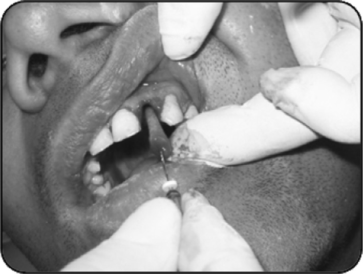
Extraction of root
Fig. 3.

Implant with abutment in place
Fig. 4.

IOPA of implant in position: a 1 week post-op, b At 4 months, c At 5 years
Fig. 5.

Metal ceramic crown at 1 year
Fig. 6.

5 years (ceramo-metal crown)
Conclusion
Implant therapy must fulfill both functional and esthetic requirements to be considered a primary treatment modality. Aiming to reduce the process of alveolar bone resorption and treatment time, the immediate placement of endosseous implants into extraction sockets is known to achieve a high success rate of between 94 and 100 %, compared to the delayed placement.
References
- 1.Wagenberg BD, Ginsburg TR. Immediate implant placement on removal of the natural tooth: retrospective analysis of 1,081 implants. Comp Cont Educ Dent. 2001;22:399–404. [PubMed] [Google Scholar]
- 2.Cooper LF, Rahman A, Moriarty J, et al. Immediate mandibular rehabilitation with endosseous implants: simultaneous extraction, implant placement, and loading. Int J Oral Maxillofac Implants. 2002;17:517–525. [PubMed] [Google Scholar]
- 3.Douglass GL, Merin RL. The immediate dental implant. J California Dent Assoc. 2002;30:362–365. [PubMed] [Google Scholar]
- 4.Gelb DA. Immediate implant surgery: ten-year clinical overview. Comp Cont Educ Dent. 1999;20:1185–1192. [PubMed] [Google Scholar]
- 5.Cornelini R, Scarano A, Covani U, Petrone G, Piattelli A. Immediate one-stage post extraction implant: a human clinical and histologic case report. Int J Oral Maxillofac Implants. 2000;15:432–437. [PubMed] [Google Scholar]
- 6.Coppel A, Prados JC, Coppel J. Implantes post-extracción: situación actual. Gaceta Dental Sept. 2001;120:80–86. [Google Scholar]
- 7.Novaes AB, Jr, Novaes AB. Soft tissue management for primary closure in guided bone regeneration: surgical technique and case report. Int J Oral Maxillofac Implants. 1997;12:84–87. [PubMed] [Google Scholar]
- 8.Novaes AB, Jr, Novaes AB. Immediate implants placed into infected sites: a clinical report. Int J Oral Maxillofac Implants. 1995;10:609–613. [PubMed] [Google Scholar]