SUMMARY
A pectoralis major myofascial flap (PMMF) is a simple variant of the pectoralis major myocutaneous flap (PMMC), and allows avoiding some of the disadvantages of Ariyan's technique while reducing well-known, overall complications. This is a retrospective analysis of 45 hypopharyngeal reconstructions (40 immediate reconstructions after subtotal pharyngolaryngectomy and 5 performed during revision surgery) using PMMF flap, performed from February 1995 to February 2008 in the Department of Otolaryngology at the "San Camillo- Forlanini" Hospitals in Rome, in collaboration with the Department of Plastic Surgery. In our series, we observed postoperative flap-related complications in 6.7% of cases. The incidence of major flap complications requiring surgical revision was 2.2%. Two minor complications were seen: hypopharyngeal stenosis and a salivary fistula, both of which were managed without surgery. Total or partial necrosis did not occur in any case. There were four postoperative deaths, but which were not related to flap complications in any case. In the remaining cases, oesophageal X-ray imaging showed the absence of fistulas and adequate calibre of the reconstructed tract; oral intake started within postoperative day 10-12, without swallowing problems of liquid or solid food. Postoperative radiotherapy performed in 30 patients was well tolerated. The PMMF flap is safe one-step procedure with low morbidity that is particularly useful for partial hypopharyngeal reconstructions, overcoming the disadvantages of the PMMC flap and offering comparable results to fasciocutaneous free flaps.
KEY WORDS: Hypopharynx reconstruction, Pectoralis major myofascial flap, Complications
RIASSUNTO
Il lembo mio-fasciale di gran pettorale (PMMF) è una semplice variante del classico lembo mio-cutaneo di gran pettorale (PMMC) che consente di evitare alcuni degli svantaggi della tecnica di Ariyan. è stata eseguita un'analisi retrospettiva di 45 ricostruzioni ipofaringee (40 ricostruzioni immediate dopo faringolaringectomia subtotale e 5 ricostruzioni dopo chirurgia di revisione) condotte utilizzando il PMMF, tra il febbraio 1995 ed il febbraio 2008 presso l'Unità Operativa di Otorinolaringoiatria dell'Azienda Ospedaliera "San Camillo- Forlanini" di Roma, in collaborazione con l'Unità Operativa di Chirurgia Plastica. Nella nostra casistica è stato registrato un tasso globale di complicanze post-operatorie, riconducibili a problematiche specifiche del lembo utilizzato, pari al 6.7%. Il tasso di complicanze maggiori che hanno richiesto una revisione chirurgica è stato del 2,2%. Sono state inoltre evidenziate 2 complicanze minori (una stenosi ipofaringea ed una fistola salivare) entrambe risolte senza necessità di chirurgia di revisione. Non sono state evidenziate necrosi totali o parziali del lembo in nessuno dei pazienti trattati. Quattro pazienti sono deceduti nel post-operatorio per cause non riconducibili a problematiche del lembo. In tutti gli altri casi, lo studio radiografico esofageo ha mostrato l'assenza di fistole ed un calibro adeguato del tratto ricostruito; l'alimentazione orale è stata ristabilita al massimo in X-XII giornata post-operatoria, senza problemi di deglutizione per i liquidi o per i solidi. Il trattamento radioterapico postoperatorio, a cui sono stati sottoposti 30 pazienti, è stato ben tollerato. In conclusione il PMMF rappresenta una procedura sicura con una bassa morbidità, particolarmente utile nelle ricostruzioni ipofaringee parziali, che consente, in un solo tempo chirurgico, di ottenere un alto tasso di successo funzionale in assenza delle limitazioni del PMMC e dei lembi liberi fascio-cutanei.
Introduction
The surgical treatment of malignant neoplasms involving the hypopharynx (either at the primary tumour site or as advanced disease from other sites) continues to be a difficult task for the head and neck surgeon. Many patients present with advanced T-stage disease that requires radical resection such as total laryngectomy in combination with total or partial pharyngectomy and cervical oesophagectomy via a cervical approach. Bilateral neck dissection is generally associated. The resulting post-surgical defect presents a further challenge for the surgeon; an adequate reconstructive procedure should guarantee an early restoration of speech and swallowing functions 1.
The pectoralis major myocutaneous flap (PMMC), initially described by Ariyan in 1979 2, is currently considered an excellent method for hypopharyngeal reconstruction after subtotal pharyngolaryngectomy. During the past years, modifications to the original technique have been proposed and adopted by some authors 3-5 in order to avoid some disadvantages of PMMC, such as hair growth, poor pliability and bulk of the skin island, poor cosmetic results at the donor site, shortness and bulk of the pedicle.
Robertson in 1985 6 and Shindo in 1992 7 reported the use of a pectoralis major flap without skin island in head and neck reconstruction; the pectoralis major myofascial flap (PMMF) appeared to overcome some of the disadvantages of PMMC. In this retrospective case review, we report 45 successful hypopharyngeal reconstructions using PMMF.
Materials and methods
Herein, we report a retrospective analysis of hypopharyngeal reconstructions using PMM F flap performed in the Department of Otolaryngology at the San Camillo Hospital in Rome, in collaboration with the Department of Plastic Surgery, from February 1995 to February 2008.
Patient and tumour-related characteristics are reported in Table I. The clinical charts of all patients were reviewed. Tumours were restaged according to the TNM classification of the Union Internationale Contre le Cancer (UICC) 2010 criteria. A descriptive analysis of the results was performed. According with literature, flap-related complications were classified as major, if additional surgical revision was required, or minor, if only conservative wound care was necessary. Major complications were considered outcome failures. All surgical procedures were carried out by the same 4 surgeons, working as an interdisciplinary team.
Table I.
Patient-related and tumour-related characteristics.
| No. patients (%) | |
|---|---|
| Sex: male | 36 (81.8%) |
| female | 8 (18.2%) |
| Age (years) | 39-81 (mean 62.5) |
| T classification | |
| T1/T2 | 0 |
| T3/T4 | 41 |
| Tx* | 3 |
Tx: T classification could not be evaluated.
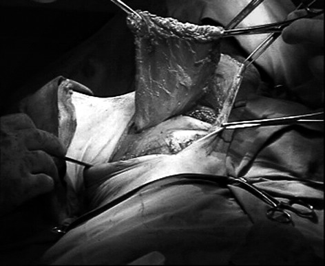
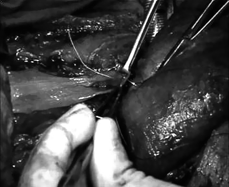
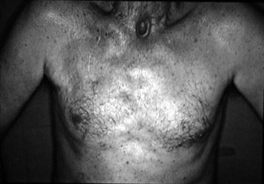
All patients underwent total laryngectomy and partial pharyngectomy with a remaining posterior pharyngeal mucosal strip of about 1.5-2 cm. The PMMF flap was harvested similarly to the PMMC flap, with the exclusion of the skin paddle and subcutaneous fat. The skin incision in female patients was made along the inframammary fold, as described by Shindo 7, while in male patients an incision along the middle clavicular line was used. Through the skin incision, the pectoralis major fascia was dissected from subcutaneous tissue, paying attention to avoid any damage to its surface; subsequent dissection of the pectoralis major muscle from the thoracic wall was performed, up to the vascular pedicle (Fig. 1). The muscular flap was brought up and placed into the cervical region through a tunnel created over the clavicle. A nasogastric feeding tube was positioned, and the fascial plane of the flap was then sutured using 3/0 absorbable monofilament separated stitches to the borders of residual hypopharynx, giving an adequate calibre to the new structure (Fig. 2). The free lateral areas of the flap were used to cover the deep cervical vessels. The donor site was repaired by direct closure (Fig. 3).
Fig. 1.

The pectoralis major myofascial flap harvested and ready to be placed in the cervical area.
Fig. 2.

The flap is sutured to the residual hypopharyngeal wall.
Fig. 3.

The donor site a few months after intervention.
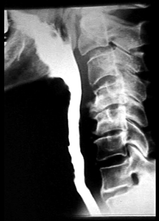
All patients underwent contrast oesophageal X-ray imaging on postoperative day 10 to evaluate the absence of fistulas and the calibre of the reconstructed hypopharynx (Fig. 4); if no fistula was found, oral intake would start on the same day. All patients without complications were discharged between postoperative days 10-12; patients experiencing minor complications were discharged as soon as complications healed, with the longest in-hospital stay of 21 days in one patient.
Fig. 4.

Postoperative oesophageal X-ray imaging showing a new well functioning hypopharynx.
Results
We reviewed the medical charts of 44 patients who had received PMMF flap procedures; 40 patients underwent subtotal pharyngolaryngectomy and immediate reconstruction, while in 5 patients hypopharyngeal reconstruction was performed during revision surgery for recurrent disease. In one patient, a second PMMF flap was needed during revision surgery. The histological diagnosis was squamous cell carcinoma in 43 cases (97.7%). All tumours were at an advanced clinical stage at presentation (T3 and T4). No patient had undergone previous radiotherapy; 30 patients received radiotherapy postoperatively (total 50- 65 Gy, 2 gy/day in 5-7 weeks, depending on the patient), starting between postoperative days 30-60. The follow-up period after PMMF flap reconstruction ranged from 3 to 120 months.
Total necrosis was not observed in any of the 45 PMM F flaps performed in our group of 44 hypopharyngeal/laryngeal cancer patients. Three postoperative flap-related complications were seen, with an overall complications rate of 6.7%. The only major complication was a salivary fistula treated with surgical revision. Two minor complications occurred: hypopharyngeal stenosis and a salivary fistula, both of which were managed without surgery.
There were four postoperative deaths, although none were related to flap complications. In the remaining cases, oesophageal X-ray imaging showed the absence of fistulas and adequate calibre of the reconstructed tract. In these patients, oral intake started within postoperative day 10-12, without swallowing problems for liquid or solid foods.
In all 30 patients who underwent postoperative radiotherapy, the treatment was very well tolerated, with no side effects that compromised regular oral feeding. Oesophageal X-ray imaging and endoscopy performed at the end of the treatment showed the persistence of an ample and stretchable new hypopharynx.
A very good cosmetic result was obtained at the donor site without deformities of the chest wall. None of the women reported disappointment because of postoperative breast contour. Furthermore, no functional defects were found at the ipsilateral arm after muscular grading and mobility evaluation, performed by using visual analogue scales (VAS) 8.
Discussion
Hypopharynx reconstruction continues to represent a clinical challenge for head and neck surgeons; since the reconstructive technique affects postoperative morbidity, as reported by Clark et al. 9, its choice becomes a crucial moment in surgical management of the hypopharynx. The goal of reconstruction is a safe, one-stage procedure that allows early functional recovery and adequate ability to tolerate postoperative radiation. The choice of reconstructive technique depends on tumour-related characteristics, the patient's medical condition, availability of microvascular experience and costs.
Contemporary reconstructive options include the use of pedicled flaps and free flaps (enteric and fasciocutaneous) 1. The fasciocutaneous free flaps most commonly used for pharyngoesophageal reconstruction are the radial forearm free flap (RFFF) and the anterolateral thigh flap (ALT). Traditionally, enteric free flaps have been used for circumferential hypopharyngeal reconstruction; the free jejunal transfer has been used for hypopharyngeal and oesophageal defects above the thoracic inlet, whereas the gastric pull-up has been the standard technique for lesions extending below the thoracic inlet. In fact, the free intestinal flaps have been surpassed by fasciocutaneous free flaps, even if gastric pull-up remains the procedure of choice for lesions extending beyond the thoracic inlet. In comparison with jejunal free flaps, fasciocutaneous free flaps avoid the added morbidity of a laparotomy and bowel anastomosis, and guarantee better tracheo-esophageal speech quality, comparable stricture (12-27%) and fistula rates (0-13%) 1.
Concerning partial pharyngeal defects, the most common reconstructive choice is represented by the use of PMMC or fasciocutaneous free flaps, as reported by Clark et al. in their reconstructive algorithm 9. Between PMMC and fasciocutaneous free flaps, Clark et al. do not consider any particular flap to be preferred in partial reconstructions, and suggest that the choice must be driven by donor site factors.
Our study suggests that the PMMF flap is a valid procedure for partial hypopharyngeal reconstruction associated with low morbidity, overcoming the disadvantages of the PMMC flap and offering comparable results with fasciocutaneous free flaps. No other studies on "pure" hypopharyngeal reconstruction with PMMF have been performed. Interesting, similar reports on the use of PMMF, published in last decades, are all biased by the fact that they show "mixed" reconstructive results, combining reconstructions of different head and neck subsites 10-13.
The myofascial flap, introduced with satisfactory results by Robertson 4 6 and Shindo 7, is a simple variant of PMMC, consisting in the use of pectoralis major muscle alone with overlying fascia. Prior to the free flap era, the tubed pectoralis major myocutaneous flap was the standard technique for one-stage immediate hypopharyngeal reconstruction with acceptable functional results. However, the high stenosis rates reported for this flap led several authors to introduce modifications in surgical technique. Direct suture of the PMMC to the prevertebral fascia in a U-shape as described by Fabian 14 and its further modifications proposed in literature 15-17 showed a decreased stenosis rate. Nonetheless, the overall rate of complication reported in the literature for PMMC in the past 20 years ranges from 16-63% with an incidence of partial and total necrosis ranging from 4-29% and 0-7%, respectively 18-22. In 2004, Vartanian and colleagues reported a total complication rate of 36.1%, an incidence of partial necrosis of 9.7%, total necrosis of 2.4% and salivary fistula of 11.8% 11.
To avoid the drawbacks of PMMC, we used the myofascial flap, performing 45 partial hypopharyngeal reconstructions in 44 patients subjected to subtotal pharyngolaryngectomy; in these patients, the remaining pharyngeal mucosal strip was about 1.5-2 cm in width.
In comparison with PMMC, the absence of the skin paddle makes PMMF flap thinner, more stretchable and easy to tube, allowing the surgeon to easily create a physiologic thin-walled pharynx with no need for previous flap shape planning. Furthermore, the absence of the cutaneous island, the most "high-risk" zone for vascular supply in the flap, decreases the risk of necrosis and fistula, avoids hair growth and provides a better cosmetic result at the donor site. Increased thinness of myofascial compared to myocutaneous flap allows closing the cervical surgical incision without tension, gaining a better aesthetic result. For this reason, using a different skin incision along the inframammary fold, the myofascial flap can also be applied easily in female patients, where harvesting of myocutaneous flap is complicated by the presence of mammary gland 7. Nonetheless, when a portion of the PMMF flap requires skin coverage, a split-thickness skin graft may be sutured to the muscle to provide either internal or external coverage.
Compared to the fasciocutaneous free flaps which are most commonly used for partial hypopharyngeal reconstruction (ALT and RFFF), the PMMF flap is technically easier and much faster to harvest, and does not require microvascular experience. This is particularly important in elderly patients and in those affected by severe medical comorbidities in which the extended surgical time and stress of free flap reconstruction are contraindicated. Furthermore, harvesting of the RFFF leads to higher donor site morbidity with a limited quantity of tissue for reconstruction; on the other hand, the ALT flap offers the advantage of a large skin paddle and a low donor site morbidity, but is often difficult to harvest and in the Western population, especially females, it can be thick and difficult to tube, leaving a prominent scar 1 9.
Considering flap-related complications, the rates obtained in this study are more favourable compared to literature data for the same flap procedure and PMMC or fasciocutaneous free flap procedures. In our series, the overall flap-related complication rate was 6.7%; the incidence of major flap complications requiring surgical revision was 2.2%. Total or partial necrosis did not occur in any case and the preoperative goal of the PMMF flap procedure was met in 44 of 45 (98%) cases. Zbar et al. 23 used the PMMF flap for primary closure of small pharyngeal defects in 2 patients and success in both cases. On the other hand, the reported rates of stenosis and fistula for PMMC flaps range from 6-16% and from 0-47%, respectively, whereas the same rates range from 10-36% and from 17- 28%, respectively, for RFFF flaps, and from 12-27% and 0-13% for ALT flaps 1.
Radiation therapy was very well tolerated, as demonstrated by the absence of stenosis in endoscopic and oesophageal X-ray imaging, even after several months or years. A reasonable explanation of our surprisingly positive results could be the absence of the flap skin paddle, which allowed better and early healing of pharyngeal walls. According to previous findings, the PMM F flap completed epithelization within 4 weeks postoperatively.
The pectoralis major myofascial flap (PMMF) has also been used for other reconstructive tasks 10-13 24: oral cavity and oropharynx reconstruction, coverage of exposed carotid artery or microvascular anastomosis, neck resurfacing and mediastinal tracheostomy formation in patients requiring radical laryngotracheal resection. In the cases reported in literature, the PMMF flap met reconstructive goals.
Conclusions
In the light of the above considerations, we conclude that in partial hypopharyngeal reconstruction the PMMF flap is associated with a high overall success rate, avoiding some of limitations of the PMMC flap and offering comparable results to fasciocutaneous free flaps.
The PMMF flap is a safe, quick, one-step procedure, not requiring microvascular experience, that is particularly indicated in elderly patients and/or those with severe medical comorbidities; the ease of harvesting, a very low rate of complications and donor site morbidity make this flap, in our opinion, a valid option for reconstruction of partial hypopharyngeal defects.
References
- 1.Richmon JD, Brumund KT. Reconstruction of the hypopharynx: current trends. Curr Opin Otolaryngol Head Neck Surg. 2007;15:208–212. doi: 10.1097/MOO.0b013e32825b0852. [DOI] [PubMed] [Google Scholar]
- 2.Ariyan S. The pectoralis major myocutaneous flap. A versatile flap for reconstruction in the head and neck. Plast Reconstr Surg. 1979;63:73–82. doi: 10.1097/00006534-197901000-00012. [DOI] [PubMed] [Google Scholar]
- 3.Murakami Y, Saito S, Ikari T, et al. Esophageal reconstruction with a skin-grafted pectoralis major muscle flap. Arch Otolaryngol. 1982;108:719–722. doi: 10.1001/archotol.1982.00790590041012. [DOI] [PubMed] [Google Scholar]
- 4.Robertson MS, Robinson JM. Immediate pharyngoesophageal reconstruction. Use of a quilted skin-grafted pectoralis major muscle flap. Arch Otolaryngol. 1984;110:386–387. doi: 10.1001/archotol.1984.00800320040009. [DOI] [PubMed] [Google Scholar]
- 5.Palmer JH, Batchelor AG. The functional pectoralis major musculocutaneous island flap in the head and neck reconstruction. Plast Reconstr Surg. 1990;85:363–367. doi: 10.1097/00006534-199003000-00004. [DOI] [PubMed] [Google Scholar]
- 6.Robertson MS, Robinson JM. Pharyngoesophageal reconstruction. Is a skin-lined pharynx necessary? Arch Otolaryngol. 1985;111:375–376. doi: 10.1001/archotol.1985.00800080061006. [DOI] [PubMed] [Google Scholar]
- 7.Shindo ML, Costantino PD, Friedman CD, et al. The pectoralis major myofascial flap for intraoral and pharyngeal reconstruction. Acta Otolaryngol Head Neck Surg. 1992;118:707–711. doi: 10.1001/archotol.1992.01880070037007. [DOI] [PubMed] [Google Scholar]
- 8.Kendall FP, McCreary EK, Provance PG. Muscles-Testing and Function. 4th ed. Baltimore: Williams & Wilkins; 1993. [Google Scholar]
- 9.ClarK JR, Gilbert R, Irish J, et al. Morbidity after flap reconstruction of hypopharyngeal defects. Laryngoscope. 2006;116:173–181. doi: 10.1097/01.mlg.0000191459.40059.fd. [DOI] [PubMed] [Google Scholar]
- 10.Gras R, Bouvier C, Guelfucci B, et al. The pectoralis myofascial flap in oropharyngeal and pharyngolaryngeal reconstruction in salvage surgery. Ann Otolaryngol Chir Cervicofac. 2000;117:378–382. [PubMed] [Google Scholar]
- 11.Vartanian JG, Carvalho AL, Carvalho SM, et al. Pectoralis major and other myofascial/myocutaneous flaps in head and neck cancer reconstruction: experience with 437 cases at a single institution. Head Neck. 2004;26:1018–1023. doi: 10.1002/hed.20101. [DOI] [PubMed] [Google Scholar]
- 12.Righi PD, Weisberger EC, Slakes SR, et al. The pectoralis major myofascial flap: clinical applications in head and neck reconstruction. Am J Otolaryngol. 1998;19:96–101. doi: 10.1016/s0196-0709(98)90102-8. [DOI] [PubMed] [Google Scholar]
- 13.Ethier JL, Trites J, Taylor SM. Pectoralis major myofascial flap in head and neck reconstruction: indications and outcomes. J Otolaryngol Head Neck Surg. 2009;38:632–641. [PubMed] [Google Scholar]
- 14.Fabian RL. Reconstruction of the laryngopharynx and cervical esophagus. Laryngoscope. 1984;94:1334–1350. doi: 10.1288/00005537-198410000-00015. [DOI] [PubMed] [Google Scholar]
- 15.Spriano G, Pellini R, Roselli R. Pectoralis major myocutaneous flap for hypopharyngeal reconstruction. Plast Reconstr Surg. 2002;110:1408–1413. doi: 10.1097/01.PRS.0000029350.61515.39. [DOI] [PubMed] [Google Scholar]
- 16.Jegoux F, Ferron C, Malard O, et al. Reconstruction of circumferential pharyngolaryngectomy using a 'horseshoeshaped' pectoralis major myocutaneous flap. J Laryngol Otol. 2007;121:483–488. doi: 10.1017/S0022215106004154. [DOI] [PubMed] [Google Scholar]
- 17.Suassez S, Cuno A, Urbain F, et al. Reconstruction of circumferential oro and hypopharyngeal defects with U-shaped pectoralis major myocutaneous flap. Otolaryngol Head Neck Surg. 2006;134:823–829. doi: 10.1016/j.otohns.2006.01.004. [DOI] [PubMed] [Google Scholar]
- 18.Schuller DE. Pectoralis myocutaneous flap in head and neck cancer reconstruction. Arch Otolaryngol. 1983;109:185–189. doi: 10.1001/archotol.1983.00800170051013. [DOI] [PubMed] [Google Scholar]
- 19.Shah JP, Haribhakti V, Loree TR, et al. Complications of the pectoralis major myocutaneous flap in head and neck reconstruction. Am J Surg. 1990;160:352–355. doi: 10.1016/s0002-9610(05)80541-0. [DOI] [PubMed] [Google Scholar]
- 20.Pompei S, Caravelli G, Palma O, et al. Morbidity and morphofunctional aspects of Myocutaneous Flaps in Head and Neck Reconstruction. Acta Otorhinol Ital. 1993;13:147–159. [PubMed] [Google Scholar]
- 21.Liu R, Gullane P, Brown D, et al. Pectoralis major myocutaneous pedicled flap in head and neck reconstruction: retrospective review of indications and results in 244 consecutive cases at the Toronto General Hospital. J Otolaryngol. 2001;30:34–40. doi: 10.2310/7070.2001.21011. [DOI] [PubMed] [Google Scholar]
- 22.Dedivitis RA, Guimaraes AV. Pectoralis major musculocutaneous flap in head and neck cancer reconstruction. World J Surg. 2002;26:67–71. doi: 10.1007/s00268-001-0183-4. [DOI] [PubMed] [Google Scholar]
- 23.Zbar RIS, Funk GF, McCulloch TM, et al. Pectoralis major myofascial flap: a valuable tool in contemporary head and neck reconstruction. Head Neck. 1997;19:412–418. doi: 10.1002/(sici)1097-0347(199708)19:5<412::aid-hed8>3.0.co;2-2. [DOI] [PubMed] [Google Scholar]
- 24.Leemans CR, Balm AJM, Gregor RT, et al. Management of carotid artery exposure with pectoralis major myofascial flap transfer and split thickness coverage. J Laryngol Otol. 1995;109:1176–1180. doi: 10.1017/s0022215100132360. [DOI] [PubMed] [Google Scholar]


