Abstract
Purpose.
To elucidate the factors in tear production, this study examined the role of endogenous opioids and opioid receptors in spontaneous episodic reduced tear volume.
Methods.
A model of spontaneous episodic decreases in the quantity of tears was characterized in otherwise normal Sprague-Dawley rats using Schirmer's test. A single eye drop of 10−5 M naltrexone (NTX), 10−5 M [Met5]-enkephalin, or sterile vehicle was administered to one eye. Tear secretion, corneal sensitivity, and corneal morphology were examined in both eyes.
Results.
At any given time period, otherwise normal rats were found to have Schirmer test scores with a bimodal distribution (6.5 mm or less, or 7.0 mm or greater). Decreased tear production was detected in male and female rats aged 4 to 24 weeks at least once per animal. The episodes of reduced tear volume ranged from 1 to 7 days. No changes in corneal sensitivity or corneal morphology were observed in any rat. One drop of NTX given to rats with a decrease in tear volume raised levels of tears to scores of 7.0 mm or greater within 1 hour, and increased tear production persisted for at least 48 hours. NTX had no effect on rats with Schirmer scores of 7.0 mm or higher. Topical application of [Met5]-enkephalin depressed tear secretion from baseline scores of 9.8 ± 0.6 mm to as low as 4.5 ± 0.7 mm.
Conclusions.
Normal rats experience fluctuations in tear production that can be modulated by opioidergic signaling pathways.
Rats undergo spontaneous episodic decreases in tear production that are regulated by opioid signaling pathways.
Introduction
Tears are important to the health of the cornea, and function in lubrication, nourishment for the cornea, gas exchange, removal of debris from the corneal surface, and prevention of bacterial and viral infections.1 In addition to maintenance of the surface homeostatic environment, the precorneal tear film serves to maintain an optically uniform corneal surface essential for normal vision. The pathophysiology of reduced tear film may involve tear production deficiency that can increase corneal inflammation, impair vision, and even have complications leading to blindness.2–4 Alternatively, changes in environmental factors such as temperature and humidity may lead to transient increases in tear film evaporation.5,6 Although the components of tears have been identified and characterized,2,5–10 the regulation of tear production is multifaceted involving not only physiology but environmental stimuli.6
Previous studies in our laboratory have established a relationship between opioidergic systems (i.e., endogenous opioid peptides and opioid receptors) and tear production in diabetic rats.11 A decrease in tear volume in animals with type 1 diabetes (T1D) was reversed within 1 hour of administration following the topical administration of the opioid antagonist naltrexone hydrochloride (NTX); the effect persisted for up to 3 days. During these studies we noted that normal rats occasionally had a marked reduction in Schirmer test scores, suggesting that normal tear production may fluctuate. The present investigation validates and characterizes this novel observation of spontaneous episodic fluctuations in tear secretion in normal Sprague-Dawley rats. We then addressed the question of whether changes in tear volume are related to endogenous opioid systems by examining the repercussions of blocking native opioid-opioid receptor interactions with NTX. Finally, studies were conducted to test whether topical application of an opioid peptide, [Met5]-enkephalin, suppressed tear secretion.
Methods
Animals
Male and female Sprague-Dawley rats were obtained from Charles River Laboratories (Wilmington, MA) or bred at Pennsylvania State University College of Medicine. The animals were housed in standard laboratory conditions with temperature regulated at 21 ± 0.5°C, and an air exchange of 10 to 15 times daily. Of particular interest to this study, humidity was maintained over the entire room with no stratification. All investigations conformed to the regulations of the Association for Research in Vision and Ophthalmology (ARVO) Statement for the Use of Animals in Ophthalmic and Vision Research, the National Institutes of Health, and the guidelines of the Department of Comparative Medicine of Pennsylvania State University.
Animals were numbered, and the investigators recording the measurements were masked to treatment.
Measurement of Tear Volume
Tear volume was measured with Schirmer strips (Alcon Laboratories, Inc., Fort Worth, TX); all tests were performed with Schirmer strips from the same manufacturer. A 1-mm wide by 17-mm long Schirmer strip was prepared from standard size strips and inserted into the lower lid cul-de-sac for 1 minute.11–14 The strip wetting length was measured to the nearest half millimeter. Testing began 1 hour after the last drop of NTX or vehicle was administered, and measurements were made 24, 48, and 72 hours later in the right eye. Evaluation of possible systemic effects or irritant effects of the NTX or vehicle was assessed by Schirmer testing of the fellow eye 1 hour after drug administration to the treated eye. Additionally, the scores of animals at 72 hours were required to be similar to their baseline levels to rule out a spontaneous endogenous reversal of tear production during the test period.
Corneal Sensitivity and Blink Rate
Corneal sensitivity was determined by two tests: an aesthesiometer and blink rate using von Frey hairs. Measurements of sensitivity were conducted prior to the Schirmer test. Four measurements were taken with a Cochet-Bonnet Aesthesiometer (Boca Raton, FL) for each animal and averaged; the end point was a blink response. The values (g/mm2) were determined directly from the protocol (and conversion table) supplied by the manufacturer. The Touch-Test Sensory Evaluator (North Coast Medical Inc., Morgan Hill, CA) , consisting of a series of von Frey hairs, was used to determine mechanical sensitivity.
Blink rate was determined by an observer masked to the identity of the treatment groups. The number of blinks in 1 minute was counted. The animals (4–6 per group) were unrestrained.
Topical Administration of Naltrexone or [Met5]-Enkephalin
Naltrexone hydrochloride (Sigma-Aldrich, Indianapolis, IN) was prepared in moxifloxacin hydrochloride ophthalmic solution (Vigamox; Alcon, Inc., Fort Worth, TX) at a 10−5 M concentration. One drop of 10−5 M NTX in Vigamox, or Vigamox alone, was administered at 0800 hours as a single drop (0.05 mL) to the central cornea of the right eye, with the lower eyelid held away from the eye for 15 seconds to avoid overflow. The left eye was untreated.
To begin to ascertain which native opioid peptide may be involved in spontaneous episodic fluctuations in tear volume, an experiment was conducted in which [Met5]-enkephalin (Sigma-Aldrich), a known opioid neurotransmitter, was topically applied as a single drop to the central cornea 3 times over an 8-hour period at a concentration of 10−5 M in Vigamox or Vigamox alone. Tear production was measured 1, 24, and 48 hours after the last exposure to [Met5]-enkephalin. The left eye was untreated.
Statistical Analysis
Data were evaluated using a one-way analysis of variance with Newman-Keuls post-tests. In some cases, a two-tailed t test (e.g., body weights) was applied to data.
Results
Spontaneous Episodic Reduction in Tear Secretion
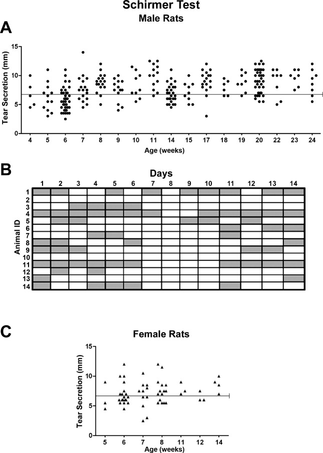
In initial studies, we noted that Schirmer scores for male rats (n = 26) of 14 weeks of age were distributed into two populations: above (8.2 ± 0.3 mm) and equal to or below (5.6 ± 0.2 mm) 6.5 mm, and the difference between the two means was statistically significant (P < 0.0001). To examine whether this difference in Schirmer scores was either special to this group of animals or to a specific age, an independent experiment was performed in which rats from 4 to 24 weeks of age (Fig. 1A) were studied. Once again, at any given time period, a bimodal distribution of tear production could be distinguished on the basis of tear volume of 7.0 mm or greater, or 6.5 mm or less. Nevertheless, every rat (n = 21) had a Schirmer score of 6.5 or below at least once during the 20 week testing period. The mean score of those rats with scores of 6.5 mm or below was 5.3 ± 0.1 mm, whereas the mean Schirmer score for rats with values of 7.0 mm and greater was 8.8 ± 0.2 mm; the difference between these groups was statistically reliable (P < 0.0001). Environmental factors such as temperature and/or humidity varied across the 24-week period as well with no trend to support dehydration of tear film.
Figure 1. .
A scattergram of Schirmer scores for male (n = 21) (A) and female (n = 15) (C) rats on a weekly basis; some rats were measured more than once per week. Both male and female animals with scores of 6.5 mm and lower differed significantly (P < 0.0001) from counterparts with scores of 7.0 mm or greater. (B) Fourteen male rats monitored daily on weeks 14 and 15, and rats with scores below 6.5 mm differed significantly (P < 0.0001) from animals with scores of 7.0 mm and greater. Shaded blocks represent ≤ 6.5 mm, white blocks represent ≥ 7.0.
To further characterize the daily variation in Schirmer scores, another investigation monitored a group of 14 male rats, age 14 weeks for 14 days (Fig. 1B). The incidence and duration of Schirmer test scores above and below 6.5 mm varied from animal to animal across the 2-week period. The mean scores were 5.0 ± 0.1 and 8.2 ± 0.1 mm, respectively, and this difference was significant at P < 0.0001. Within this period, some animals exhibited scores of 6.5 mm or below for up to 7 days, and other rats for as little as 1 day. Two of these 14 animals had Schirmer scores 7.0 mm or greater for the entire 2-week period.
All rats used in this study were healthy and showed no signs of eye disorders. Examination of body weights revealed no difference between animals with scores of 6.5 mm or below (n = 24) or above 7.0 mm (n = 45) (520 ± 7.4 g and 508 ± 10.6 g, respectively) when measured at 14 weeks. Food and water consumption, as well as husbandry (e.g., number of rats per cage, position of cages), did not differ between rats with scores above and below 6.5 mm. In fact, rats with scores above or below 6.5 mm were often housed in the same cage. Observations with a slit-lamp also disclosed no abnormal findings between groups. Finally, differences in Schirmer scores were observed in rats purchased directly from the supplier, as well as laboratory-bred animals. Analyses of humidity measurements collected by the Department of Comparative Medicine revealed that humidity was controlled on a given day to within 1% to 2%, and analysis of day-to-day variation revealed that humidity values were 33 ± 11% during our experimental period.
An examination of the confidence and repeatability of the Schirmer test scores also was conducted. Animals with Schirmer scores of 6.5 mm and below were investigated every hour for 3 hours and had average scores of 5.4 ± 0.2, 5.7 ± 1.2, and 5.7 ± 0.2 mm, respectively; no differences within the 3-hour range were detected. Examination of rats with scores of 7.0 mm or greater were studied every hour for 3 hours, and had average scores of 8.6 ± 0.5, 7.8 ± 0.6, and 7.4 ± 0.6 mm, respectively; no differences were noted across the 3-hour time period. Finally, both right and left eyes of these animals had similar Schirmer scores. Reproducibility of these observations was confirmed through 10 studies over a period of 3 years.
To address whether the differences in Schirmer scores observed in male rats was dependent on sex, tear production was monitored in female rats of 5 to 14 weeks of age (Fig. 1C). A Schirmer score of 6.5 or below (5.5 ± 0.1 mm) was detected at least once in all (n = 15) but 2 female rats during the 9-week testing period; the mean score differed significantly (P < 0.0001) from the mean (8.5 ± 0.3 mm) for females with scores of 7.0 mm and above.
Given our confidence in the Schirmer test and the repeatability of our findings, we concluded that animals with scores of 6.5 mm or below had decreased tear volume and this was termed “spontaneous episodic decreased tear production.” Those animals with scores of 7.0 mm and above were “normal” with respect to tear volume.
Topical Naltrexone Treatment and Tear Fluid Volume
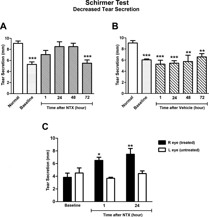
Male rats (ages 15–24 weeks) with spontaneous episodic decreased tear secretion were administered one drop of 10−5 M NTX (Fig. 2A) or an equivalent volume of vehicle alone (Fig. 2B). On the day of the experiment, baseline values of all rats with decreased tear production were 42% lower than those of normal animals. Rats with reduced tear volume and receiving 1 drop of NTX had values comparable to normal animals when measured at 1, 24, and 48 hours. However, at 72 hours, the effect of NTX was no longer observed, and Schirmer scores of these NTX-treated rats were similar to baseline values and reduced 39% from the values of animals observed to have normal tear volume. Tear volume in the left (untreated) eye of NTX-exposed rats was reduced throughout the experiment. The effects of NTX in reversing reductions in tear secretion were repeated in 10 individual experiments.
Figure 2. .
Schirmer test of rats with dry eye given 1 drop of topical (A) NTX (n = 7) or (B) vehicle (n = 5) to the cornea. Significantly different from Normal rats at P < 0.01 (**) or P < 0.001 (***). (C) Schirmer test scores on the right and left eyes of rats with dry eye given one drop of NTX (right eye only) and tested at 1 and 24 hours. Significantly different from left eye at P < 0.05 (*) and P < 0.01 (**). Values represent means ± SEM.
Male rats (ages 6–24 weeks) with reduced tear volume (Fig. 2B) were administered one drop of vehicle. The baseline value for these rats was 5.9 ± 0.2 mm. Animals receiving vehicle did not differ from rats at baseline (or from each other) when examined at 1, 24, 48, or 72 hours, with mean scores ranging from 5.0 to 6.1 mm.
To investigate the effects of NTX (right eye) on the eye opposite to that receiving drug (left eye), measures of tear volume were made in both eyes. The results (Fig. 2C) show that despite an increased tear production in the NTX-treated right eye, a persistent reduced tear secretion was noted in the left eye of the same rat.
Male rats (ages 9–24 weeks) with normal tear volume were administered one drop of 10−5 M NTX in vehicle (Fig. 3A). Schirmer scores for animals receiving NTX were similar to baseline (9.2 ± 0.5 mm) at 1, 24, 48, and 72 hours after treatment. Moreover, male rats (9–24 weeks of age) with normal tear secretion and receiving vehicle only did not have Schirmer scores that differed from baseline values (10.3 ± 0.3 mm) (Fig. 3B).
Figure 3. .
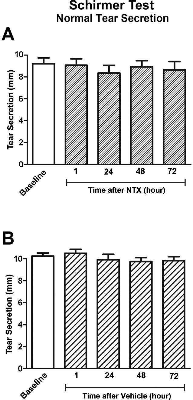
Schirmer test of rats with normal tear production and receiving 1 drop of topical NTX (A) or vehicle (B). Data are expressed as means ± SEM for rats receiving NTX (n = 7) and rats receiving vehicle (n = 6).
Corneal Sensitivity, Blink Rate, and Spontaneous Episodic Reductions in Tear Volume
Corneal sensitivity measured with the Bonnet-Cochet aesthesiometer was comparable between male rats, ages 8 to 24 weeks, with decreased tear secretion and those with normal tear volume (Figs. 4A–C). Rats with normal tear secretion had a mean corneal sensitivity of 0.42 ± 0.01 g/mm2 and rats with reduced tear production had a mean corneal sensitivity of 0.41 ± 0.01 g/mm2. Using von Frey hairs, another measure of mechanical sensitivity, rats with a reduced tear volume had a similar response to animals with normal tear secretion across a 20-fold difference in force ranging from 0.008 g to 0.16 g.
Figure 4. .
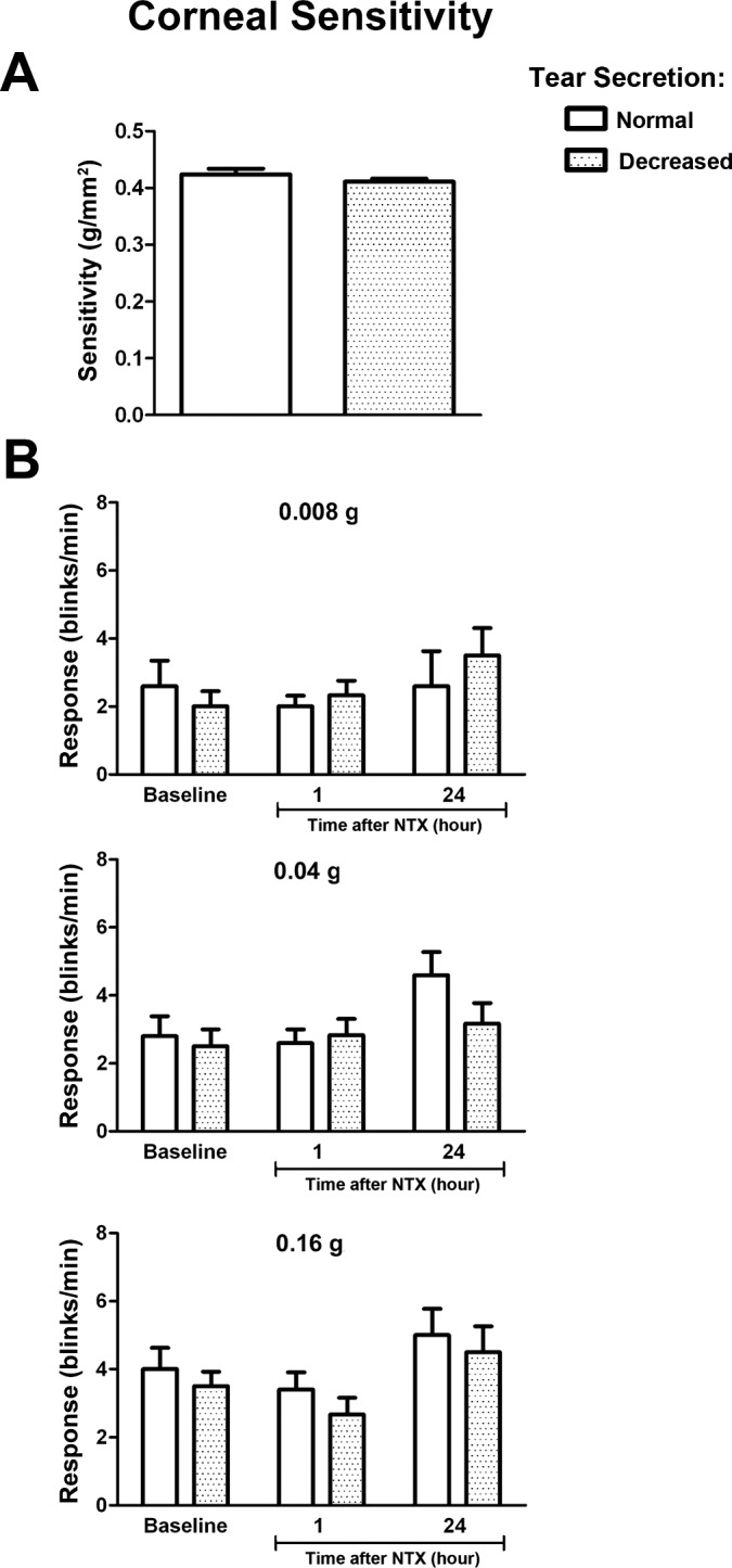
(A) Corneal sensitivity of rats with Schirmer scores of 7.0 mm or greater (Normal) (n = 10) and 6.5 mm or less (Dry) (n = 11) determined with a Cochet-Bonnet aesthesiometer. (B) Blink response to stimulus intensity of 0.008, 0.04, and 0.16 g using von Frey filaments in rats with Schirmer scores of 7.0 mm or greater (n = 4) and 6.5 mm or less (n = 4). Values represent means ± SEM.
Blink rate in unrestrained rats documented to have normal and decreased tear volumes was 2.0 ± 0.4 and 1.5 ± 0.5 blinks/minute; no statistical difference between groups was recorded.
Topical [Met5]-Enkephalin and Tear Volume
The native opioid agonist, [Met5]-enkephalin, at a dosage of 10−5 M was administered topically three times to normal rats (n = 6) over an 8-hour period, and Schirmer tests were conducted at baseline, and 1, 24, and 48 hours later (Fig. 5). In comparison to baseline values of Schirmer scores of 9.8 ± 0.6 mm, scores were reduced 23.5% (P < 0.05), 43% (P < 0.001), and 54% (P < 0.001) at 1, 24, and 48 hours, respectively. The left (untreated) eye of those rats receiving [Met5]-enkephalin had normal tear volumes.
Figure 5. .
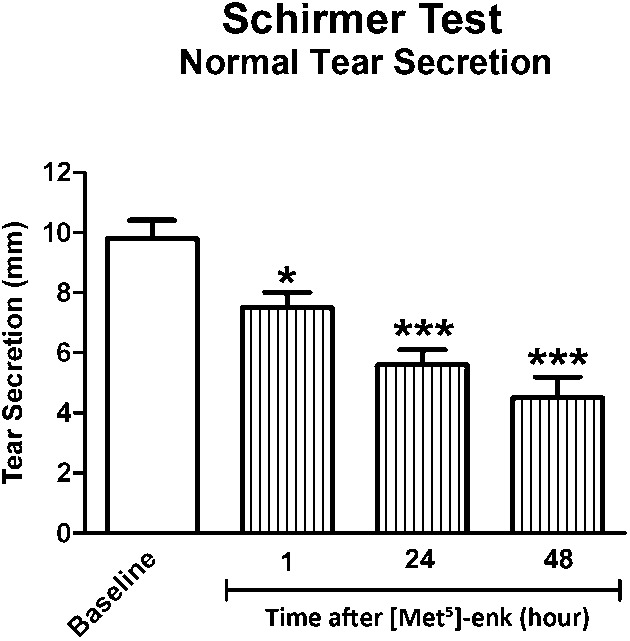
Schirmer tests of rats with normal tear production (baseline) receiving one drop of the endogenous opioid [Met5]-enkephalin (10−5 M) and tested at 1, 24, and 48 hr later. Values represent means ± SEM (n = 6).
Discussion
The present study makes the novel observation that otherwise normal rats can experience marked fluctuations in tear volumes. To date, animal studies5,6,15–19 exploring decreased tear secretion have utilized a number of models, including experimental immune dacryoadenitis, pharmacologic blockade of cholinergic muscarinic receptors, mechanical control of tear secretion, and environmental regulation of humidity and evaporation. These models could not only cause changes in tear film composition, but also may achieve their effects in a manner that could be unrelated to the homoeostatic mechanisms of tear production. The present study did not involve any physical, metabolic, pharmacologic, or mechanical factors that would overtly confound our understanding of the physiology of tear secretion. The episodic reductions in tear volume recorded herein were spontaneous and occurred simultaneously in both eyes, but were not associated with changes in body weights, food and water consumption, animal husbandry, clinical signs of ocular surface disease, corneal sensitivity, age, or sex. Therefore, there were no indicators predicting whether an animal would experience an episode of reduced tear volume, or the duration of such an occurrence. Moreover, transient depression in tear production was found in rats obtained directly from the breeder as well as those laboratory bred. Nevertheless, over a 20-week observation period, every rat displayed at least one period of reduced tear secretion.
Episodes of reduced tear volume were not an isolated occurrence, but were documented in at least 10 independent experiments conducted over a 3-year period. Fluctuations in humidity and/or temperature in the controlled environment in which rats were housed did not exert an overall influence on tear volume as rats showed no standard pattern of change in tear volume indicating evaporation. Whether other strains of rats experience transient reductions in tear secretion will require future investigation. Thus, we submit that spontaneous episodic fluctuations in tear volume occur in normal Sprague-Dawley rats, and that this characteristic offers the potential to study the physiology of tear production.
A second major finding in this investigation was the discovery that opioid signaling pathways regulate tear volume. Treatment with topical NTX restored tear volume in rats with spontaneous decreased tear production, and did so in as little as 60 minutes following NTX administration, indicating an extraordinarily rapid onset of activity by this opioid antagonist. The normalization of the tear film with only one eye drop of NTX had a considerable duration, extending for at least 2 days after treatment. Restoration of normal tear volume in animals with spontaneous episodic reduction in tear secretion was not due to the vehicle itself, because there was no change in Schirmer test scores noted after treatment with the vehicle alone. NTX also did not have any effect on rats with normal tear production, suggesting that opioid-receptor interaction does not tonically regulate tear production. Rather, opioid production most likely is physiologically suppressed/depressed during periods of maximal tear secretion. Finally, the effects of NTX were localized only to the eye that received topical administration and did not alter tear volume in the opposite eye, indicating that NTX was not acting through the systemic circulation or as an ocular irritant.
A matter of potential concern is whether the animals with decreased tear secretion and treated with NTX appeared to recover when in fact they reverted to a state of normal fluid volume. However, by monitoring the untreated eye of each animal, requiring animals to be at baseline after 72 hours of experimentation, and because NTX did not enter the systemic circulation, only those rats that demonstrated an episodic reduction in tear secretion were included for evaluation. Thus, to our knowledge, we have demonstrated for the first time that opioid signaling pathways can modulate tear volume in healthy rats with spontaneous decreased tear quantity. Future studies are needed to define whether a more extensive regimen using additional drops of NTX over a longer period of application, or different concentrations of NTX, would extend the period of restoration of tear production.
A third observation in these studies was the reduction in tear volume produced in animals with initially normal levels of tear volume produced by topical application of an opioid an opioid peptide, [Met5]-enkephalin, which is known to function as a neuromodulator.20 Exposure to this peptide was observed to induce a rapid reduction in tear secretion rapidly (within 60 minutes), and the reduced tear production persisted for at 2 days. Thus, an excess of at least one native opioid peptide given topically can reproduce the decreased tear volume recorded spontaneously and reversed by NTX. Moreover, this finding lends support to the hypothesis that increased levels of endogenous opioids are involved with spontaneous episodic decreased tear production.
Finally, we have demonstrated that reduced tear production occurs without recording alterations in corneal sensitivity. One might postulate either that 1) opioids involved with tear production do not colocalize with opioids that may modulate corneal sensitivity, or 2) the specific opioid(s)/opioid receptor(s) involved with tear production is(are) not the same as those mediating corneal sensitivity or pain. Further studies are necessary to determine the mechanisms underlying this separation of physiologic processes.
The observations in this study in regard to episodic reductions in tear secretion have relied on Schirmer test scores. Although Schirmer's test has been cited as a “practical, objective, clinical test available to most clinicians”21 that is used frequently as an adjunct to the differential diagnosis of dry eye in patients with ocular discomfort,22–24 others25–27 have stated that this test is unreliable and variable, with excessive scatter in a given day and/or on different days in the same individual. The same objection could be raised that perhaps similar variability occurs in rats administered Schirmer's test, and therefore is the basis for our observations of spontaneous episodic decreased tear volume. First, it should be recognized that many authors have used Schirmer's test reliably in rats.12–14,28,29 Second, to address this concern even further, we examined rats with the Schirmer test every hour over a 3-hour period and found no significant inter- or intra-group variation in the scores of subjects with episodic reduced tear secretion or, for that matter in rats with normal tear volume. Third, many experiments performed in an independent manner, and across a 3-year period, produced similar results, supporting the reliability of Schirmer's test to reflect systematic responses in rats. Fourth, both the right and left eyes of a single rat did not differ in baseline values with respect to detection of tear volume.
The ocular surface, tear secreting glands, central nervous system, and interconnecting reflex neural pathways function as an integrated functional unit.9,30,31 Tear flow is engendered through stimulation of ocular surface, eyelids, and nasal mucosal trigeminal sensory afferent nerves.8,32,33 With respect to opioid peptides, gene expression for preproenkephalin (the precursor of [Met5]- and [Leu5]-enkephalin) has been detected in cornea, limbus, and conjunctiva,34 and either or both peptides have been reported in tears and the conjunctiva, corneal epithelium, corneal and conjunctival stroma, Harderian gland or lacrimal gland.34–42 At least one opioid receptor, the δ opioid receptor, has been reported in monkey cornea and limbus.43 In the case of the lacrimal gland, enkephalins and enkephalin analogues have been reported to inhibit secretion.44,45 Placing these previous findings in perspective with the current report, endogenous opioid systems appear to play a critical role in modulating tear production. One might postulate that an elevation in endogenous opioids such as [Met5]-enkephalin, perhaps provoked by a response to stress or emotional state (e.g., environmental, metabolic), depresses secretory activity20,46 and is related to the observed spontaneous episodic decreased tear volume. NTX, which is known to be a pure biological agent47–49 that blocks the interaction of opioids with opioid receptors, and enters cells by passive diffusion within 1 minute,50 can rapidly restore tear production to normal. Moreover, we have found that the addition of exogenous topical [Met5]-enkephalin quickly depresses tear secretion. Clearly, in view of the role of opioid systems in the regulation of tear production demonstrated herein, further research at the cellular and molecular levels is needed to define the role of endogenous opioid peptides and opioid receptors in the secretion and regulation of tears.
Acknowledgments
The authors thank Jessica Immonen for technical assistance.
Footnotes
Supported by National Institutes of Health Grant EY16666.
Disclosure: I.S. Zagon, None; A.M. Campbell, None; J.W. Sassani, None; P.J. McLaughlin, None
References
- 1. Maurice D. The Charles Prentice Award Lecture 1989: the physiology of tears. Optom Vis Sci. 1990;67:391–399 [DOI] [PubMed] [Google Scholar]
- 2. Johnson ME, Murphy PJ. Changes in the tear film and ocular surface from dry eye syndrome. Prog Retin Eye Res. 2004;23:449–474 [DOI] [PubMed] [Google Scholar]
- 3. Miljanovic B, Dana R, Sullivan DA, Schaumberg DA. Impact of dry eye syndrome on vision-related quality of life. Am J Ophthalmol. 2007;143:409–415 [DOI] [PMC free article] [PubMed] [Google Scholar]
- 4. Stern ME, Beuerman RW, Fox RI, Gao J, Mircheff AK, Pflugfelder SC. The pathology of dry eye: the interaction between the ocular surface and lacrimal glands. Cornea. 1998;17:584–589 [DOI] [PubMed] [Google Scholar]
- 5. Barabino S, Rolando M, Chen L, Dana MR. Exposure to a dry environment induces strain-specific responses in mice. Exp Eye Res. 2007;84:973–977 [DOI] [PubMed] [Google Scholar]
- 6. Barabino S, Shen LL, Chen L, Rasbid S, Rolando M, Dana MR. The controlled-environment chamber: a new mouse model of dry eye. Invest Ophthalmol Vis Sci. 2005;46:2766–2771 [DOI] [PubMed] [Google Scholar]
- 7. Crooke A, Guzman-Arangues A, Peral A, Abdurrahman MKA, Pintor J. Nucleotides in ocular secretions: their role in ocular physiology. Pharmacol Ther. 2008;119:55–73 [DOI] [PubMed] [Google Scholar]
- 8. Gillan WDH. Tear biochemistry: a review. S Afr Optom. 2010;69:100–106 [Google Scholar]
- 9. Tsubota K. Tear dynamics and dry eye. Prog Retin Eye Res. 1998;17:565–596 [DOI] [PubMed] [Google Scholar]
- 10. Dartt DA. Neural regulation of lacrimal gland secretory processes: relevance in dry eye diseases. Prog Retin Eye Res. 2009;28:155–177 [DOI] [PMC free article] [PubMed] [Google Scholar]
- 11. Zagon IS, Klocek MS, Sassani JW, McLaughlin PJ. Dry eye reversal and corneal sensation restoration with topical naltrexone in diabetes mellitus. Arch Ophthalmol. 2009;127:1468–1473 [DOI] [PMC free article] [PubMed] [Google Scholar]
- 12. Fujihara T, Murakami T, Fujita H, Nakamura M, Nakata K. Improvement of corneal barrier function by the P2Y2 agonist INS365 in a rat dry eye model. Invest Ophthalmol Vis Sci. 2001;42:96–100 [PubMed] [Google Scholar]
- 13. Nagano T, Nakamura M, Nakata K, et al. Effects of substance P and IGF-1 in corneal epithelial barrier function and wound healing in a rat model of neurotrophic keratopathy. Invest Ophthalmol Vis Sci. 2004;44:3810–3815 [DOI] [PubMed] [Google Scholar]
- 14. Wakuta M, Morishige N, Chikama T, Seki K, Nagano T, Nishida T. Delayed wound closure and phenotypic changes in corneal epithelium of the spontaneously diabetic Goto-Kakizaki rat. Invest Ophthalmol Vis Sci. 2007;48:590–596 [DOI] [PubMed] [Google Scholar]
- 15. Dursun D, Wang M, Monroy D, et al. A mouse model of keratoconjunctivitis sicca. Invest Ophthalmol Vis Sci. 2002;43:632–638 [PubMed] [Google Scholar]
- 16. Hemady R, Chu W, Foster CS. Keratoconjunctivitis sicca and corneal ulcers. Cornea. 1990;9:170–173 [PubMed] [Google Scholar]
- 17. Hirata H, Meng ID. Cold-sensitive corneal afferents respond to a variety of ocular stimuli central to tear production: implications for dry eye disease. Invest Ophthalmol Vis Sci. 2010;51:3969–3976 [DOI] [PMC free article] [PubMed] [Google Scholar]
- 18. Honygok T, Chae JJ, Shin YJ, Na D, Li L, Chuck RS. Effect of chitosan-N-acetylcysteine conjugate in a mouse model of botulinum toxin B-induced dry eye. Arch Ophthalmol. 2009;127:525–532 [DOI] [PubMed] [Google Scholar]
- 19. Barabino S, Dana MR. Tear film and ocular surface tests in animal models of dry eye: uses and limitations. Invest Ophthalmol Vis Sci. 2004;79:613–621 [DOI] [PubMed] [Google Scholar]
- 20. Akil H, Watson SJ, Young E, Lewis ME, Khachaturian H, Walker JM. Endogenous opioids: biology and function. Annu Rev Neurosci. 1984;7:223–255 [DOI] [PubMed] [Google Scholar]
- 21. Clinich TE, Benedetto DA, Felberg NT, Laibson PR. Schirmer's test. A closer look. Arch Ophthalmol. 1983;101:1383–1386 [DOI] [PubMed] [Google Scholar]
- 22. Cho P, Yap M. Schirmer test. I. A review Optom Vis Sci. 1993;70:152–156 [DOI] [PubMed] [Google Scholar]
- 23. Serin D, Karsloğlu S, Kyan A, Alagöz G. A simple approach to the repeatability of the Schirmer test without anesthesia. Cornea. 2007;26:903–906 [DOI] [PubMed] [Google Scholar]
- 24. Han SB, Hyon JY, Woo SJ, Lee JJ, Kim TH, Kim KW. Prevalence of dry eye disease in an elderly Korean population. Arch Ophthalmol. 2011;129:633–638 [DOI] [PubMed] [Google Scholar]
- 25. Sullivan BD, Whitmer D, Nichols KK, et al. An objective approach to dry eye disease severity. Invest Ophthalmol Vis Sci. 2010;51:6126–6130 [DOI] [PubMed] [Google Scholar]
- 26. Nichols KK, Mitchell GL, Zadnik K. The repeatability of clinical measurements of dry eye. Cornea. 2004;23:272–285 [DOI] [PubMed] [Google Scholar]
- 27. Lee JH, Hyun PM. The reproducibility of the Schirmer test. Korean J Ophthalmol. 1988;2:5–8 [DOI] [PubMed] [Google Scholar]
- 28. Nakamura S, Shibuya M, Nakashima H, Imagawa T, Uehara M, Tsubota K. D-beta-hydroxybutyrate protects against corneal epithelial disorders in a rat dry eye model with jogging board. Invest Ophthalmol Vis Sci. 2005;46:2379–2387 [DOI] [PubMed] [Google Scholar]
- 29. Nemet A, Belkin M, Rosner M. Transplantation of newborn lacrimal gland cells in a rat model of reduced tear secretion. Isr Med Assoc J. 2007;9:94–98 [PubMed] [Google Scholar]
- 30. Mathers WK. Why the eye becomes dry: a cornea and lacrimal gland feedback model. CLAO J. 2000;26:159–165 [PubMed] [Google Scholar]
- 31. Stern ME, Gao J, Siemasko KF, Beuerman RW, Pflugfelder SC. The role of the lacrimal functional unit in the pathophysiology of dry eye. Exp Eye Res. 2004;78:409–416 [DOI] [PubMed] [Google Scholar]
- 32. Pflugfelder SC. Tear fluid influence on the ocular surface. Adv Exp Med Biol. 1998;438:611–617 [DOI] [PubMed] [Google Scholar]
- 33. Diebold Y, Ríos JD, Hodges RR, Rawe I, Dartt DA. Presence of nerves and their receptors in mouse human conjunctival goblet cells. Invest Ophthalmol Vis Sci. 2001;42:2270–2282 [PubMed] [Google Scholar]
- 34. Zagon IS, Sassani JW, Wu Y, McLaughlin PJ. The autocrine derivation of the opioid growth factor, [Met5]-enkephalin, in ocular surface epithelium. Brain Res. 1998;792:72–78 [DOI] [PubMed] [Google Scholar]
- 35. Kashi SD, Lee VHL. Hydrolysis of enkephalins in homogenates of anterior segment tissues of the albino rabbit eye. Invest Ophthalmol Vis Sci. 1986;27:1300–1303 [PubMed] [Google Scholar]
- 36. Tervo T, Haltia M, Tervo K, Eranko L, Vannas A. Conjunctival nerve pathology in mutiple endocrine neoplasia. A case report. Acta Ophthalmol. 1987;65:37–42 [DOI] [PubMed] [Google Scholar]
- 37. Ito H, Maeda S, Hayakawa T, Seki M. A parasympathetic ganglion innervating the harderian gland and lacrimal gland of the musk shrew (Suncus murinus): fluorescent tracing and immunohistochemical studies. Exp Anim. 1999;48:145–152 [DOI] [PubMed] [Google Scholar]
- 38. Cripps MM, Bennett DJ. Proenkephalin A derivatives in lacrimal gland: occurrence and regulation of lacrimal function. Exp Eye Res. 1992;54:829–834 [DOI] [PubMed] [Google Scholar]
- 39. Lehtosalo JI, Uusitalo H, Mahrberg T, Panula P, Palkama A. Nerve fibers showing immunoreactivities for proenkephalin A-derived peptides in the lacrimal glands of the guinea pig. Graefes Arch Clin Exp Ophthalmol. 1989;227:455–458 [DOI] [PubMed] [Google Scholar]
- 40. Weihe E, Leibold A, Nohr D, Fink T, Gauweiler B. Co-existence of prodynorphin-opioid peptides and substance P in primary sensory afferents of guinea-pigs. NIDA Res Monogr. 1986;75:295–298 [PubMed] [Google Scholar]
- 41. Muller LJ, Marfurt CF, Kruse F, Tervo TMT. Corneal nerves: structure, contents and function. Exp Eye Res. 2003;76:521–542 [DOI] [PubMed] [Google Scholar]
- 42. Jones MA, Marfurt CF. Peptidergic innervation of the rat cornea. Exp Eye Res. 1998;66:421–435 [DOI] [PubMed] [Google Scholar]
- 43. Wenk HN, Honda CN. Immunohistochemical localization of delta opioid receptors in peripheral tissues. J Comp Neurol. 1999;408:567–579 [DOI] [PubMed] [Google Scholar]
- 44. Cripps MM, Patchen-Moor K. Inhibition of stimulated lacrimal secretion by [D-Ala2]Met-enkephalinamide. Am J Physiol. 1989;257:G151–G156 [DOI] [PubMed] [Google Scholar]
- 45. Meneray MS, Fields TY, Bennett DJ. Gi2 and Gi3 couple met-enkephalin to inhibition of lacrimal secretion. Invest Ophthalmol Vis Sci. 1998;39:1339–1345 [PubMed] [Google Scholar]
- 46. Brown CH, Russell JA, Long G. Opioid modulation of magnocellular neuroscretory cell activity. Neurosci Res. 2000;36:97–120 [DOI] [PubMed] [Google Scholar]
- 47. Blumberg H, Dayton HB. Naloxone, naltrexone, and related noroxymorphones : Braude MC, LS Harris, EL May, JP Smith, Villareal JE. Narcotic Antagonists. New York City, NY: Raven Press; 1974:33–43 [PubMed] [Google Scholar]
- 48. Gutstein HB, Akil H. Opioid analgesics : Hardman JG, Limbird LE. The Pharmacological Basis of Therapeutics. 10th ed. New York City, NY: : McGraw-Hill; 2001:569–619 [Google Scholar]
- 49. Jasinski DR, Martin WR, Haertzen CA. The human pharmacology and abuse potential of N-allylnoroxymorphone (naloxone). J Pharmacol Exp Ther. 1967;157:420–426 [PubMed] [Google Scholar]
- 50. Cheng F, McLaughlin PJ, Banks WA, Zagon IS. Passive diffusion of naltrexone into human and animal cells and upregulation of cell proliferation. Am J Physiol. 2009;297:R844–R852 [DOI] [PubMed] [Google Scholar]


