SUMMARY
Osteomas are benign tumours characterized by proliferation of compact or cancellous bone. The most common site is the mandible, followed by the sinuses. These tumours are slow-growing, usually asymptomatic, and are generally discovered as incidental radiological findings. Osteomas occur commonly in frontal sinus, followed by the ethmoid and maxillary sinus, and very rarely in the sphenoid sinus. Symptoms arise when osteomas obstruct the ostium of the sinus or impinge on adjacent orbital or intracranial structures. Two cases of maxillary sinus osteoma are presented along with a review of the literature.
KEY WORDS: Maxillary sinus, Osteoma
RIASSUNTO
Gli osteomi sono tumori benigni caratterizzati da proliferazione dell'osso compatto. Il sito più frequente di origine è rappresentato dalla mandibola e a seguire dai seni paranasali. Si tratta di tumori a lenta crescita, generalmente asintomatici, diagnosticati di solito incidentalmente grazie ad esami radiologici. Gli osteomi dei seni paranasali si localizzano più frequentemente a livello del seno frontale, e a seguire in ordine di frequenza decrescente a livello dell'etmoide, del seno mascellare, e solo raramente nel seno sfenoidale. Diventano sintomatici in caso di ostruzione degli osti sinusali oppure in caso di compressione delle strutture orbitali o intracraniche adiacenti. Presentiamo due casi di osteoma del seno mascellare con revisione della letteratura.
Introduction
Osteomas are common benign osteogenic lesions of the paranasal sinuses 1. They are benign tumours that consist mainly of mature compact or cancellous bone 2. The most common site is the mandible (particularly the angle), followed by the sinuses, of which the frontal is involved in 96%, the ethmoid in 2%, and the maxillary in 2% of cases. The sphenoid sinus is rarely affected 2 3. Osteomas grow slowly and they may extend to surrounding structures, which can result in severe complications such as orbital involvement or intracranial invasion 1 4. Many patients diagnosed with an osteoma of the paranasal sinuses are asymptomatic. These lesions are generally discovered incidentally during radiographic evaluation for unrelated problems such as minor head trauma 5.
Case series
Case 1
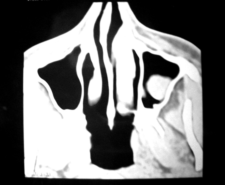
A 25-year-old male presented with a complaint of intermittent localized pain over the left cheek for the past nine months. It was not associated with fever or nasal discharge. There was no history of trauma. On examination, the left cheek was normal and on palpation there was no tenderness. Examination of the nose showed anterior deviation of the nasal septum to the right and normal nasal mucosa. On palpation paranasal sinuses were non-tender. Digital X-ray of the paranasal sinuses showed a bony mass in the left maxillary antrum. CT scan revealed a pedunculated bony mass arising from the lateral wall of the maxillary antrum (Fig. 1). Routine blood and urine examinations were within normal limits. Under anaesthesia bony mass arising from the lateral wall was removed via a Caldwell- Luc approach. The resected specimen was submitted for histopathological evaluation and diagnosed as osteoma (Fig. 2). The postoperative period was uneventful. Following surgery, the patient referred relief from cheek pain. One year after surgery, digital X-ray of paranasal sinuses did not show any recurrence of growth.
Fig. 1.
CT scan showing a bony mass in the left maxillary antrum.
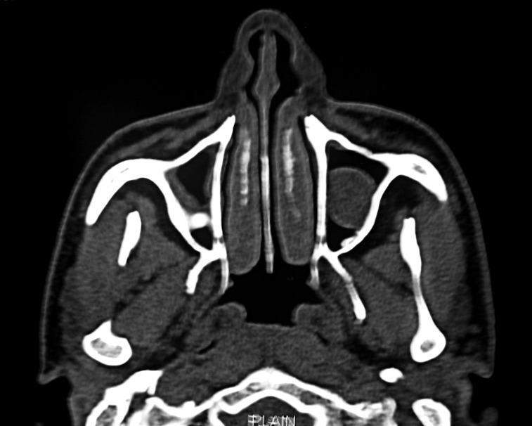
Fig. 2.
CT scan showing an osteoma and mucosal thickening in the right maxillary antrum and polypoidal mass in the left maxillary antrum.
Case 2
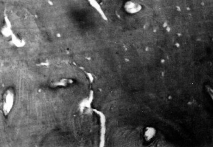
A 40-year-old male was referred to our specialty clinic for treatment of chronic bilateral sinusitis. The patient had recurrent bilateral nasal discharge and headache for the past nine months, and did not have relief from antibiotic treatment. Nasal examination showed a thick yellow bilateral nasal discharge with congested nasal mucosa. Postnasal discharge was present. On palpation nasal sinuses were nontender. CT scan showed a polypoidal soft tissue mass in the left maxillary antrum. In the right maxillary antrum, thickened mucosa and a small bony mass were present in the lateral wall (Fig. 3). Routine blood and urine examinations were within normal limits. The patient underwent endoscopic sinus surgery consisting in bilateral middle meatus antrostomy. The polypoidal mass was removed from the left maxillary antrum, and the thickened mucosa with the bony mass was removed from the right maxillary antrum. Histopathological examination of the removed masses confirmed the diagnosis of inflammatory polyp and osteoma. Following surgery, the patient had relief from nasal discharge and has not had any recurrence after one year.
Fig. 3.
Photomicrograph showing dense bone (H&E high power).
Discussion
Osteomas are the most common fibro-osseous lesions in the paranasal sinuses 5. They may be classified as peripheral, central or extraskeletal 6-9. Peripheral osteoma occurs mainly in the head and neck region 6. Sinus osteomas are infrequent, with an incidence of 0.43%, and are seen in up to 3% of sinus CT series 5. Other reported sites of osteoma formation in the skull include the external ear canal, orbital bones, temporal bone, pterygoid plates, mandible, sphenoid and occipital bones 5 10 11.
The pathogenesis of osteoma remains controversial. There are three accepted theories of the aetiology of paranasal sinus osteoma: developmental, traumatic and infectious. No single theory adequately explains all osteomas 5. Varboncover et al. 12 hypothesized that osteomas arise either from embryonic cartilaginous remnants or from a persistent embryological periosteum. The inflammatory theory suggests that chronic inflammations of the paranasal sinuses stimulate the proliferation of the periosteum-related osteogenetic cells 6 13 14. It has been suggested that osteoma is a product of a post-traumatic or post-inflammatory process. A possible aetiological factor includes the stimulation of embryologic cartilaginous remnants 15. Kaplan et al. 16 suggested that a combination of trauma and muscle traction may play a role in its development. Histopathological appearance includes abnormal bone structure, dense compact bone and the absence of Haversian systems 6.
Osteomas can occur at any age, but are more common in young adults. According to some authors, they are more common in males 16, whereas others report that they are more common in females 9 17. The maxillary sinus is involved in less than 2% of all cases, usually on the lateral wall of the sinus 2. Both of our patients were male and the osteoma arose from the lateral wall.
Many patients diagnosed with an osteoma of the paranasal sinuses are asymptomatic. They are discovered incidentally during radiographic evaluation for unrelated problems such as minor head trauma 5. The majority of osteomas are asymptomatic at an early stage and usually found on routine radiological examination 4. Delay in diagnosis has been attributed to the fact that these lesions are asymptomatic when they are small 1.
The clinical signs, symptoms and complications depend on the location, size and growth direction of the lesion 4. Symptoms related directly to an osteoma generally arise from a "mass effect" as the lesion impinges on normal structures 5.
Maxillary sinus osteomas are slow growing and usually asymptomatic, but they may be symptomatic depending on the location and onset 15 18. Thus, an anterior extension may lead to facial deformity. Continued growth may completely obstruct the sinus ostia or nasal cavity and lead to the development of mucoceles 1 4. Presenting symptoms are pain, swelling, sinusitis and nasal discharge. Rarely, they may expand into the orbit causing diplopia, ptosis and decreased visual acuity 15 18. Complications arise when an osteoma has grown large enough to impinge on surrounding structures. One of our patients had chronic sinusitis and the other had intermittent pain over the left cheek.
Pain may or may not be related to an osteoma, especially if the location of the pain and the osteoma are not congruent 5. When a patient complains of headache in the vicinity of an osteoma, and other pathologies leading to headache has been ruled out, excision is indicated 19. One of our patients had pain over the left cheek that was relieved after surgical excision of the osteoma.
Osteomas may be solitary or multiple. Multiple osteomas of the facial skeleton may occur in cases of Gardner syndrome. This autosomal dominant disorder is characterized by intestinal polyposis, multiple osteomas, cutaneous fibromas, epidermal cysts and impacted teeth 20. No such lesions were found in our patients.
Diagnosis and evaluation of the extension of the tumour in all three dimensions can be achieved with radiographs and CT 21, although the latter is more precise in delineating an osteoma. They appear as circumscribed dense masses attached to the originating bone by either a broad- or narrow-based pedicle 5. The surrounding bone is normal and does not have a lytic or moth-eaten appearance. Even for extensive osteomas, the surrounding bone is thinned and moved by pressure rather than by direct invasion 5. Magnetic resonance imaging may be used to establish the condition of adjacent soft tissues and mucoperiosteum of the sinus 21.
Asymptomatic osteomas may not require intervention, while symptomatic osteomas definitely require surgical excision 1. Since most osteomas are asymptomatic, many investigators advocate periodic imaging to follow their growth and intervene before the development of complications 22. Osteomas that do not cause symptoms, but are shown in serial radiographs to be fast growing and have the likelihood of producing symptoms in the future may require removal 23. Koivunen et al. 18 noted that paranasal sinus osteomas should be removed if the lesion fills 50% of the volume of the sinus.
Indications for surgical treatment include serious cosmetic disfigurement, limitation or loss of function, significant growth rate or need for definitive histopathological diagnosis 6. Treatment of osteoma consists of complete surgical removal at the base where it unites with the cortical bone 24. The surgical procedure depends on the location, extent and existing complications 4. There are no reports of osteomas undergoing malignant transformation 6 24.
Both of our patients had symptomatic osteomas. In the first case, the osteoma was large in size and was removed by a Caldwell-Luc procedure. It is difficult to remove large osteomas endoscopically, and they are best excised with the procedure we used. In the second case, the patient had bilateral chronic maxillary sinusitis and unilateral osteoma. Bilateral endoscopic middle meatus antrostomy was performed, and the osteoma was removed from the right maxillary antrum.
Conclusions
Maxillary sinus osteomas are benign osteogenic lesions. Symptomatic osteomas require surgical intervention. The type of surgical procedure depends on the location and extent of the osteoma as well as existing complications. Small osteomas can be removed endoscopically, while large osteomas require Caldwell-Luc surgery.
References
- 1.Mesolella M, Galli V, Testa D. Inferior turbinate osteoma: A rare cause of nasal obstruction. Otolaryngol Head Neck Surg. 2005;133:989–991. doi: 10.1016/j.otohns.2005.03.045. [DOI] [PubMed] [Google Scholar]
- 2.Zouloumis L, Lazaridis N, Maria P, et al. Osteoma of the ethmoidal sinus: a rare case of recurrence. Br J Oral Maxillofac Surg. 2005;43:520–522. doi: 10.1016/j.bjoms.2005.01.014. [DOI] [PubMed] [Google Scholar]
- 3.Gillman GS, Lampe HB, Allen LH. Orbito ethmoid osteoma: case report of an uncommon presentation of an uncommon tumor. Otolaryngol Neck Surg. 1997;117:218–220. doi: 10.1016/S0194-59989770107-4. [DOI] [PubMed] [Google Scholar]
- 4.Lin C, Lin Y, Kang B. Middle turbinate osteoma presenting with ipsilateral facial pain, epiphora and nasal obstruction. Otolaryngol Head Neck Surg. 2003;128:282–283. doi: 10.1067/mhn.2003.29. [DOI] [PubMed] [Google Scholar]
- 5.Eller R, Sillers M. Common fibro-osseous lesions of the paranasal sinuses. Otolaryngol Clin North Am. 2006;39:585–600. doi: 10.1016/j.otc.2006.01.013. [DOI] [PubMed] [Google Scholar]
- 6.Stelios D, Christos B, Ioannis T. Peripheral osteoma of the maxilla: Report of an unusual case. Oral Surg Oral Med Oral Pathol Oral Radiol Endod. 2005;100:E19–E24. doi: 10.1016/j.tripleo.2005.03.011. [DOI] [PubMed] [Google Scholar]
- 7.Bodner L, Gatot A, Sion-Vardy N, et al. Peripheral osteoma of the mandibular ascending ramus. J Oral Maxillofac Surg. 1998;56:1446–1449. doi: 10.1016/s0278-2391(98)90414-1. [DOI] [PubMed] [Google Scholar]
- 8.Ertas U, Tozoglu S. Uncommon peripheral osteoma of the mandible:report of cases. J Contemp Dent Pract. 2003;4:98–104. [PubMed] [Google Scholar]
- 9.Longo F, Califano L, Maria G, et al. Solitary osteoma of the mandibular ramus: report of a case. J Oral Maxillofac Surg. 2001;59:698–700. doi: 10.1053/joms.2001.23408. [DOI] [PubMed] [Google Scholar]
- 10.Sayan NB, Ucok C, Karasu HA, et al. Peripheral osteoma of the oral and maxillofacial region: a study of 35 new cases. J Oral Maxillofac Surg. 2002;60:1299–1301. doi: 10.1053/joms.2002.35727. [DOI] [PubMed] [Google Scholar]
- 11.Viswanatha B. A case of osteoma with cholesteatoma of the external auditory canal and cerebellar abscess. Int J Pediatr Otolaryngol Extra. 2007;2:34–39. [Google Scholar]
- 12.Varboncoeur AP, Vanbelois HJ, Bowen LL. Osteoma of the maxillary sinus. J Oral Maxillofac Surg. 1990;48:882–883. doi: 10.1016/0278-2391(90)90351-2. [DOI] [PubMed] [Google Scholar]
- 13.Sugiyama M, Suei Y, Takata T, et al. Radiopaque mass at the mandibular ramus. J Oral Maxillofac Surg. 2001;59:1211–1214. doi: 10.1053/joms.2001.26727. [DOI] [PubMed] [Google Scholar]
- 14.Seward MHE. An osteoma of the maxilla. Br Dent J. 1965;5:27–30. [PubMed] [Google Scholar]
- 15.Park W, Kim HS. Osteoma of the maxillary sinus: case report. Oral Surg Oral Med Oral Pathol Oral Radiol Endod. 2006;102:e26–e27. doi: 10.1016/j.tripleo.2006.05.005. [DOI] [PubMed] [Google Scholar]
- 16.Kaplan I, Calderon S, Buchner A. Peripheral osteoma of the mandible: a study of 10 new cases and analysis of literature. J Oral Maxillo Fac Surg. 1994;52:467–470. doi: 10.1016/0278-2391(94)90342-5. [DOI] [PubMed] [Google Scholar]
- 17.Denia A, Perez F, Canalis R, et al. Extracanalicular osteoma of the temporal bone. Arch Otolaryngol. 1979;105:706–709. doi: 10.1001/archotol.1979.00790240020005. [DOI] [PubMed] [Google Scholar]
- 18.Koivunen P, Lopponen H, Fors AP, et al. The growth rate of osteomas of paranasal sinuses. Clin Otolaryngol Allied Sci. 1997;22:111–114. doi: 10.1046/j.1365-2273.1997.00869.x. [DOI] [PubMed] [Google Scholar]
- 19.Schick B, Steigerwald C, el Rahman el Tahan A, et al. The role of endonasal surgery in the management of frontoethmoidal osteomas. Rhinology. 2001;39:66–70. [PubMed] [Google Scholar]
- 20.Payne M, Anderson JA, Cook J. Gardner's syndrome – a case report. Br Dent J. 2002;193:383–384. doi: 10.1038/sj.bdj.4801571. [DOI] [PubMed] [Google Scholar]
- 21.Theodorou DJ, Theodorou SJ, Sartoris DJ. Primary nonodontogenic tumors of the jawbones: an overview of essential radiographic findings. Clin Imaging. 2003;27:59–70. doi: 10.1016/s0899-7071(02)00518-1. [DOI] [PubMed] [Google Scholar]
- 22.Johnson D, Tan L. Intraparenchymal tension pneumatocele complicating frontal sinus osteoma: case report. Neurosurgery. 2002;50:878–879. doi: 10.1097/00006123-200204000-00038. [DOI] [PubMed] [Google Scholar]
- 23.Namdar I, Edelstein DR, Huo J, et al. Management of osteomas of the paranasal sinuses. Am J Rhinol. 1998;12:393–398. doi: 10.2500/105065898780707955. [DOI] [PubMed] [Google Scholar]
- 24.Nejat BS, Cahit U, Hakan AK, et al. Peripheral osteoma of the oral and maxillofacial region: A study of 35 new cases. J Oral Maxillofac Surg. 2002;60:1299–1301. doi: 10.1053/joms.2002.35727. [DOI] [PubMed] [Google Scholar]


