

Abstract
This article presents the novel method of training of creating cartilage framework for total ear reconstruction in microtia. Replica of costal cartilage harvested for real surgery was simulated by silicon dental impression material. Carving of framework was done with wood carving instruments. Silicon dental impression material gives the consistency and texture almost comparable to real costal cartilage. Sequential steps similar to actual surgery were simulated to create the three-dimensional framework.
By using this novel technique, novice surgeons can practice creating ear framework and improvise their results in the actual surgery.
KEY WORDS: Cartilage framework, microtia, silicon ear model
INTRODUCTION
Creating the ear framework for total auricular reconstruction is technically challenging procedure. For novice surgeons it becomes difficult and time consuming. So, a new technique is introduced to practice creating ear framework with silicon impression material. By this one can have the idea of sculpting the real costal cartilage.
MATERIALS AND METHODS
To start with we will require following materials and armamentarium:
Impression Compound [Figure 1]
Silicon Impression Material
5/0 Stainless Steel Sutures (Ear Set, Steelex, B/BRAUN) [Figure 2]
Wood Carving Instruments [Figure 3]
Thin Plastic Sheet - to prepare template
Rapid Cure Acrylic Material
Modeling Wax
Blue and Red Marker
Glass Slab
Figure 1.
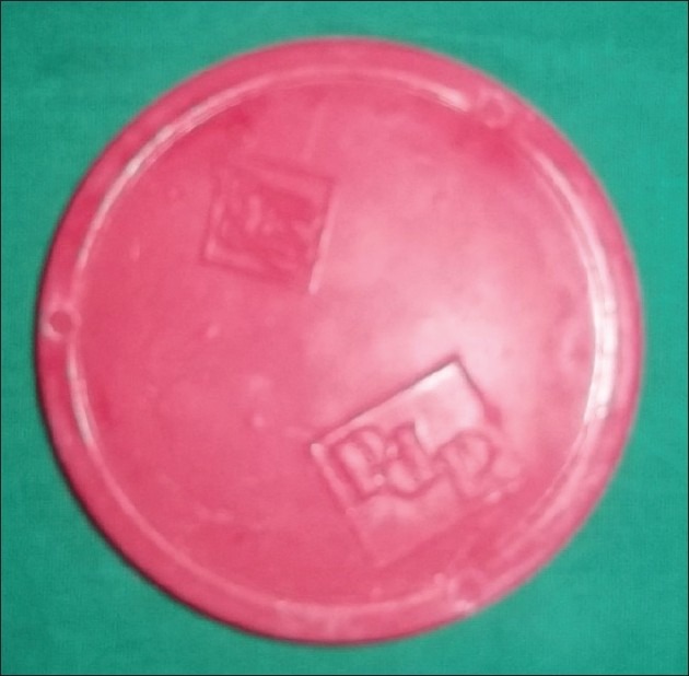
Impression compound cake
Figure 2.
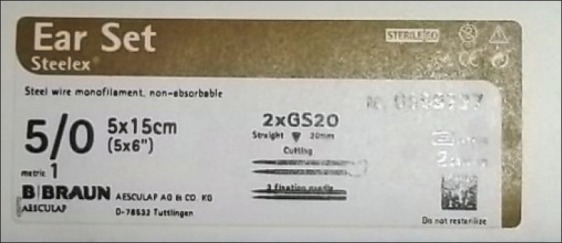
5/0 Stainless Steel Sutures(Ear Set, Steelex, B/BRAUN)
Figure 3.
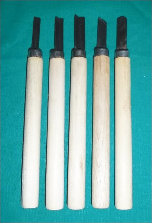
Wood carving instruments
Preparation of silicon rib model
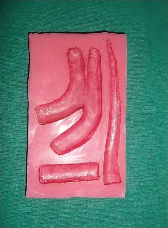
With the help of impression compound, model of costal cartilage harvested for actual surgery is prepared. This model is positive replica of sixth, seventh and eighth costal cartilage [Figure 4]. As this is for training we have made an extra small piece of cartilage. Then a mould is prepared with rapid cure acrylic, which is negative replica of this model, on a glass slab [Figure 5]. Silicon impression material is used to take the impression using the mould, which yields positive replica of sixth, seventh and eighth costal cartilage [Figure 6]. This silicon rib model is now used for sculpting ear framework.
Figure 4.
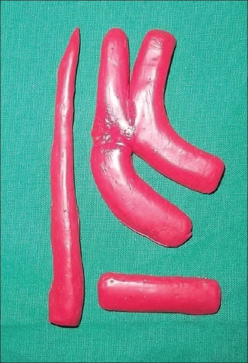
3-D model of harvested costal cartilage prepared from impression compound cake
Figure 5.
Mould prepared with rapid cure acrylic for making silicon cartilage model
Figure 6.
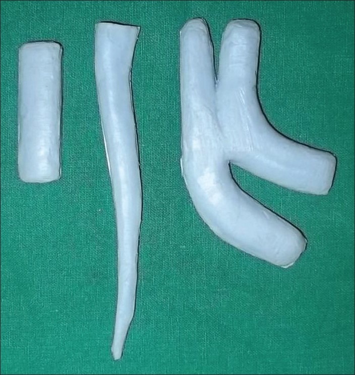
Silicon model of harvested costal cartilage
Preparation of surgical template
With the help of thin plastic sheet, surgical template is prepared keeping in mind the design of auricular cartilage. In case of unilateral microtia, a template is made following opposite ear's anatomy [Figure 7].
Figure 7.
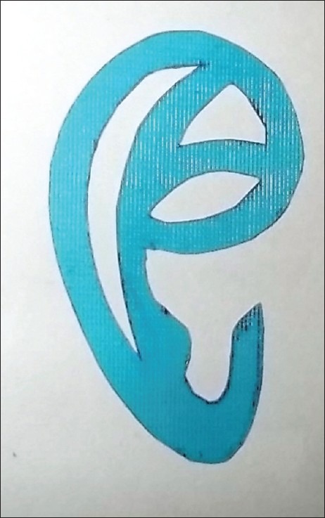
Surgical template
Sculpting ear framework
Sculpting a 3D model of ear cartilage is initiated with marking the base frame. This is done with placing a surgical template[1–5] on sixth and seventh rib cartilage in such a way that whole of helical rim is placed on the cartilage except crus helicis [Figure 8]. Now the base frame is cut with scalpel and no. 15 blade. Then scaphoid fossa, fossa triangularis and concha are carved in the base frame with the help of wood carving instruments [Figure 9]. Helical rim and antihelix portion are sculpted from eighth costal cartilage [Figure 10]. Helical rim and antihelix are now fixed to base frame with the help of stainless steel sutures in a way that tags end in the posterior portion of the framework. Now the tragus portion is carved from the remaining pieces of sixth and seventh cartilage and fixed to base frame. After this all the edges of framework should be rounded so that final framework should not look ‘boxy type’ and all suture tags submerged in framework to prevent extrusion. With this a 3D ear cartilage framework is ready for placement [Figure 11].
Figure 8.
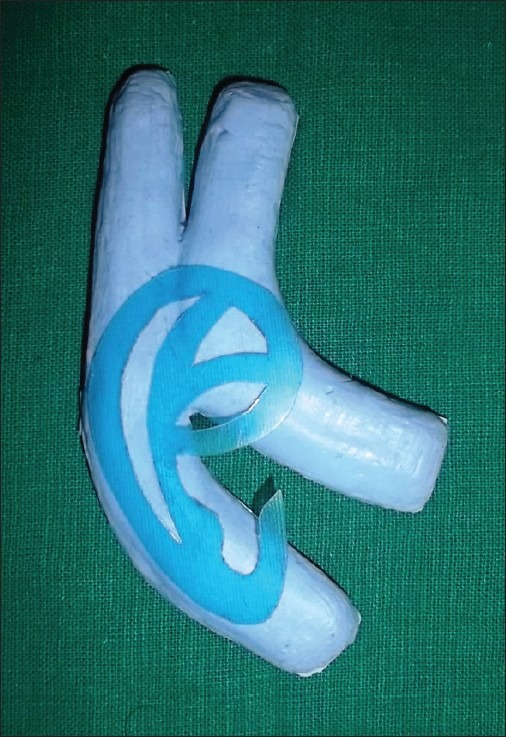
Placement of surgical template on sixth and seventh costal cartilage in such a way that whole helical rim is located on base frame
Figure 9.
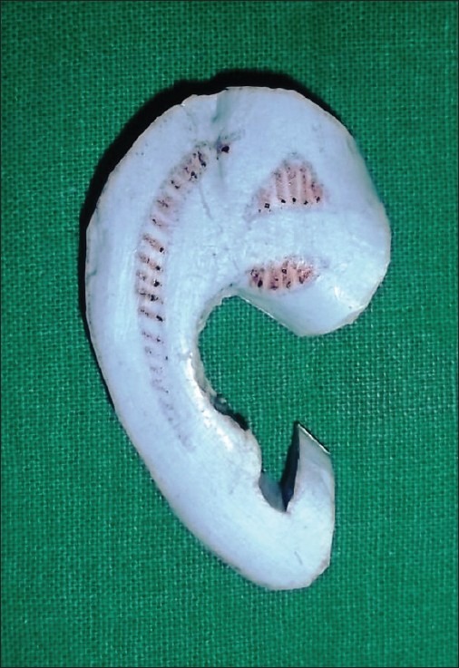
Marked area's for scaphoid fossa, fossa triangularis and concha- to be carved
Figure 10.
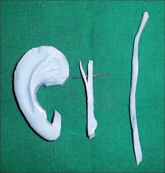
Base frame with carved scaphoid fossa, fossa triangularis and concha and helical rim and antihelix
Figure 11.
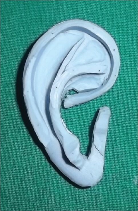
Completed 3-dimensional model of ear cartilage
RESULTS
The technique was successful in simulating the ear framework, and the consistency of the silicon material with relatively blunt carving instruments was almost similar to real costal cartilage with sharp instruments.
Hence with this technique one can access the difficulties that may be encountered in real surgery and may reduce surgical time.
DISCUSSION
A total ear reconstruction with autogenous tissues is one of the greatest technical challenges to a reconstructive surgeon. Major part of difficulty in surgery is creating a 3D cartilage framework out of costal cartilage. This difficulty lies in the fact that costal cartilage structure is different in each individual. Differences even lie between children and adults. So, novice surgeons might find it difficult and time consuming to locate future parts of 3D ear in costal cartilage.
It was recommended by Brent[6] to practice framework creation in cadaver's in volume. But due to less availability of cadavers, it is practically difficult for surgeon to practice on it. So, Brent used potatoes and carrots for practicing cartilage framework designing.[7] Advantages of this technique are 1) Easy availability and 2) low cost. But the technique had disadvantage that consistency and configuration of potatoes and carrots was far different from costal cartilage.
To sculpt the framework in one piece from resin block was suggested by Chen.[8] However this practice does not help novice surgeons carving 3D model from costal cartilage.
Akira Yamada et al[9] suggested technique for preparing silicone rib model by taking impression of harvested costal cartilage and pouring with silicon impression material and fabricating framework with exactly same instruments that were used for real surgery. But taking impression of harvested cartilage might cause contamination of it. Silicon model is less stiff than real costal cartilage. So, working with surgical instruments on silicone model will not simulate real surgery.
Considering these challenges, we herewith present a technique which will be helpful for teaching novice surgeons, and large number of silicone models can be made from a single mould which can be helpful in teaching in large number like in training workshops. Practicing with less sharp instruments on silicon model will simulate working with surgical instruments on costal cartilage. We believe that our method will help novice surgeons to create framework that will simulate real surgery in a cost-effective way.
CONCLUSION
By using this novel technique, novice surgeons can practice creating ear framework and improvise their results in the actual surgery.
Footnotes
Source of Support: Nil
Conflict of Interest: None declared.
REFERENCES
- 1.Nagata S. Chapter 66: microtia (auricular reconstruction) In: Achauer M, Eriksson E, editors. Plastic Surgery: Indications, Operations, Outcomes. Vol. 2. St Louis MO: Mosby; 2000. pp. 1023–56. [Google Scholar]
- 2.Nagata S. New method for total reconstruction of the auricle for microtia. Plast Reconstr Surg. 1993;92:187–201. doi: 10.1097/00006534-199308000-00001. [DOI] [PubMed] [Google Scholar]
- 3.Nagata S. The modification stages involved in the total reconstruction of the auricle: part I. The modification in the grafting of the three-dimensional costal cartilage framework (3-D frame) for the lobule type microtia. Plast Reconstr Surg. 1994;93:221–30. [PubMed] [Google Scholar]
- 4.Nagata S. The modification stages involved in the total reconstruction of the auricle: part II.The modification in the grafting of the three-dimensional costal cartilage framework (3-D frame) for the concha type microtia. Plast Reconstr Surg. 1994;93:231–42. [PubMed] [Google Scholar]
- 5.Nagata S. The modification stages involved in the total reconstruction of the auricle: part III. The modification in the grafting of the three-dimensional costal cartilage framework (3-D frame) for the small concha type microtia. Plast Reconstr Surg. 1994;93:243–53. [PubMed] [Google Scholar]
- 6.Brent BD. Reconstruction of the auricle. In: Mathes S, editor. Plastic Surgery. 2nd ed. Vol. 3. Philadelphia, PA: Saunders Elsevier; 2006. pp. 633–98. [Google Scholar]
- 7.UT Southwestern Medical Center. [last Accessed on 2009 Apr 08]. Available from: http://www.utsouthwestern.edu/utsw/cda/dept16543/files/93303.html .
- 8.Chen ZC, Chen PK, Hung KF, Lo LJ, Chen YR. Microtia reconstruction with adjuvant 3-dimensional template model. Ann Plast Surg. 2004;53:282–7. doi: 10.1097/01.sap.0000106434.69246.29. [DOI] [PubMed] [Google Scholar]
- 9.Yamada A, Imai K, Fujimoto T, Morimoto K, Niitsuma K, Matsumoto H. New Training Method of Creating Ear Framework byUsing Precise Copy of Costal Cartilage. J Craniofac Surg. 2009;20:899–902. doi: 10.1097/scs.0b013e3181a2ef97. [DOI] [PubMed] [Google Scholar]